Berg J.M., Tymoczko J.L., Stryer L. Biochemistry
Подождите немного. Документ загружается.


subsequent binding of β-arrestin.
II. Transducing and Storing Energy 15. Signal-Transduction Pathways: An Introduction to Information Metabolism
15.2. The Hydrolysis of Phosphatidyl Inositol Bisphosphate by Phospholipase C
Generates Two Messengers
Cyclic AMP is not the only second messenger employed by 7TM receptors and the G proteins. We turn now to another
ubiquitous second-messenger cascade that is used by many hormones to evoke a variety of responses. The
phosphoinositide cascade, like the adenylate cyclase cascade, converts extracellular signals into intracellular ones. The
intracellular messengers formed by activation of this pathway arise from the cleavage phosphatidyl inositol 4,5-
bisphosphate (PIP
2
), a phospholipid present in cell membranes. The binding of a hormone such as vasopressin to a 7TM
receptor leads to the activation of the β isoform of phospholipase C. The G
α
protein that activates phospholipase C is
called G
α
q
. The activated enzyme then hydrolyzes the phosphodiester bond linking the phosphorylated inositol unit to
the acylated glycerol moiety. The cleavage of PIP
2
produces two messengers: inositol 1,4,5-trisphosphate, a soluble
molecule that can diffuse from the membrane, and diacylglycerol, which stays in the membrane.
Comparison of the amino acid sequences of different isoforms of phospholipase C as well as examination of the known
three-dimensional structures of phospholipase components reveal an intriguing modular structure (Figure 15.11).
This analysis reveals the basis for both phospholipase enzymatic activity and its regulation by signal-transduction
pathways. The catalytic core of these enzymes has an α β barrel structure similar to the catalytic core of triose phosphate
isomerase and other enzymes (Section 16.1.4). This domain is flanked by domains that interact with membrane
components. At the amino terminus is a pleckstrin homology (PH) domain. This ~120-residue domain binds a lipid head
group such as that of PIP
2
(Figure 15.12). The PH domain is joined to the catalytic domain by a set of four EF-hand
domains. Although EF-hand domains often take part in calcium-binding (Section 15.3.2), the EF-hand domains of
phospholipase C lack many of the calcium-binding residues. On the carboxyl-terminal side of the catalytic domain is a
C2 domain (for protein kinase C domain 2). This ~130-residue domain is a member of the immunoglobulin domain
superfamily (Chapter 33) and plays a role in binding phospholipid headgroups. Such interactions, often but not always,
require the presence of bound calcium ions.
The binding of a G protein brings the enzyme into a catalytically active position. The β isoform of phospholipase C has
an additional domain at its carboxyl terminus
a domain that interacts with the α subunit of G
q
in its GTP form.
Because this G protein is linked to the membrane by its fatty acid anchor, this interaction helps pull the β isoform of
phospholipase to the membrane. This interaction acts in concert with the binding of the PH and C2 domains of
phospholipase C to membrane components to bring the active site in the catalytic core into a position against a
membrane surface that is favorable for cleavage of the phosphodiester bond of PIP
2
(Figure 15.13). Some of these
interactions and the enzymatic reaction itself also depend on the presence of calcium ion. Phospholipase isoforms that
lack the carboxyl-terminal regulatory domain do not respond to these signal-transduction pathways. The two products of
the cleavage reaction, inositol 1,4,5-trisphosphate and diacylglycerol, each trigger additional steps in the signal-

transduction cascades.
15.2.1. Inositol 1,4,5-trisphosphate Opens Channels to Release Calcium Ions from
Intracellular Stores
What are the biochemical effects of the second messenger inositol 1,4,5-trisphosphate? These effects were delineated by
microinjecting IP
3
molecules into cells or by allowing IP
3
molecules to diffuse into cells whose plasma membranes had
been made permeable. Michael Berridge and coworkers found that IP
3
causes the rapid release of Ca
2
+
from
intracellular stores
the endoplasmic reticulum and, in smooth muscle cells, the sarcoplasmic reticulum. The elevated
level of Ca
2
+
in the cytosol then triggers processes such as smooth muscle contraction, glycogen breakdown, and
vesicle release. In Xenopus oocytes, the injection of IP
3
suffices to activate many of the early events of fertilization.
Inositol 1,4,5-trisphosphate is able to increase Ca
2+
concentration by associating with a membrane protein called the IP
3
-gated channel or IP
3
receptor. This receptor, which is composed of four large, identical subunits, forms an ion channel.
The ion-conducting channel itself is likely similar to the structurally characterized K
+
channel (Section 13.5.6). At least
three molecules of IP
3
must bind to sites on the cytosolic side of the membrane protein to open the channel and release
Ca
2+
. The highly cooperative opening of calcium channels by nanomolar concentrations of IP
3
enables cells to detect
and amplify very small changes in the concentration of this messenger. The cooperativity of IP
3
binding and channel
opening is another example of the role of allosteric interactions.
How is the IP
3
-initiated signal turned off? Inositol 1,4,5-trisphosphate is a short-lived messenger because it is rapidly
converted into derivatives that do not open the channel (Figure 15.14). Its lifetime in most cells is less than a few
seconds. Inositol 1,4,5-trisphosphate can be degraded to inositol by the sequential action of phosphatases or it can be
phosphorylated to inositol 1,3,4,5-tetrakisphosphate, which is then converted into inositol by an alternative route.
Lithium ion, widely used to treat bipolar affective disorder, may act by inhibiting the recycling of inositol 1,3,4-
trisphosphate.
15.2.2. Diacylglycerol Activates Protein Kinase C, Which Phosphorylates Many Target
Proteins
Diacylglycerol, the other molecule formed by the receptor-triggered hydrolysis of PIP
2
, also is a second messenger and
it, too, activates a wide array of targets. We will examine how diacylglycerol activates protein kinase C (PKC), a protein
kinase that phosphorylates serine and threonine residues in many target proteins. The amino acid sequences and the
results of three-dimensional structural studies of protein kinase C isozymes reveal an elegant modular architecture
(Figure 15.15). The α, β, and γ isozymes of protein kinase C have in common a structurally similar catalytic domain,
homologous to that in PKA, at the carboxyl terminus. Adjacent to the catalytic domain is a C2 domain that is related to
the C2 domain of phospholipase C and that interacts with membrane phospholipids. On the other side of the C2 domain
is a pair of C1 domains, each organized around two bound zinc ions. These two domains, particularly the second (C1B)
domain, bind diacylglycerol (Figure 15.16A). Finally, at the amino terminus is a sequence of the form -A-R-K-G-A-L-R-
Q-K-. This sequence is striking because the consensus sequence for PKC substrates is X-R-X-X-(S,T)-Hyd-R-X in
which Hyd refers to a large, hydrophobic residue. This sequence from PKC is referred to as a pseudosubstrate sequence
because it resembles the substrate sequence except that it has an alanine residue in place of the serine or threonine
residue; so it cannot be phosphorylated. Recall that an analogous pseudosubstrate sequence is present in the regulatory
chain of PKA and that it prevents activity by occluding the active site in the R
2
C
2
tetramer of PKA (Section 10.4.2).
Similarly, the pseudosubstrate sequence of PKC binds to that enzyme's active site and prevents substrate binding.
With this structure in mind, we can now understand how PKC is activated on PIP
2
hydrolysis (Figure 15.16B). Before
activation, PKC is free in solution. On PIP
2
hydrolysis in the membrane by phospholipase C, the C1B domain of PKC
binds to diacylglycerol. This binding and the interaction of the C2 domain with membrane phospholipids anchors the
enzyme to the membrane. The interaction between the C2 domain and the phospholipids, especially phosphatidyl serine,
requires calcium ions. The binding of the C1A domain to diacylglycerol pulls the pseudosubstrate out of the active site;
the released pseudosubstrate, which is quite positively charged, probably interacts with the negatively charged
membrane surface as well. The result of the conformational transitions is the binding of PKC to diacylglycerol-rich
regions of the membrane in an active state, ready to phosphorylate serine and threonine residues in appropriate sequence
contexts. Note that diacylglycerol and IP
3
work in tandem: IP
3
increases the Ca
2+
concentration, and Ca
2+
facilitates
PKC activation as well as phospholipase C activity.
Diacylglycerol, like IP
3
, acts transiently because it is rapidly metabolized. It can be phosphorylated to phosphatidate
(Section 26.1) or it can be hydrolyzed to glycerol and its constituent fatty acids (Figure 15.17). Arachidonate, the C
20
polyunsaturated fatty acid that usually occupies the position 2 on the glycerol moiety of PIP
2
, is the precursor of a series
of 20-carbon hormones, including the prostaglandins (Section 22.6.2). Thus, the phosphoinositide pathway gives rise to
many molecules that have signaling roles.
II. Transducing and Storing Energy 15. Signal-Transduction Pathways: An Introduction to Information Metabolism 15.2. The Hydrolysis of Phosphatidyl Inositol Bisphosphate by Phospholipase C Generates Two Messengers
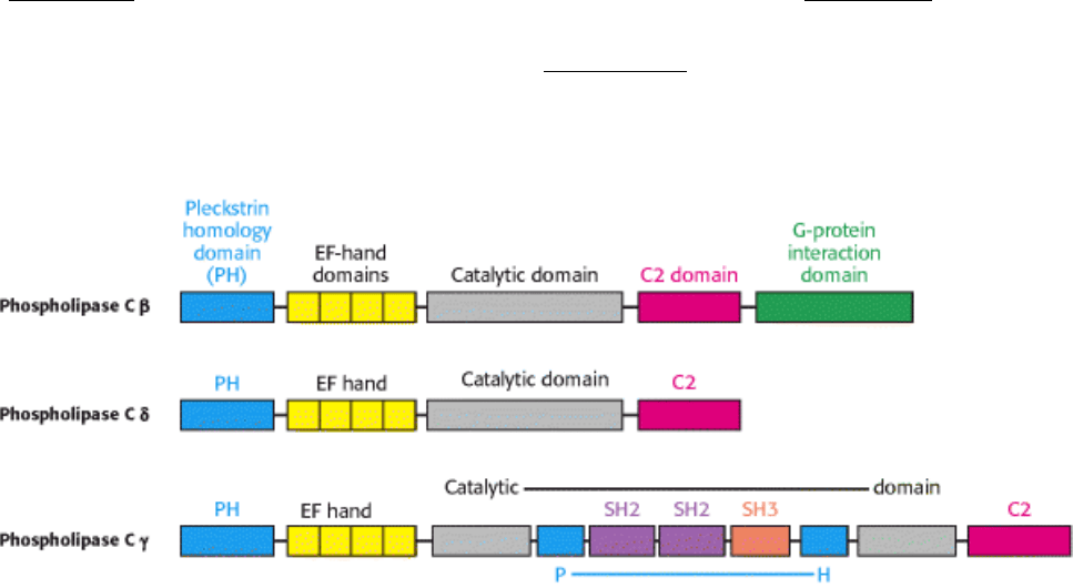
Figure 15.11. Modular Structure of Phospholipase C. The domain structures of three isoforms of phospholipase C
reveal similarities and differences among the isoforms. Only the β isoform, with the G-protein-binding domain, can be
stimulated directly by G proteins. For phospholipase Cγ, the insertion of two SH2 (Src homology 2) domains and one
SH3 (Src homology 3) domain splits the catalytic domain and a PH domain into two parts.
II. Transducing and Storing Energy 15. Signal-Transduction Pathways: An Introduction to Information Metabolism 15.2. The Hydrolysis of Phosphatidyl Inositol Bisphosphate by Phospholipase C Generates Two Messengers
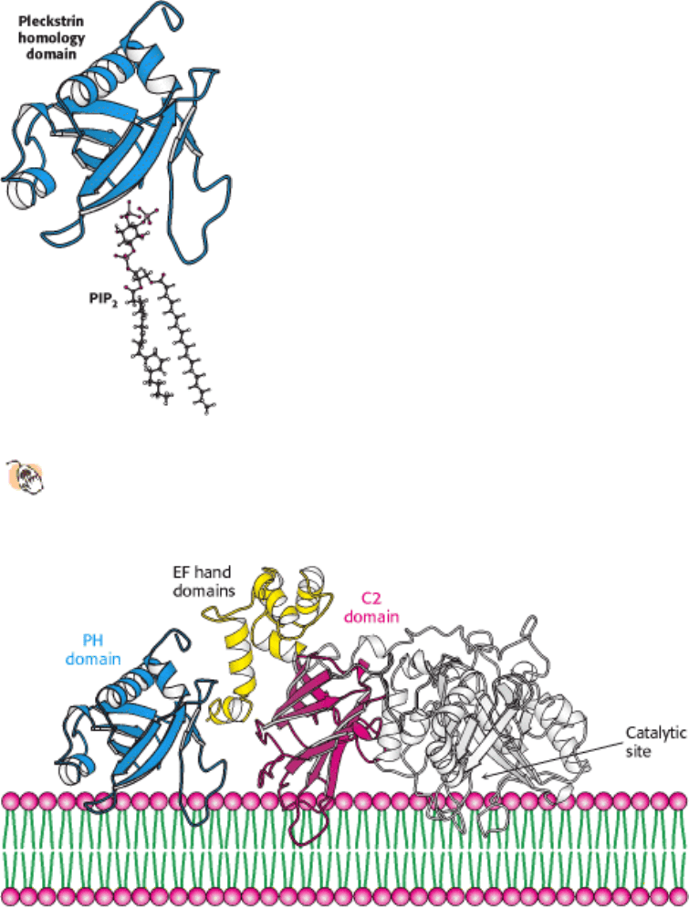
Figure 15.12. Pleckstrin Homology Domain.
PH domains facilitate the binding of proteins to membrane lipids,
particularly PIP
2
. In regard to phospholipase C, the PH domains help to localize the enzyme near its substrate,
PIP
2
.
II. Transducing and Storing Energy 15. Signal-Transduction Pathways: An Introduction to Information Metabolism 15.2. The Hydrolysis of Phosphatidyl Inositol Bisphosphate by Phospholipase C Generates Two Messengers
Figure 15.13. Phospholipase C Acts at the Membrane Surface. The PH and C2 domains of phospholipase help to
position the enzyme's catalytic site for ready access to the phosphodiester bond of the membrane lipid substrate, PIP
2
.
II. Transducing and Storing Energy 15. Signal-Transduction Pathways: An Introduction to Information Metabolism 15.2. The Hydrolysis of Phosphatidyl Inositol Bisphosphate by Phospholipase C Generates Two Messengers

Figure 15.14. Metabolism of IP
3
. The IP
3
signal is terminated by the metabolism of the compound into derivatives
lacking second-messenger capabilities.
II. Transducing and Storing Energy 15. Signal-Transduction Pathways: An Introduction to Information Metabolism 15.2. The Hydrolysis of Phosphatidyl Inositol Bisphosphate by Phospholipase C Generates Two Messengers
Figure 15.15. Modular Structure of Protein Kinase C. Seven isozymes of PKC can be divided into two classes on the
basis of their domain organization.

II. Transducing and Storing Energy 15. Signal-Transduction Pathways: An Introduction to Information Metabolism 15.2. The Hydrolysis of Phosphatidyl Inositol Bisphosphate by Phospholipase C Generates Two Messengers
Figure 15.16. Protein Kinase C Activation.
(A) The C1 domain of PKC, structurally organized around two bound zinc
ions, binds diacylglycerol. (B) When the C1 domains bind to diacylglycerol in the membrane, the pseudosubstrate
is pulled from the active site, permitting catalysis. Calcium-binding C2 domains help to localize PKC to the
membrane.
II. Transducing and Storing Energy 15. Signal-Transduction Pathways: An Introduction to Information Metabolism 15.2. The Hydrolysis of Phosphatidyl Inositol Bisphosphate by Phospholipase C Generates Two Messengers

Figure 15.17. Metabolism of Diacylglycerol. Diacylglycerol may be (1) phosphorylated to phosphatidate or (2)
hydrolyzed to glycerol and fatty acids.
II. Transducing and Storing Energy 15. Signal-Transduction Pathways: An Introduction to Information Metabolism
15.3. Calcium Ion Is a Ubiquitous Cytosolic Messenger
We have already seen that Ca
2+
is an important component of one signal-transduction circuit, the phosphoinositide
cascade. Indeed, Ca
2+
is itself an intracellular messenger in many eukaryotic signal-transducing pathways. Why is this
ion commonly found to mediate so many signaling processes? First, an apparent drawback is in fact an advantage:
calcium complexes of phosphorylated and carboxylated compounds are often insoluble, but such compounds are
fundamental to many biochemical processes in the cell. Consequently, the intracellular levels of Ca
2+
must be kept low
to prevent precipitation of these compounds. These low levels are maintained by transport systems for the extrusion of
Ca
2+
. In eukaryotic cells, two in particular the Ca
2+
ATPase (Section 13.2.1) and the sodium-calcium exchanger are
especially important. Because of their actions, the cytosolic level of Ca
2+
in unexcited cells is typically 100 nM, several
orders of magnitude lower than the concentration in the blood, which is approximately 5 mM. This steep concentration
gradient presents cells with a matchless opportunity: the cytosolic Ca
2
+
concentration can be abruptly raised for
signaling purposes by transiently opening calcium channels in the plasma membrane or in an intracellular membrane.
A second property of Ca
2+
that makes it a highly suitable intracellular messenger is that it can bind tightly to proteins
(Figure 15.18). Negatively charged oxygen atoms (from the side chains of glutamate and aspartate) and uncharged
oxygen atoms (main-chain carbonyl groups and side-chain oxygen atoms from glutamine and asparagine) bind well to
Ca
2+
. The capacity of Ca
2
+
to be coordinated to multiple ligands from six to eight oxygen atoms enables it to cross-
link different segments of a protein and induce significant conformational changes.
15.3.1. Ionophores Allow the Visualization of Changes in Calcium Concentration
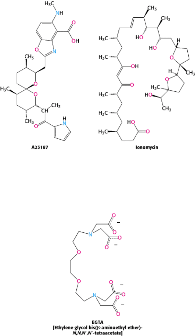
Our understanding of the role of calcium in cellular processes has been greatly enhanced by the use of calcium-specific
reagents. Ionophores such as A23187 and ionomycin can traverse a lipid bilayer because they have a hydrophobic
periphery.
They can be used to introduce Ca
2+
into cells and organelles. Many physiological responses that are normally triggered
by the binding of hormones to cell-surface receptors can also be elicited by using calcium ionophores to raise the
cytosolic calcium level. Conversely, the concentration of unbound calcium in a cell can be made very low (nanomolar or
less) by introducing a calcium-specific chelator such as EGTA. The concentration of free Ca
2+
in intact cells can be
monitored by using polycyclic chelators such as Fura-2.
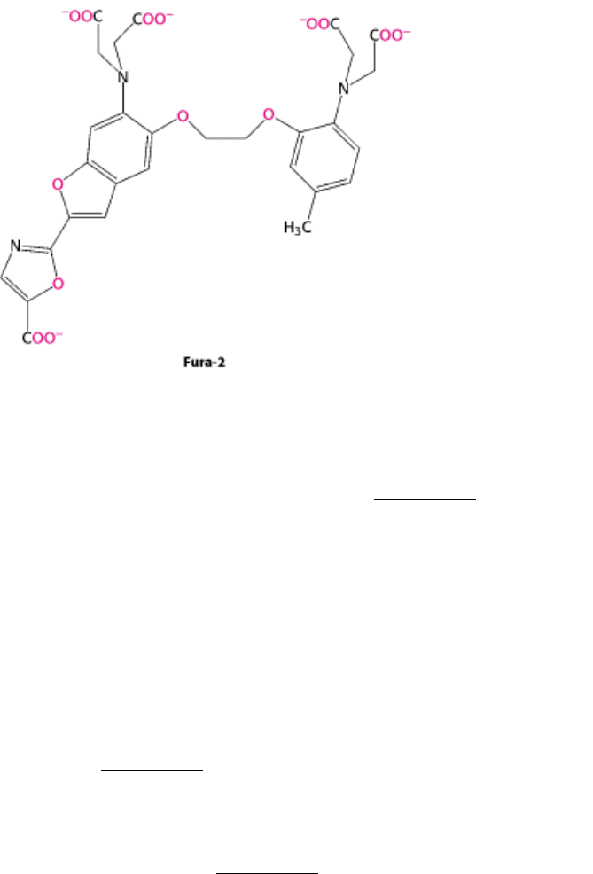
The fluorescence properties of this and related indicators change markedly when Ca
2+
is bound (Figure 15.19). These
compounds can be introduced into cells to allow measurement of the available Ca
2+
concentration in real time through
the use of fluorescence microscopy. Such methods allow the direct detection of calcium fluxes and diffusion within
living cells in response to the activation of specific signal-transduction pathways (Figure 15.20).
15.3.2. Calcium Activates the Regulatory Protein Calmodulin, Which Stimulates Many
Enzymes and Transporters
Calmodulin (CaM), a 17-kd protein with four calcium-binding sites, serves as a calcium sensor in nearly all eukaryotic
cells. Calmodulin is activated by the binding of Ca
2
+
when the cytosolic calcium level is raised above about 500 nM.
Calmodulin is a member of the EF-hand protein family. The EF hand is a Ca
2+
-binding motif that consists of a helix, a
loop, and a second helix. This motif, originally discovered in the protein parvalbumin, was named the EF hand because
helices designated E and F in parvalbumin form the calcium-binding motif and because the two helices are positioned
like the forefinger and thumb of the right hand (Figure 15.21). Seven oxygen atoms are coordinated to each Ca
2+
: six
from the protein and one from a bound water molecule.
Let us consider how calmodulin changes conformation in response to Ca
2+
binding and how these conformational
changes enable the calmodulin to interact with other proteins (Figure 15.22). In the absence of bound Ca
2+
, calmodulin
consists of two domains, each consisting of a pair of EF-hand motifs joined by a flexible helix. Many of the residues that
typically participate in calcium binding are on the surface of these domains, oriented in a manner inappropriate for Ca
2+
binding. On the binding of one Ca
2+
to each EF hand, these units change conformation: the Ca
2+
-binding sites turn
inward to bind the Ca
2+
, moving hydrophobic residues from the inside to the outside of the domains. These
conformational changes generate hydrophobic patches on the surface of each domain that are suitable for interacting
with other proteins.
The Ca
2
+
-calmodulin complex stimulates a wide array of enzymes, pumps, and other target proteins. Two targets are
especially noteworthy: one that propagates the signal and another that abrogates it. Calmodulin-dependent protein
kinases (CaM kinases) phosphorylate many different proteins. These enzymes regulate fuel metabolism, ionic
permeability, neurotransmitter synthesis, and neurotransmitter release. The binding of Ca
2+
-calmodulin to CaM kinase
activates the enzyme and enables it to phosphorylate target proteins. In addition, the activated enzyme phosphorylates
itself and is thus partly active even after Ca
2+
concentration falls and calmodulin is released from the kinase. In essence,
autophosphorylation of CaM kinase is the memory of a previous calcium pulse. The plasma membrane Ca
2
+
-ATPase
pump is another important target of Ca
2+
-calmodulin. Stimulation of the pump by Ca
2+
-calmodulin drives the calcium
level down to restore the lowcalcium basal state, thus helping to terminate the signal.

Are there structural features common to all calmodulin target proteins? Comparisons of the amino acid sequences of
calmodulin-binding domains of target proteins suggests that calmodulin recognizes positively charged, amphipathic α
helices. The results of structural studies fully support this conclusion and reveal the details of calmodulin-target
interactions (Figure 15.23). The two domains of calmodulin surround the amphipathic helix and are linked to it by
extensive hydrophobic and ionic interactions. The α helix linking the two EF hands folds back onto itself to facilitate
binding to the target helix. How does this binding of calmodulin to its target lead to enzyme activation? In regard to CaM
kinase I, calmodulin targets a peptide present at the carboxyl terminus of the kinase. This region interacts with a loop
that is crucial for ATP binding and holds it in a conformation inappropriate for ATP binding. Calmodulin surrounds this
peptide and extracts it, freeing the kinase active site to bind ATP and phosphorylate appropriate substrates.
II. Transducing and Storing Energy 15. Signal-Transduction Pathways: An Introduction to Information Metabolism 15.3. Calcium Ion Is a Ubiquitous Cytosolic Messenger
Figure 15.18. Mode of Binding of CA
2+
to Calmodulin. Calcium is coordinated to six oxygen atoms from the protein
and one (top) of water.
II. Transducing and Storing Energy 15. Signal-Transduction Pathways: An Introduction to Information Metabolism 15.3. Calcium Ion Is a Ubiquitous Cytosolic Messenger
Figure 15.19. Calcium Indicator. The fluorescence spectra of the calcium-binding dye Fura-2 can be used to measure
available calcium ion concentrations in solution and in cells. [After S. J. Lippard and J. M. Berg, Principles of
Bioinorganic Chemistry. (University Science Books, 1994), p. 193.]