Cook R.A., Stewart B. Colour Atlas of Anatomical Pathology
Подождите немного. Документ загружается.

ALIMENTARY
SYSTEM
Fig. 4.109
Fig.
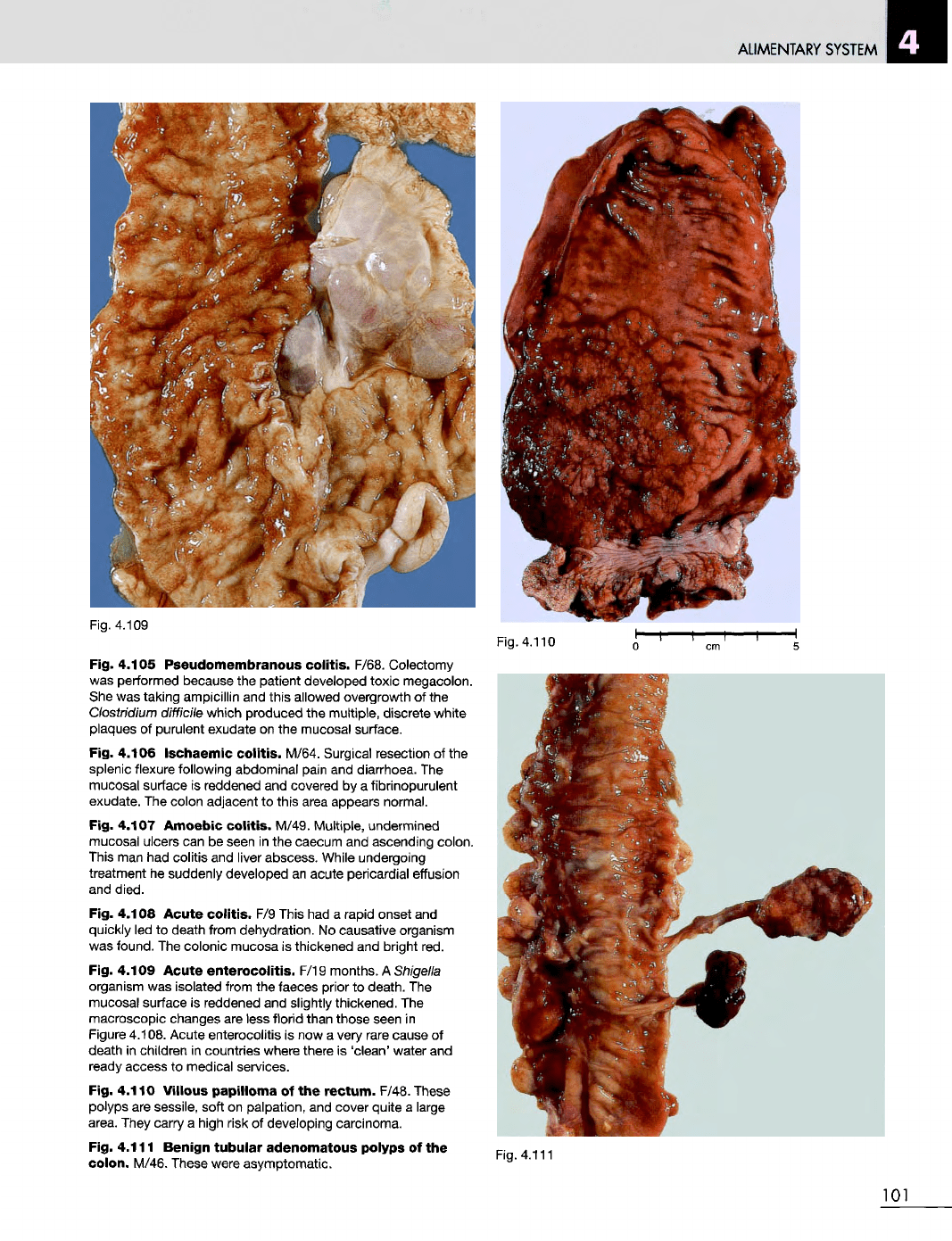
4.105
Pseudomembranous
colitis.
F/68.
Colectomy
was
performed because
the
patient developed toxic megacolon.
She
was
taking ampicillin
and
this allowed overgrowth
of the
Clostridium
difficile
which
produced
the
multiple,
discrete
white
plaques
of
purulent exudate
on the
mucosal surface.
Fig.
4.106
Ischaemic
colitis. M/64.
Surgical resection
of the
splenic flexure following abdominal pain
and
diarrhoea.
The
mucosal surface
is
reddened
and
covered
by a
fibrinopurulent
exudate.
The
colon adjacent
to
this area appears normal.
Fig.
4.107
Amoebic
colitis. M/49.
Multiple,
undermined
mucosal ulcers
can be
seen
in the
caecum
and
ascending colon.
This
man had
colitis
and
liver abscess. While undergoing
treatment
he
suddenly developed
an
acute pericardial effusion
and
died.
Fig.
4.108
Acute
colitis.
F/9
This
had a
rapid onset
and
quickly
led to
death from dehydration.
No
causative organism
was
found.
The
colonic mucosa
is
thickened
and
bright
red.
Fig.
4.109
Acute
enterocolitis.
F/19 months.
A
Shigella
organism
was
isolated from
the
faeces prior
to
death.
The
mucosal surface
is
reddened
and
slightly thickened.
The
macroscopic changes
are
less florid than those seen
in
Figure
4.108. Acute enterocolitis
is now a
very
rare cause
of
death
in
children
in
countries
where there
is
'clean'
water
and
ready
access
to
medical services.
Fig.
4.110
Villous
papilloma
of the
rectum.
F/48.
These
polyps
are
sessile, soft
on
palpation,
and
cover quite
a
large
area.
They carry
a
high risk
of
developing carcinoma.
Fig.
4.111
Benign
tubular
adenomatous polyps
of the
colon.
M/46.
These were
asymptomatic.
Fig.
4.110
Fig. 4.111
101
ALIMENTARY
SYSTEM
Fig. 4.113
Fig. 4.112
Tubular
adenoma
removed
via a
colonoscope.
M/73. This polyp
was
causing rectal bleeding from
the
ulcer
on
its
surface.
Fig. 4.113
Juvenile
polyp. F/7½. This
was
removed
via a
colonoscope. These polyps
are
characterized
by the
presence
of
greatly
dilated mucus glands, which
can be
seen
on its
surface.
Fig. 4.114
Hamartomatous
polyp from
the
rectum.
M/6
weeks.
This
had
caused rectal
bleeding.
It is
composed
of
vascular fibrofatty tissue.
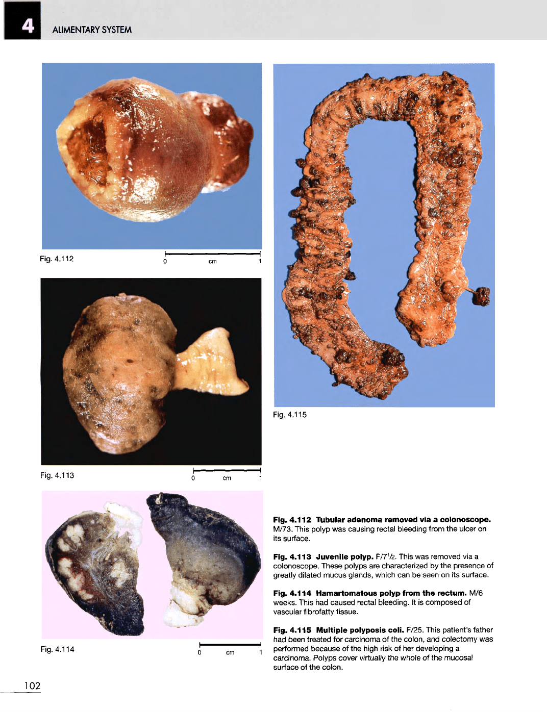
Fig. 4.115
Multiple
polyposis
coli.
F/25. This patient's father
had
been
treated
for
carcinoma
of the
colon,
and
colectomy
was
performed because
of the
high risk
of her
developing
a
carcinoma.
Polyps cover
virtually
the
whole
of the
mucosal
surface
of the
colon.
102
Fig. 4.115
Fig. 4.112
ALIMENTARY
SYSTEM
Fig.
4.117
Fig. 4.116
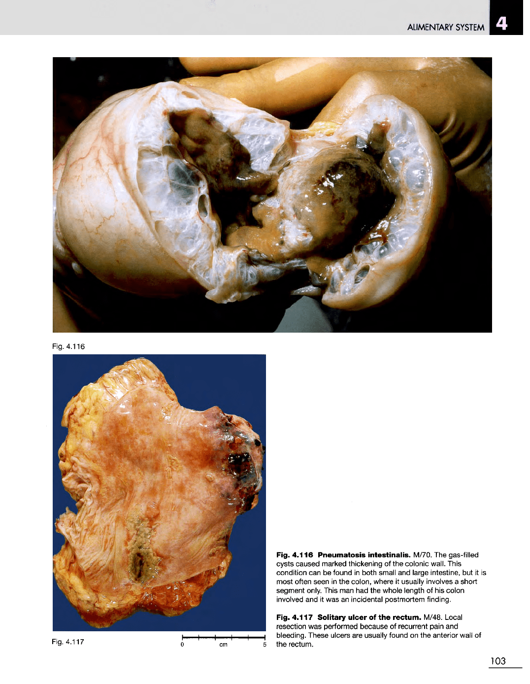
Pneumatosis
intestinalis.
M/70.
The
gas-filled
cysts caused marked thickening
of the
colonic wall. This
condition
can be
found
in
both
small
and
large intestine,
but it is
most often seen
in the
colon,
where
it
usually involves
a
short
segment only. This
man had the
whole length
of his
colon
involved
and it was an
incidental postmortem finding.
Fig. 4.117
Solitary
ulcer
of the
rectum.
M/48. Local
resection
was
performed because
of
recurrent pain
and
bleeding. These ulcers
are
usually found
on the
anterior wall
of
the
rectum.
103
Fig. 4.116
ALIMENTARY SYSTEM
Fig. 4.119
Fig.
4.120
Fig.
4.121
I
04
Fig.
4.118
ALIMENTARY
SYSTEM
Fig. 4.122
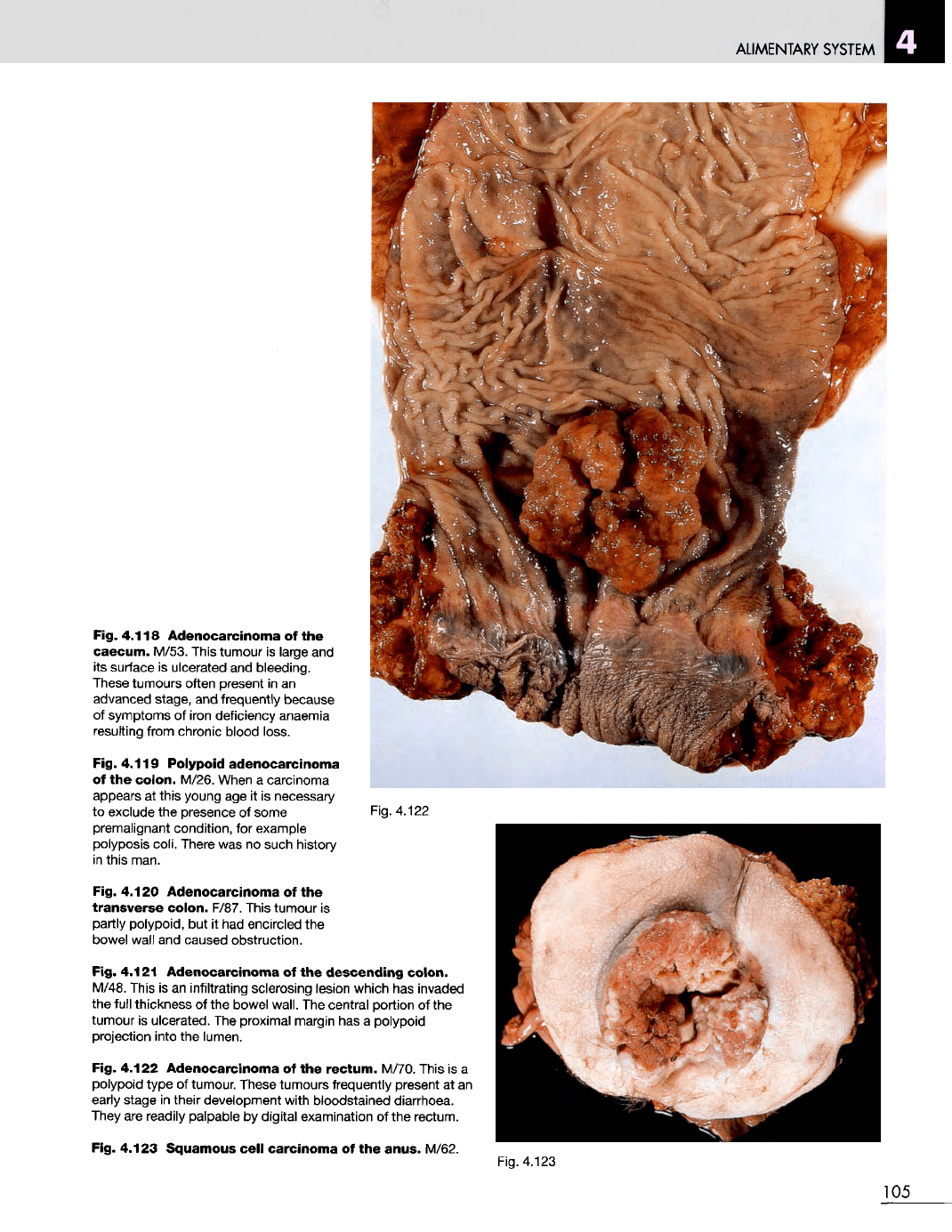
Fig. 4.118
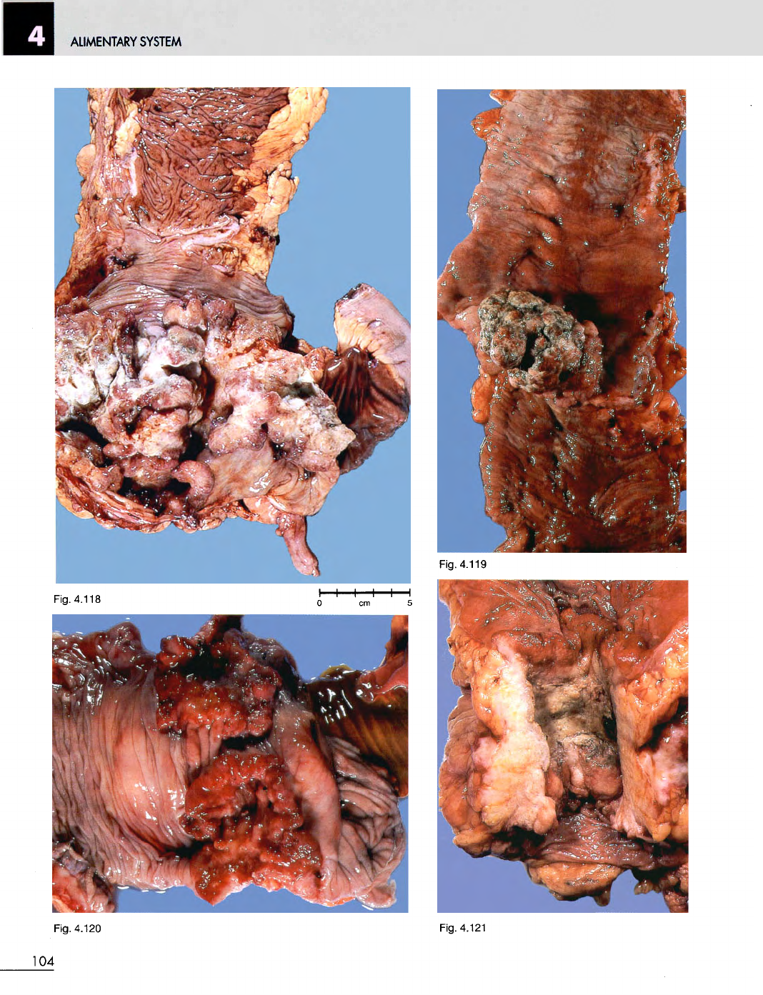
Adenocarcinoma
of the
caecum.
M/53. This tumour
is
large
and
its
surface
is
ulcerated
and
bleeding.
These
tumours often present
in an
advanced
stage,
and
frequently
because
of
symptoms
of
iron deficiency anaemia
resulting from chronic blood loss.
Fig. 4.119 Polypoid
adenocarcinoma
of
the
colon.
M/26. When
a
carcinoma
appears
at
this
young
age it is
necessary
to
exclude
the
presence
of
some
premalignant condition,
for
example
polyposis coli. There
was no
such history
in
this man.
Fig. 4.120 Adenocarcinoma
of the
transverse
colon.
F/87. This tumour
is
partly polypoid,
but it had
encircled
the
bowel wall
and
caused obstruction.
Fig. 4.121
Adenocarcinoma
of the
descending
colon.
M/48. This
is an
infiltrating sclerosing lesion which
has
invaded
the
full thickness
of the
bowel wall.
The
central portion
of the
tumour
is
ulcerated.
The
proximal margin
has a
polypoid
projection into
the
lumen.
Fig. 4.122
Adenocarcinoma
of the
rectum.
M/70. This
is a
polypoid
type
of
tumour. These tumours frequently present
at an
early
stage
in
their development with bloodstained diarrhoea.
They
are
readily palpable
by
digital
examination
of the
rectum.
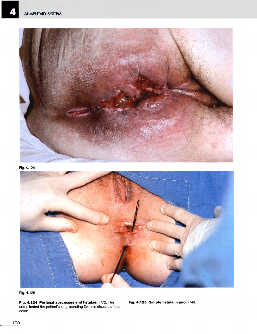
Fig. 4.123 Squamous
cell
carcinoma
of the
anus.
M/62.
Fig. 4.123
105
ALIMENTARY
SYSTEM
Fig.
4.125
Fig. 4.124
Perianal
abscesses
and
fistulae.
F/72. This
complicated
the
patient's long-standing Crohn's disease
of the
colon.
Fig. 4.125 Simple fistula
in
ano. F/40.
106
PANCREAS, BILIARY
SYSTEM
AND
LIVER
5
PANCREAS,
BILIARY
SYSTEM
AND
LIVER
Fig.
5.1
Fig.
5.1
Calculi
in the
ampulla
of
Vater. F/87. There
are
multiple calculi
impacted
in the
ampulla.
The
duodenal
surface
on the
left
is
ulcerated
as a
result
of
a
failed endoscopic attempt
to
remove
them
prior
to
death from liver failure.
Fig.
5.2
Pancreatic
atrophy
caused
by
obstruction
of the
pancreatic
duct
by
a
calculus.
M/44.
The
pancreas
has
been
sliced along
its
length.
The
pancreatic duct
in
the
middle
of the
gland
is
dilated,
and
there
is
atrophy
of the
acinar tissue. This
was
an
incidental finding
in a
patient
who
died from
a
carcinoma
of the
pharynx.
Fig.
5.3
Nodule
of
ectopic
pancreas
on
the
surface
of the
jejunum.
F/69.
An
incidental postmortem finding. Ectopic
pancreatic tissue
is
found especially
in the
pylorus, duodenum
and
jejunum.
Fig.
5.3
108
Fig.
5.2
PANCREAS,
BILIARY
SYSTEM
AND
LIVER
Fig.
5.6
Fig.
5.4
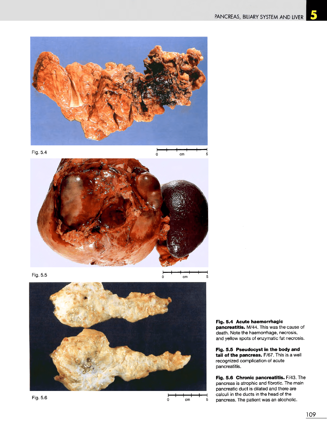
Acute
haemorrhagic
pancreatitis.
M/44. This
was the
cause
of
death. Note
the
haemorrhage, necrosis,
and
yellow spots
of
enzymatic
fat
necrosis.
Fig.
5.5
Pseudocyst
in the
body
and
tail
of the
pancreas.
F/67. This
is a
well
recognized complication
of
acute
pancreatitis.
Fig.
5.6
Chronic
pancreatitis.
F/43.
The
pancreas
is
atrophic
and
fibrotic.
The
main
pancreatic duct
is
dilated
and
there
are
calculi
in the
ducts
in the
head
of the
pancreas.
The
patient
was an
alcoholic.
109
Fig.
5.5
Fig.
5.4
PANCREAS,
BILIARY
SYSTEM
AND
LIVER
Fig.
5.7
Fig.
5.8
110