Cook R.A., Stewart B. Colour Atlas of Anatomical Pathology
Подождите немного. Документ загружается.


RENAL SYSTEM
6
RENAL
SYSTEM
Fig.
6.3
0 cm 5
Fig.
6.4
132
Fig.
6.1
Fig.
6.2
RENAL
SYSTEM
Fig.
6.5
Fig.
6.6
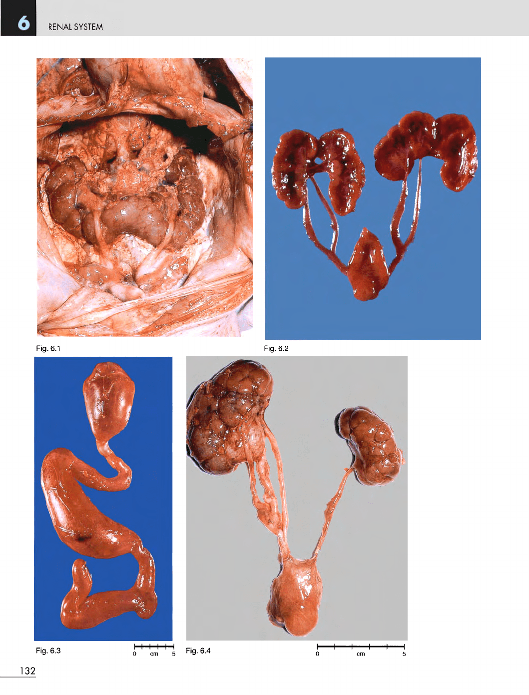
Fig.
6.1
Horseshoe
kidney.
F/9
days.
The
lower poles
of
both
kidneys
are
joined across
the
midline
and the
ureters pass
anterior
to the
renal substance.
This
child
died
as a
result
of
multiple congenital abnormalities.
Fig.
6.2
Double
ureters.
Neonate. Both ureters
are
bifid
and
join
to
form
a
single ureter before
opening
into
the
bladder.
Fig.
6.3
Megaloureter.
M/11. This
was
caused
by
stenosis
at
the
ureterovesical junction.
The
patient
had
recurrent urinary
tract infections,
and
finally
the
kidney
and
ureter were surgically
removed.
Fig.
6.4
Triple
ureter.
Neonate. Double ureters
are
fairly
common,
but
triple ureter
is
very rare.
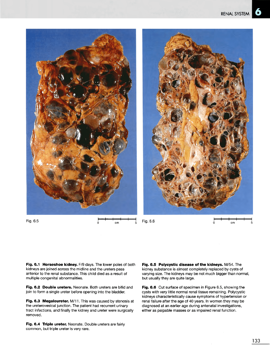
Fig.
6.5
Polycystic
disease
of the
kidneys.
M/54.
The
kidney substance
is
almost completely
replaced
by
cysts
of
varying
size.
The
kidneys
may be not
much bigger than normal,
but
usually they
are
quite
large.
Fig.
6.6 Cut
surface
of
specimen
in
Figure 6.5, showing
the
cysts
with very
little
normal renal tissue remaining. Polycystic
kidneys characteristically cause symptoms
of
hypertension
or
renal failure after
the age of 40
years.
In
women they
may be
diagnosed
at an
earlier
age
during antenatal investigations,
either
as
palpable
masses
or as
impaired renal function.
133

RENAL
SYSTEM
Fig.
6.9
134
Fig.
6.7
Fig.
6.8
Fig. 6.10
RENAL
SYSTEM
Fig. 6.11
Fig.
6.7
Cystic
dysplastic
kidney.
Stillborn. There
is
a
very
small multicystic kidney
on the
right (arrow)
and no
kidney
on the
left.
The
adrenal glands
are
relatively large
in
neonates,
but
they
are
accentuated
in
this case
because
of the
very small size
of the
kidney.
Fig.
6.8
Cystic
dysplastic
kidney.
F/24 hours.
The
right kidney
is
normal
but the
left
is
grossly cystic.
Microscopic examination showed malformed renal
substance together with areas
of
cartilage. Cystic
dysplastic kidney
may be
unilateral
or
bilateral.
Fig.
6.9
Multiple
simple
cysts
in the
kidney.
M/70.
This
was an
incidental postmortem finding.
Fig. 6.10
Sponge
kidney
in a
neonate
who
died
a few
minutes
after
birth. This type
of
cystic kidney
is
usually large
and
bilateral
and may
interfere with delivery
of the
fetus.
Fig. 6.11
Ureteritis
cystica.
M/80. Incidental autopsy
finding. This condition
may be
associated with chronic
urinary
tract infection.
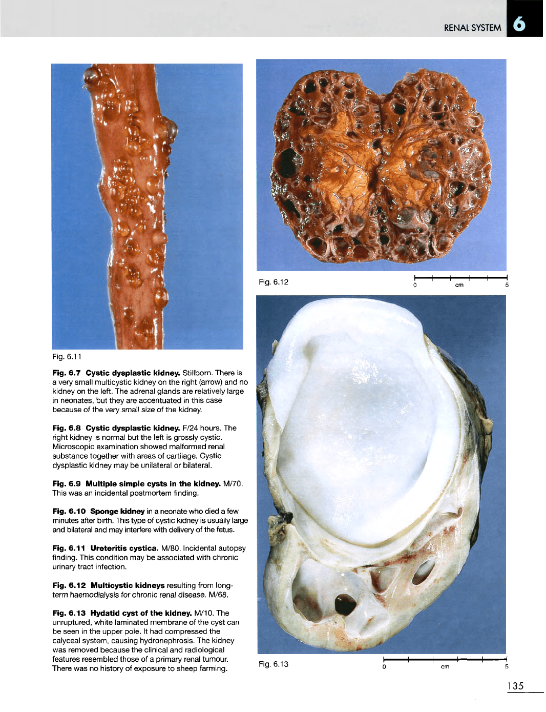
Fig. 6.12
Multicystic
kidneys
resulting from long-
term
haemodialysis
for
chronic renal
disease.
M/68.
Fig. 6.13
Hydatid
cyst
of the
kidney.
M/10.
The
unruptured, white laminated membrane
of the
cyst
can
be
seen
in the
upper pole.
It had
compressed
the
calyceal
system, causing hydronephrosis.
The
kidney
was
removed because
the
clinical
and
radiological
features resembled those
of a
primary renal tumour.
There
was no
history
of
exposure
to
sheep farming.
Fig. 6.13
135
Fig. 6.12
RENAL
SYSTEM
Fig.
6.14
Fig.
6.16
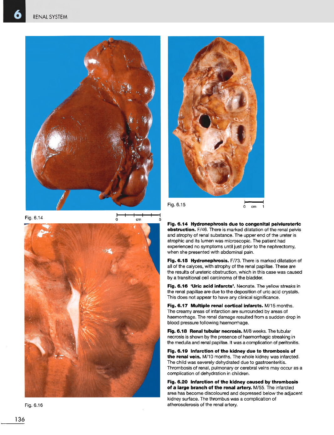
Fig. 6.14 Hydronephrosis
due to
congenital
pelviureteric
obstruction.
F/46. There
is
marked dilatation
of the
renal pelvis
and
atrophy
of
renal substance.
The
upper
end of the
ureter
is
atrophic
and its
lumen
was
microscopic.
The
patient
had
experienced
no
symptoms until just prior
to the
nephrectomy,
when
she
presented with abdominal pain.
Fig. 6.15 Hydronephrosis. F/73. There
is
marked dilatation
of
all
of the
calyces, with atrophy
of the
renal papillae. These
are
the
results
of
ureteric obstruction, which
in
this case
was
caused
by
a
transitional cell carcinoma
of the
bladder.
Fig. 6.16
'Uric
acid
infarcts'.
Neonate.
The
yellow streaks
in
the
renal papillae
are due to the
deposition
of
uric acid crystals.
This
does
not
appear
to
have
any
clinical significance.
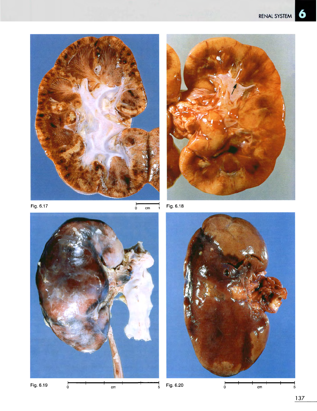
Fig. 6.17
Multiple
renal
cortical
infarcts.
M/15 months.
The
creamy areas
of
infarction
are
surrounded
by
areas
of
haemorrhage.
The
renal damage resulted from
a
sudden drop
in
blood pressure following haemorrhage.
Fig. 6.18 Renal
tubular
necrosis.
M/8
weeks.
The
tubular
necrosis
is
shown
by the
presence
of
haemorrhagic streaking
in
the
medulla
and
renal papillae.
It was a
complication
of
peritonitis.
Fig. 6.19
Infarction
of the
kidney
due to
thrombosis
of
the
renal
vein.
M/10 months.
The
whole kidney
was
infarcted.
The
child
was
severely dehydrated
due to
gastroenteritis.
Thrombosis
of
renal, pulmonary
or
cerebral veins
may
occur
as a
complication
of
dehydration
in
children.
Fig. 6.20
Infarction
of the
kidney
caused
by
thrombosis
of
a
large
branch
of the
renal
artery.
M/55.
The
infarcted
area
has
become discoloured
and
depressed below
the
adjacent
kidney surface.
The
thrombus
was a
complication
of
atherosclerosis
of the
renal artery.
136
Fig.
6.15
RENAL
SYSTEM
Fig.
6.19
137
Fig.
6.18
Fig.
6.17
RENAL
SYSTEM
Fig.
6.21
Fig.
6.22
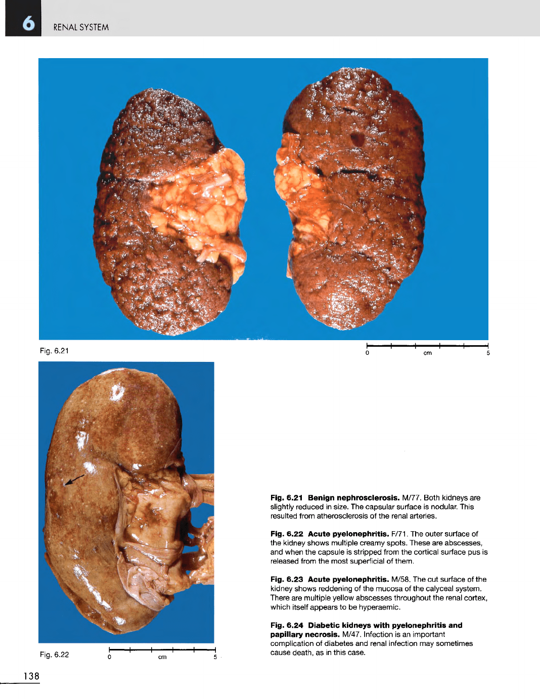
Fig. 6.21
Benign
nephrosclerosis.
M/77. Both kidneys
are
slightly reduced
in
size.
The
capsular surface
is
nodular. This
resulted from atherosclerosis
of the
renal arteries.
Fig. 6.22 Acute
pyelonephritis.
F/71.
The
outer surface
of
the
kidney
shows
multiple
creamy
spots.
These
are
abscesses,
and
when
the
capsule
is
stripped from
the
cortical surface
pus is
released
from
the
most superficial
of
them.
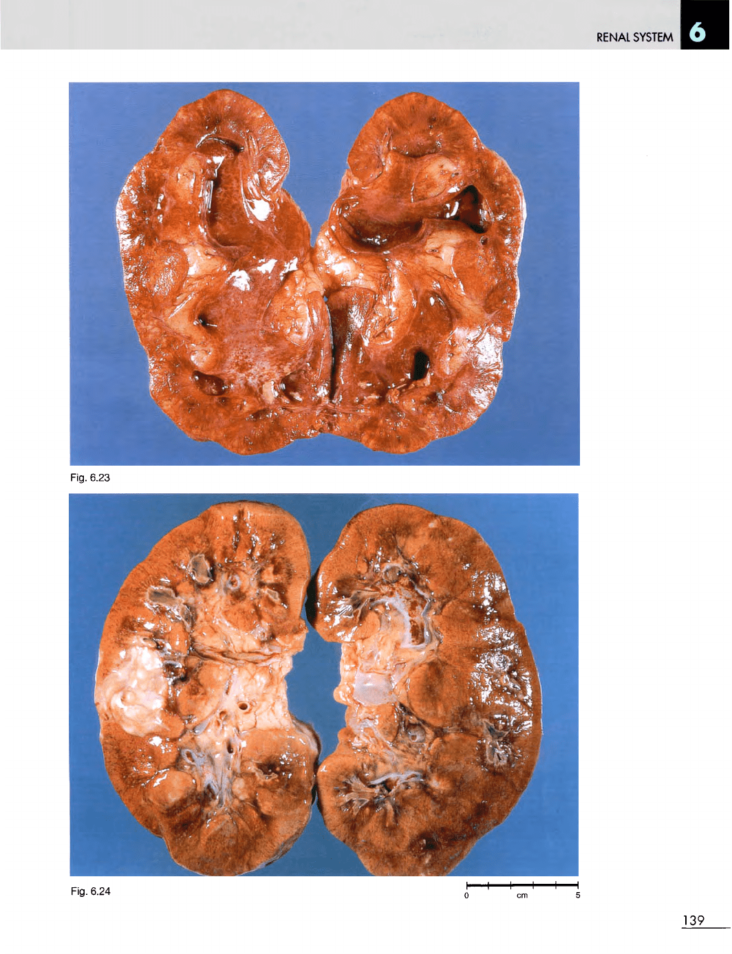
Fig. 6.23
Acute
pyelonephritis.
M/58.
The cut
surface
of the
kidney shows reddening
of the
mucosa
of the
calyceal system.
There
are
multiple yellow abscesses throughout
the
renal cortex,
which
itself appears
to be
hyperaemic.
Fig. 6.24
Diabetic
kidneys
with
pyelonephritis
and
papillary
necrosis.
M/47. Infection
is an
important
complication
of
diabetes
and
renal infection
may
sometimes
cause
death,
as in
this case.
138
RENAL
SYSTEM
Fig.
6.23
Fig.
6.24
139
RENAL SYSTEM
Fig .
6.2 7
Fig .
6.2 8
14 0
Fig .
6.2 5
Fig .
6.2 6