Cook R.A., Stewart B. Colour Atlas of Anatomical Pathology
Подождите немного. Документ загружается.

PANCREAS,
BILIARY
SYSTEM
AND
LIVER
Fig.
5.37
Fig.
5.38
Fig.
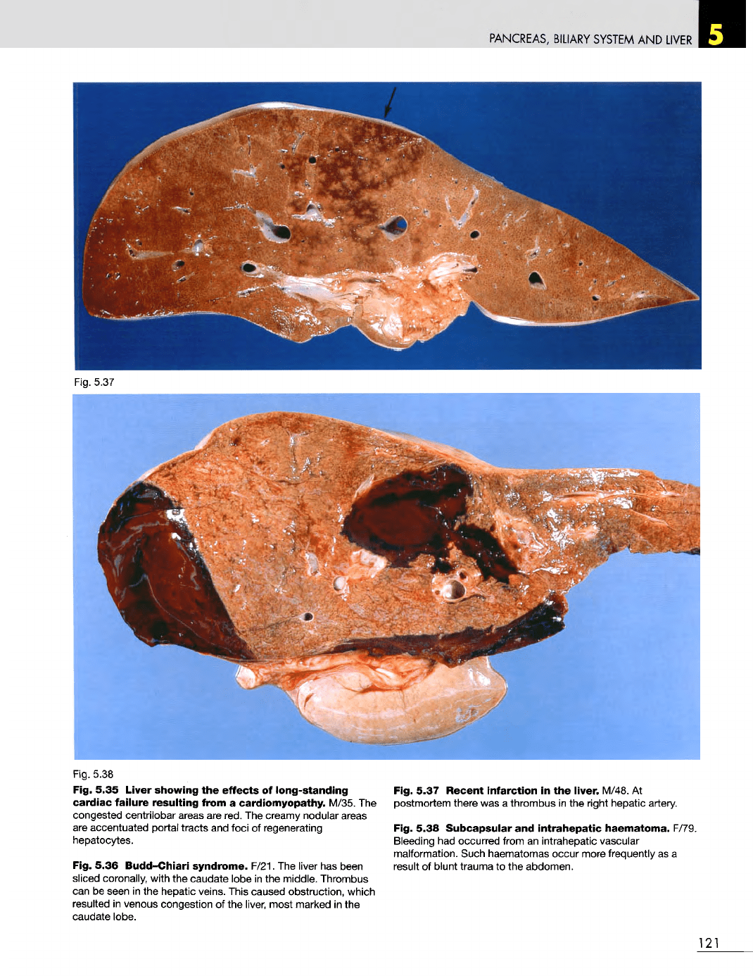
5.35
Liver showing
the
effects
of
long-standing
cardiac
failure
resulting
from
a
cardiomyopathy. M/35.
The
congested centrilobar areas
are
red.
The
creamy nodular areas
are
accentuated portal tracts
and
foci
of
regenerating
hepatocytes.
Fig.
5.36 Budd-Chiari
syndrome.
F/21.
The
liver
has
been
sliced coronally, with
the
caudate lobe
in the
middle. Thrombus
can
be
seen
in the
hepatic veins. This caused obstruction, which
resulted
in
venous congestion
of the
liver, most marked
in the
caudate lobe.
Fig.
5.37
Recent
infarction
in the
liver.
M/48.
At
postmortem there
was a
thrombus
in the
right hepatic artery.
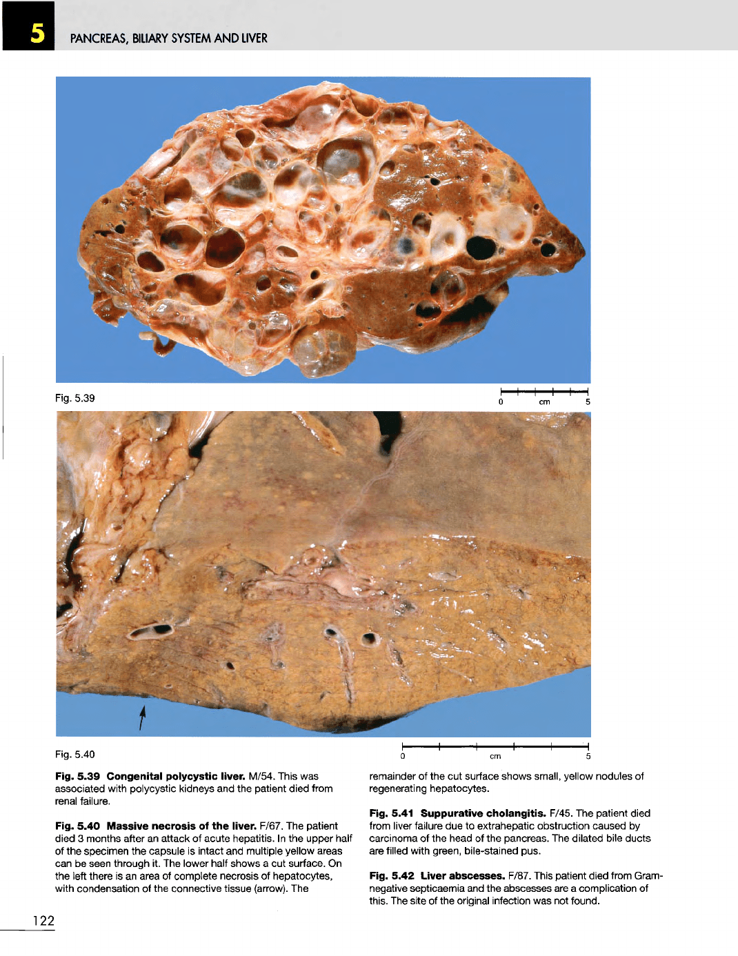
Fig.
5.38
Subcapsular
and
intrahepatic
haematoma. F/79.
Bleeding
had
occurred from
an
intrahepatic vascular
malformation. Such haematomas occur more frequently
as a
result
of
blunt trauma
to the
abdomen.
121
PANCREAS,
BILIARY
SYSTEM
AND
LIVER
Fig.
5.40
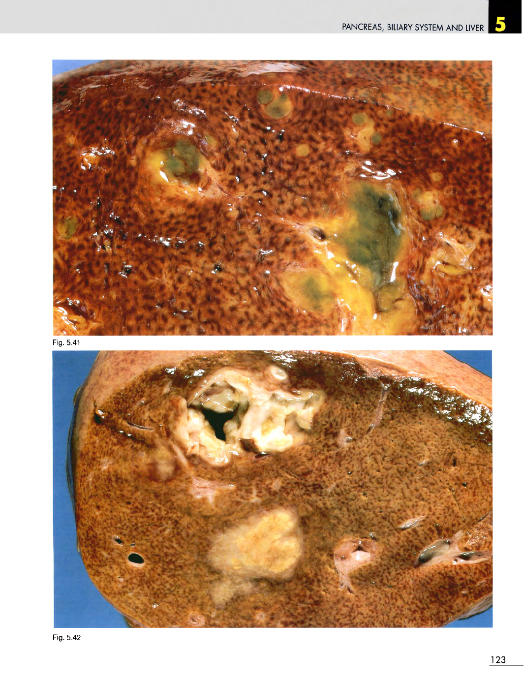
Fig. 5.39
Congenital
polycystic
liver.
M/54. This
was
associated with polycystic kidneys
and the
patient
died
from
renal failure.
Fig. 5.40
Massive
necrosis
of the
liver.
F/67.
The
patient
died
3
months after
an
attack
of
acute hepatitis.
In the
upper half
of
the
specimen
the
capsule
is
intact
and
multiple yellow areas
can be
seen through
it. The
lower half shows
a cut
surface.
On
the
left there
is an
area
of
complete necrosis
of
hepatocytes,
with condensation
of the
connective tissue (arrow).
The
remainder
of the cut
surface shows small, yellow nodules
of
regenerating hepatocytes.
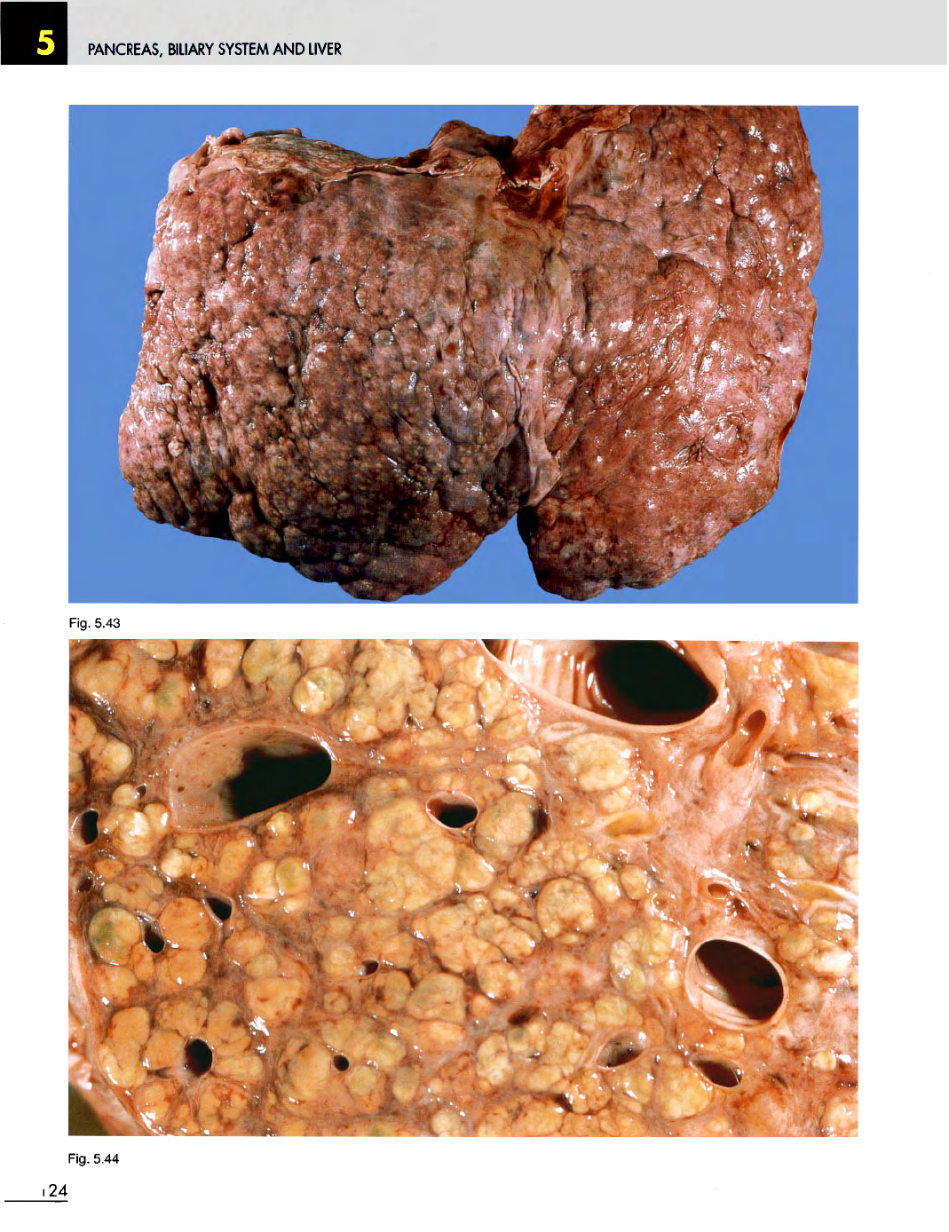
Fig. 5.41
Suppurative
cholangitis.
F/45.
The
patient died
from
liver
failure
due to
extrahepatic obstruction caused
by
carcinoma
of the
head
of the
pancreas.
The
dilated
bile ducts
are
filled with green, bile-stained pus.
Fig. 5.42 Liver
abscesses.
F/87. This
patient
died from Gram-
negative septicaemia
and the
abscesses
are a
complication
of
this.
The
site
of the
original infection
was not
found.
122
Fig.
5.39
PANCREAS,
BILIARY SYSTEM
AND
LIVER
Fig. 5.42
123
Fig.
5.41
PANCREAS,
BILIARY
SYSTEM
AND
LIVER
Fig. 5.44
124
Fig. 5.43
PANCREAS,
BILIARY
SYSTEM
AND
LIVER
Fig.
5.45
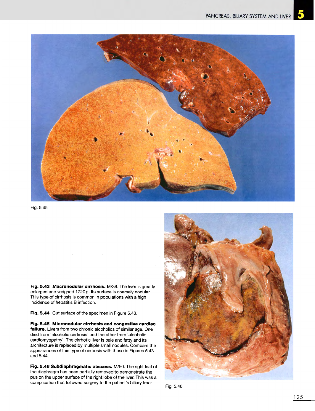
Fig. 5.43
Macronodular
cirrhosis.
M/39.
The
liver
is
greatly
enlarged
and
weighed 1720g.
Its
surface
is
coarsely nodular.
This
type
of
cirrhosis
is
common
in
populations with
a
high
incidence
of
hepatitis
B
infection.
Fig. 5.44
Cut
surface
of the
specimen
in
Figure 5.43.
Fig. 5.45
Micronodular
cirrhosis
and
congestive
cardiac
failure.
Livers from
two
chronic alcoholics
of
similar age.
One
died from 'alcoholic cirrhosis'
and the
other from 'alcoholic
cardiomyopathy'.
The
cirrhotic
liver
is
pale
and
fatty
and its
architecture
is
replaced
by
multiple small nodules. Compare
the
appearances
of
this type
of
cirrhosis with those
in
Figures 5.43
and
5.44.
Fig. 5.46
Subdiaphragmatic
abscess.
M/50.
The
right leaf
of
the
diaphragm
has
been partially removed
to
demonstrate
the
pus on the
upper surface
of the
right lobe
of the
liver. This
was a
complication that followed surgery
to the
patient's biliary tract.
Fig.
5.46
125
PANCREAS,
BILIARY
SYSTEM
AND
LIVER
Fig.
5.47
Fig.
5.48
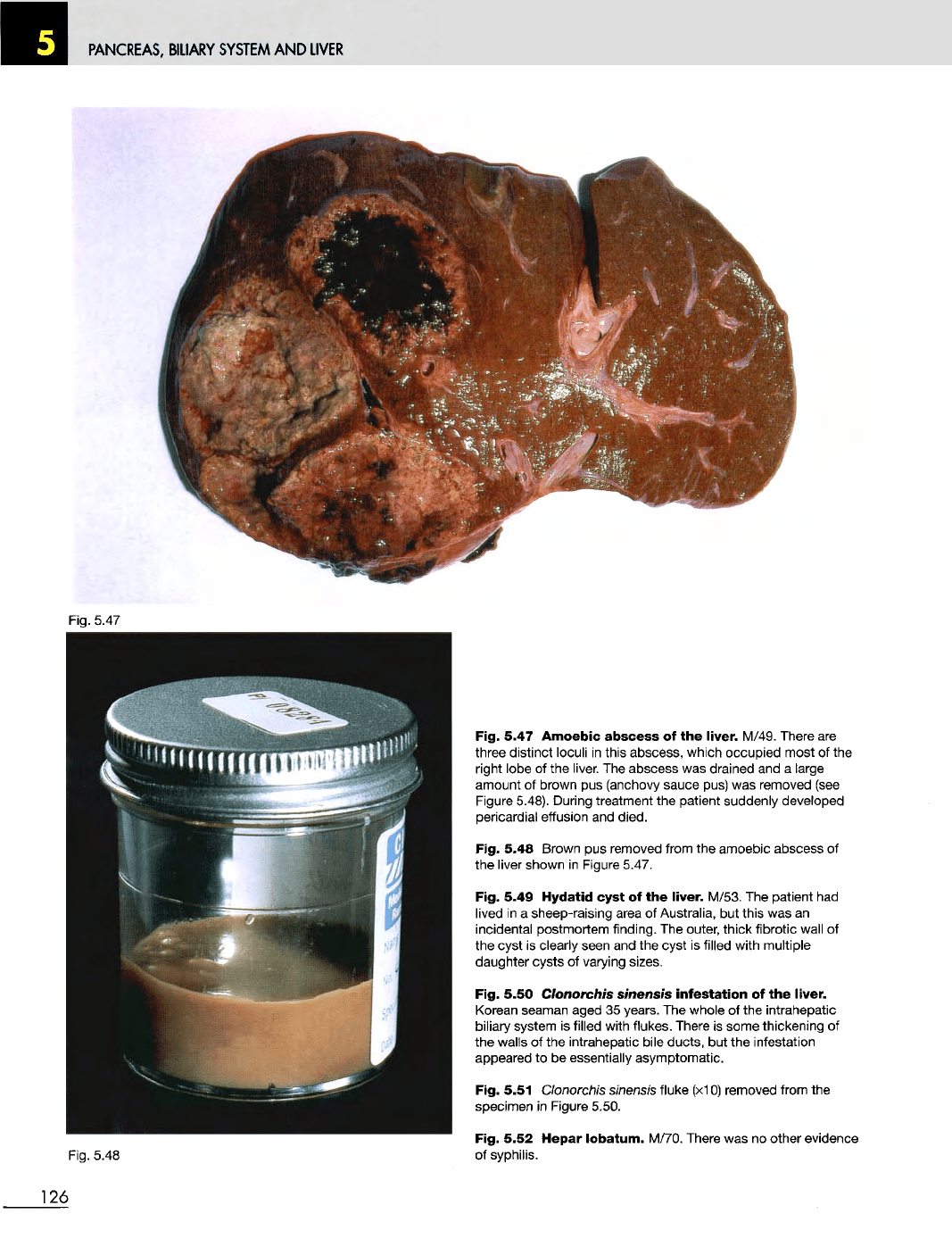
Fig. 5.47
Amoebic
abscess
of the
liver.
M/49. There
are
three distinct loculi
in
this abscess, which occupied most
of the
right lobe
of the
liver.
The
abscess
was
drained
and a
large
amount
of
brown
pus
(anchovy sauce pus)
was
removed (see
Figure
5.48). During treatment
the
patient suddenly developed
pericardial effusion
and
died.
Fig. 5.48 Brown
pus
removed from
the
amoebic abscess
of
the
liver
shown
in
Figure 5.47.
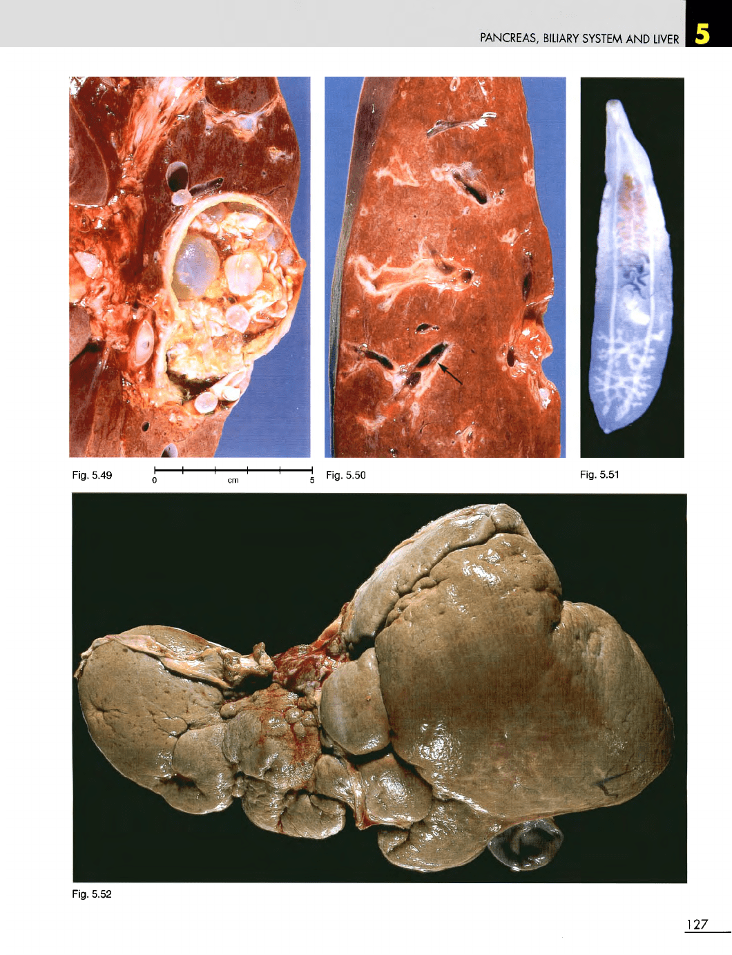
Fig. 5.49
Hydatid
cyst
of the
liver.
M/53.
The
patient
had
lived
in a
sheep-raising area
of
Australia,
but
this
was an
incidental postmortem finding.
The
outer, thick fibrotic wall
of
the
cyst
is
clearly seen
and the
cyst
is
filled with multiple
daughter cysts
of
varying sizes.
Fig. 5.50 Clonorchis sinensis
infestation
of the
liver.
Korean
seaman aged
35
years.
The
whole
of the
intrahepatic
biliary system
is
filled with flukes. There
is
some thickening
of
the
walls
of the
intrahepatic bile ducts,
but the
infestation
appeared
to be
essentially asymptomatic.
Fig. 5.51 Clonorchis
sinensis
fluke (x10) removed from
the
specimen
in
Figure 5.50.
Fig. 5.52
Hepar
lobatum.
M/70. There
was no
other evidence
of
syphilis.
126
PANCREAS,
BILIARY
SYSTEM
AND
LIVER
Fig.
5.52
127
Fig.
5.49
Fig.
5.51
Fig.
5.50
PANCREAS,
BILIARY
SYSTEM
AND
LIVER
Fig.
5.55
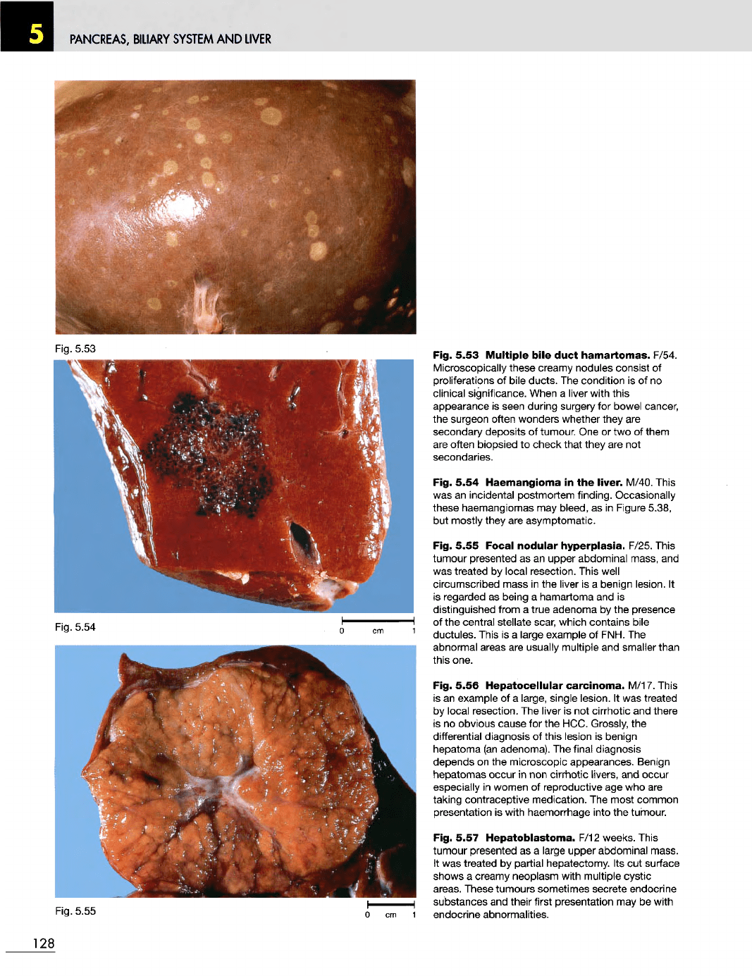
Fig. 5.53
Multiple
bile
duct
hamartomas.
F/54.
Microscopically these creamy nodules consist
of
proliferations
of
bile ducts.
The
condition
is of no
clinical significance. When
a
liver with this
appearance
is
seen during surgery
for
bowel cancer,
the
surgeon often wonders whether they
are
secondary deposits
of
tumour.
One or two of
them
are
often biopsied
to
check that they
are not
secondaries.
Fig. 5.54
Haemangioma
in the
liver.
M/40. This
was
an
incidental postmortem finding. Occasionally
these haemangiomas
may
bleed,
as in
Figure 5.38,
but
mostly they
are
asymptomatic.
Fig. 5.55 Focal
nodular
hyperplasia.
F/25. This
tumour presented
as an
upper abdominal mass,
and
was
treated
by
local resection. This well
circumscribed mass
in the
liver
is a
benign lesion.
It
is
regarded
as
being
a
hamartoma
and is
distinguished from
a
true adenoma
by the
presence
of
the
central stellate scar, which contains bile
ductules. This
is a
large example
of
FNH.
The
abnormal areas
are
usually multiple
and
smaller than
this one.
Fig. 5.56
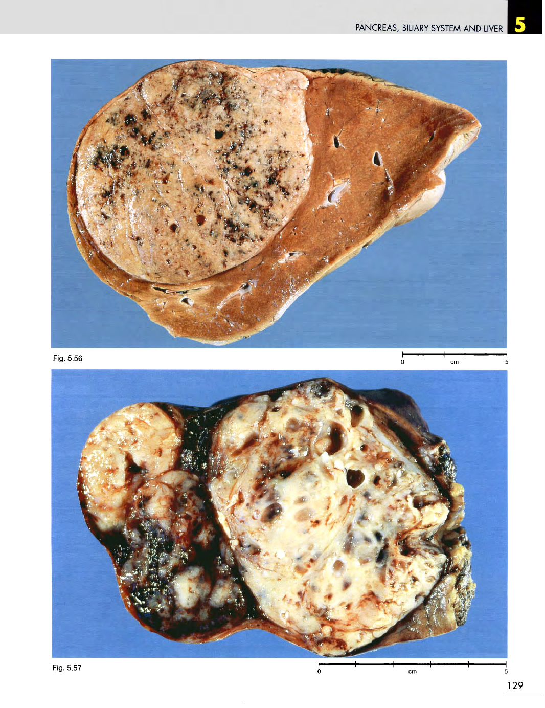
Hepatocellular
carcinoma.
M/17. This
is
an
example
of a
large, single lesion.
It was
treated
by
local resection.
The
liver
is not
cirrhotic
and
there
is
no
obvious cause
for the
HCC. Grossly,
the
differential diagnosis
of
this lesion
is
benign
hepatoma
(an
adenoma).
The
final diagnosis
depends
on the
microscopic appearances. Benign
hepatomas
occur
in non
cirrhotic
livers,
and
occur
especially
in
women
of
reproductive
age who are
taking contraceptive medication.
The
most common
presentation
is
with haemorrhage into
the
tumour.
Fig. 5.57
Hepatoblastoma.
F/12 weeks. This
tumour presented
as a
large upper abdominal mass.
It
was
treated
by
partial hepatectomy.
Its cut
surface
shows
a
creamy neoplasm with multiple cystic
areas.
These tumours sometimes secrete endocrine
substances
and
their first presentation
may be
with
1
endocrine abnormalities.
128
Fig.
5.54
Fig.
5.53
PANCREAS,
BILIARY
SYSTEM
AND
LIVER
Fig. 5.57
129
Fig. 5.56
PANCREAS,
BILIARY
SYSTEM
AND
LIVER
Fig.
5.58
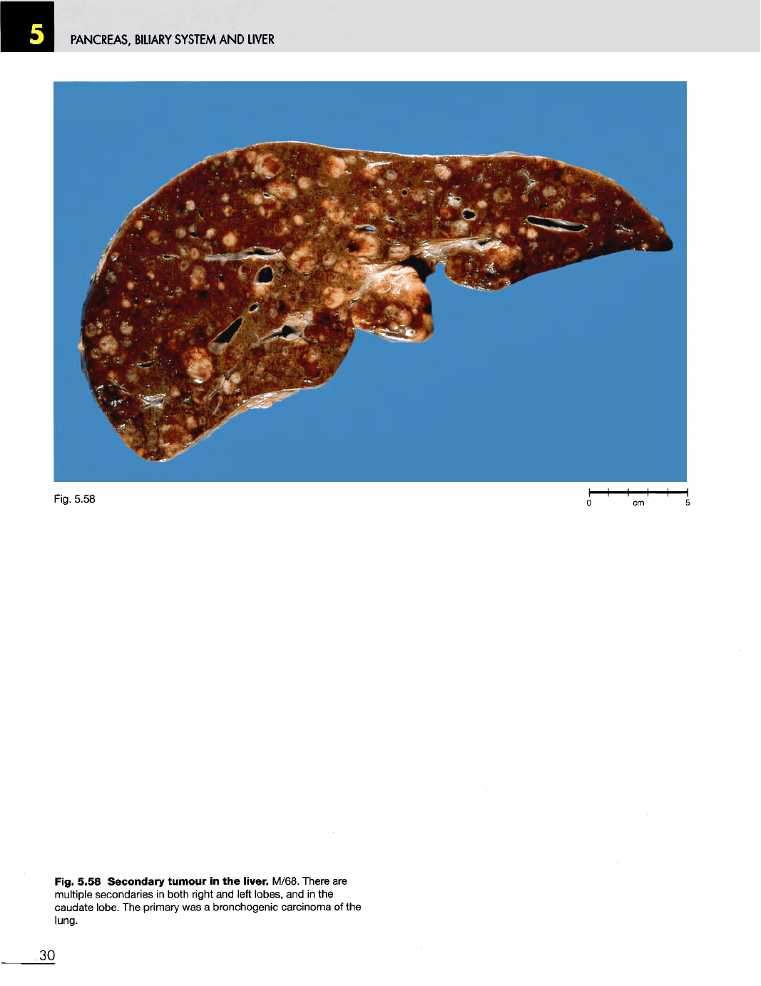
Fig. 5.58 Secondary
tumour
in the
liver. M/68. There
are
multiple secondaries
in
both right
and
left
lobes,
and in the
caudate
lobe.
The
primary
was a
bronchogenic carcinoma
of the
lung.
130