Cook R.A., Stewart B. Colour Atlas of Anatomical Pathology
Подождите немного. Документ загружается.

PANCREAS,
BILIARY
SYSTEM
AND
LIVER
Fig.
5.9
Fig.
5.7
Mucoviscidosis
(fibrocystic
disease
of the
pancreas).
M/11.
The
pancreas
is
attached
to the
second part
of
the
duodenum.
The
main pancreatic duct
in the
head
of the
pancreas
is
dilated.
The
body
and
tail
are
fibrotic,
and
numerous
cysts
can be
seen.
Fig.
5.8
Hepatic
cirrhosis
in
mucoviscidosis.
M/6.
Cirrhosis
complicates mucoviscidosis
in a
proportion
of
cases,
usually
presenting about this
age as
haematemesis.
Fig.
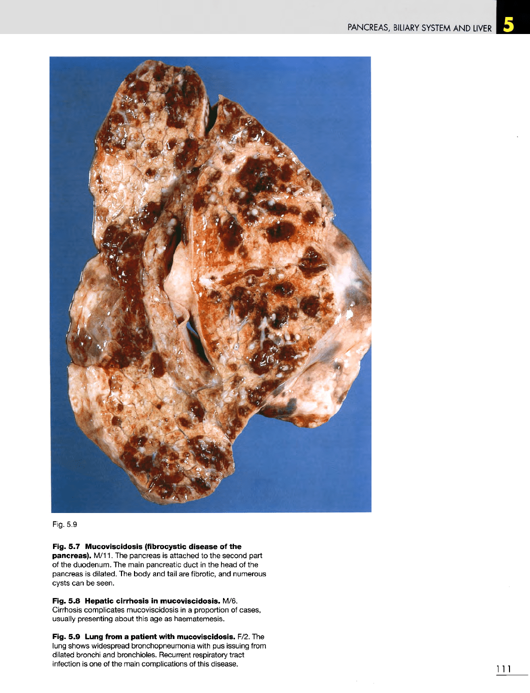
5.9
Lung from
a
patient
with
mucoviscidosis. F/2.
The
lung shows widespread bronchopneumonia with
pus
issuing from
dilated bronchi
and
bronchioles. Recurrent respiratory tract
infection
is one of the
main complications
of
this disease.
Ill
PANCREAS,
BILIARY
SYSTEM
AND
LIVER
Fig. 5.11
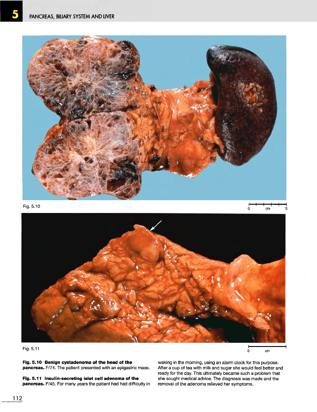
Fig. 5.10
Benign
cystadenoma
of the
head
of the
pancreas.
F/74.
The
patient presented with
an
epigastric mass.
Fig. 5.11
Insulin-secreting
islet
cell
adenoma
of the
pancreas.
F/45.
For
many years
the
patient
had had
difficulty
in
waking
in the
morning, using
an
alarm clock
for
this purpose.
After
a cup of tea
with milk
and
sugar
she
would
feel
better
and
ready
for the
day. This ultimately became such
a
problem that
she
sought medical advice.
The
diagnosis
was
made
and the
removal
of the
adenoma relieved
her
symptoms.
112
Fig. 5.10
PANCREAS,
BILIARY
SYSTEM
AND
LIVER
Fig.
5.12
Fig. 5.14
Fig.
5.15
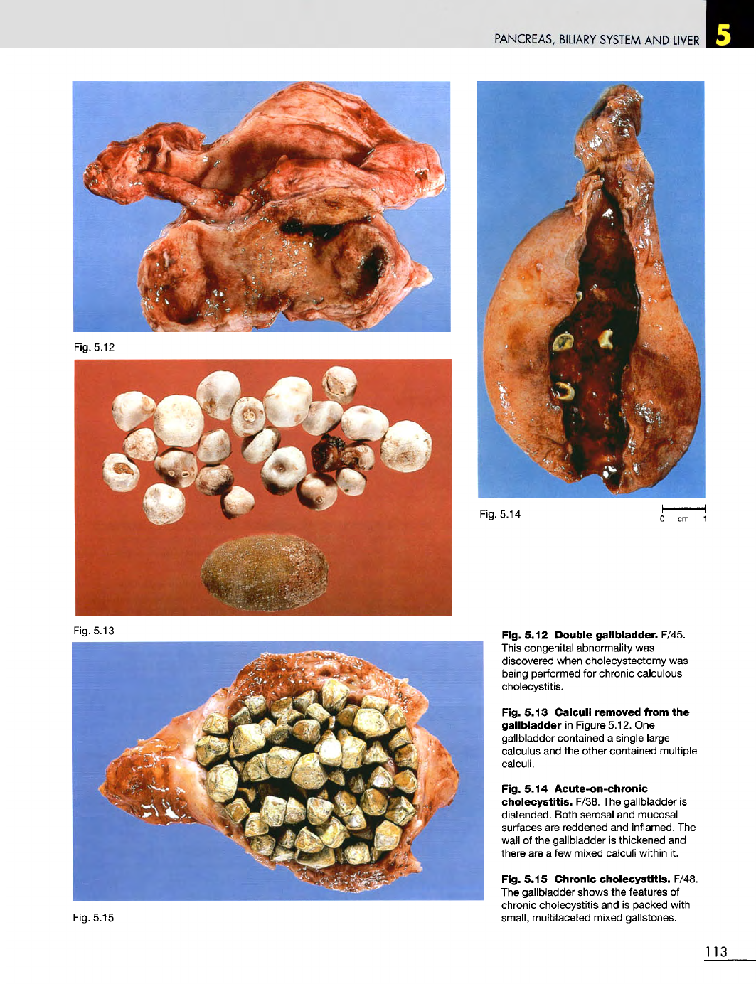
Fig. 5.12
Double
gallbladder.
F/45.
This congenital abnormality
was
discovered when cholecystectomy
was
being performed
for
chronic calculous
cholecystitis.
Fig. 5.13
Calculi
removed
from
the
gallbladder
in
Figure 5.12.
One
gallbladder contained
a
single large
calculus
and the
other contained multiple
calculi.
Fig. 5.14
Acute-on-chronic
cholecystitis.
F/38.
The
gallbladder
is
distended. Both serosal
and
mucosal
surfaces
are
reddened
and
inflamed.
The
wall
of the
gallbladder
is
thickened
and
there
are a few
mixed calculi within
it.
Fig. 5.15
Chronic
cholecystitis.
F/48.
The
gallbladder shows
the
features
of
chronic cholecystitis
and is
packed with
small, multifaceted mixed gallstones.
113
Fig.
5.13
PANCREAS,
BILIARY
SYSTEM
AND
LIVER
Fig. 5.16
Fig.
5.17
Fig.
5.18
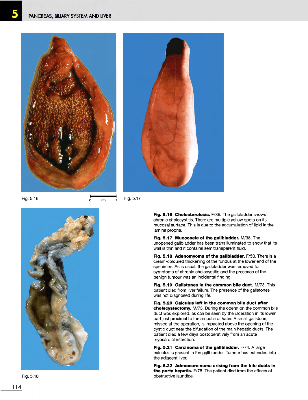
Fig. 5.16
Cholesterolosis.
F/36.
The
gallbladder shows
chronic cholecystitis. There
are
multiple yellow spots
on its
mucosal surface. This
is due to the
accumulation
of
lipid
in the
lamina propria.
Fig. 5.17
Mucocoele
of the
gallbladder.
M/38.
The
unopened gallbladder
has
been transilluminated
to
show that
its
wall
is
thin
and it
contains semitransparent fluid.
Fig. 5.18 Adenomyoma
of the
gallbladder.
F/50. There
is a
cream-coloured thickening
of the
fundus
at the
lower
end of the
specimen.
As is
usual,
the
gallbladder
was
removed
for
symptoms
of
chronic cholecystitis
and the
presence
of the
benign tumour
was an
incidental finding.
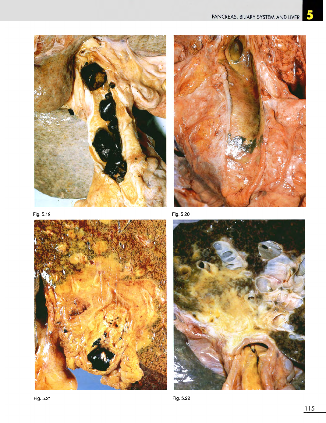
Fig. 5.19
Gallstones
in the
common
bile
duct.
M/73. This
patient died from
liver
failure.
The
presence
of the
gallstones
was
not
diagnosed during life.
Fig. 5.20
Calculus
left
in the
common
bile
duct
after
cholecystectomy.
M/73. During
the
operation
the
common bile
duct
was
explored,
as can be
seen
by the
ulceration
in its
lower
part just proximal
to the
ampulla
of
Vater.
A
small gallstone,
missed
at the
operation,
is
impacted above
the
opening
of the
cystic
duct
near
the
bifurcation
of the
main hepatic ducts.
The
patient died
a few
days postoperatively from
an
acute
myocardial infarction.
Fig. 5.21
Carcinoma
of the
gallbladder.
F/74.
A
large
calculus
is
present
in the
gallbladder. Tumour
has
extended into
the
adjacent liver.
Fig. 5.22
Adenocarcinoma
arising from
the
bile
ducts
in
the
porta
hepatis.
F/78.
The
patient died from
the
effects
of
obstructive jaundice.
114
PANCREAS,
BILIARY SYSTEM
AND
LIVER
Fig. 5.19
Fig. 5.20
Fig. 5.21
Fig. 5.22
115
PANCREAS,
BILIARY
SYSTEM
AND
LIVER
Fig.
5.23
Fig.
5.24
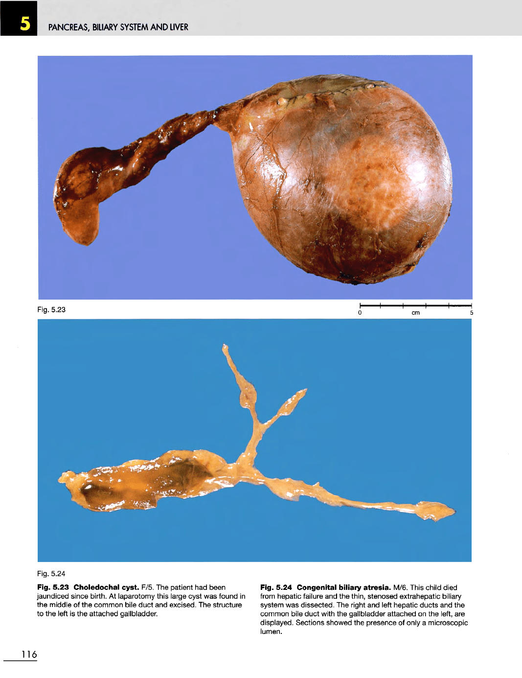
Fig. 5.23
Choledochal
cyst. F/5.
The
patient
had
been
jaundiced since birth.
At
laparotomy this large cyst
was
found
in
the
middle
of the
common bile duct
and
excised.
The
structure
to the
left
is the
attached gallbladder.
Fig. 5.24
Congenital
biliary
atresia.
M/6. This child died
from
hepatic failure
and the
thin, stenosed extrahepatic biliary
system
was
dissected.
The
right
and
left hepatic ducts
and the
common
bile duct with
the
gallbladder attached
on the
left,
are
displayed. Sections showed
the
presence
of
only
a
microscopic
lumen.
116
PANCREAS,
BILIARY
SYSTEM
AND
LIVER
Fig. 5.26
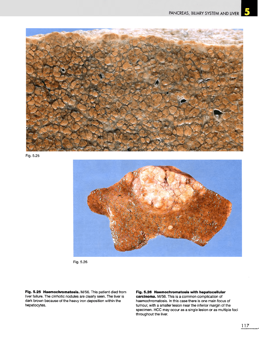
Fig. 5.25
Haemochromatosis.
M/56. This patient died from
liver
failure.
The
cirrhotic nodules
are
clearly seen.
The
liver
is
dark brown because
of the
heavy iron deposition within
the
hepatocytes.
Fig. 5.26
Haemochromatosis
with
hepatocellular
carcinoma.
M/56. This
is a
common complication
of
haemochromatosis.
In
this
case there
is one
main focus
of
tumour, with
a
smaller lesion near
the
inferior margin
of the
specimen.
HCC may
occur
as a
single lesion
or as
multiple foci
throughout
the
liver.
117
Fig.
5.25
PANCREAS,
BILIARY
SYSTEM
AND
LIVER
Fig.
5.30
Fig.
5.27
Fig.
5.28
Fig.
5.29
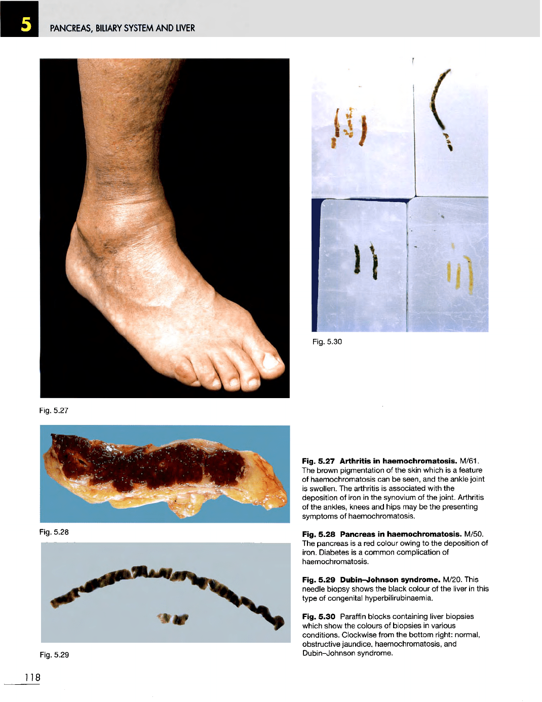
Fig. 5.27
Arthritis
in
haemochromatosis.
M/61.
The
brown pigmentation
of the
skin which
is a
feature
of
haemochromatosis
can be
seen,
and the
ankle joint
is
swollen.
The
arthritis
is
associated with
the
deposition
of
iron
in the
synovium
of the
joint. Arthritis
of
the
ankles,
knees
and
hips
may be the
presenting
symptoms
of
haemochromatosis.
Fig. 5.28
Pancreas
in
haemochromatosis.
M/50.
The
pancreas
is a red
colour owing
to the
deposition
of
iron. Diabetes
is a
common complication
of
haemochromatosis.
Fig. 5.29
Dubin-Johnson
syndrome. M/20. This
needle
biopsy shows
the
black colour
of the
liver
in
this
type
of
congenital hyperbilirubinaemia.
Fig. 5.30
Paraffin
blocks containing
liver
biopsies
which
show
the
colours
of
biopsies
in
various
conditions. Clockwise from
the
bottom right: normal,
obstructive jaundice, haemochromatosis,
and
Dubin-Johnson
syndrome.
118
PANCREAS,
BILIARY
SYSTEM
AND
LIVER
Fig.
5.31
Fig.
5.32
Fig.
5.33
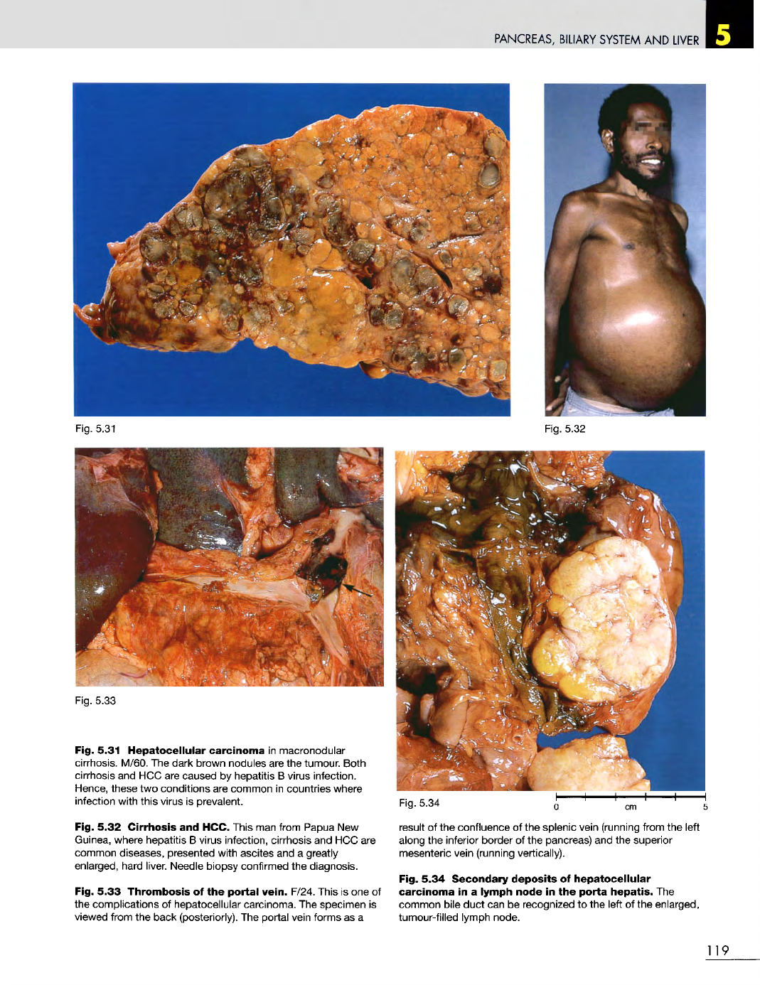
Fig. 5.31
Hepatocellular
carcinoma
in
macronodular
cirrhosis. M/60.
The
dark brown nodules
are the
tumour. Both
cirrhosis
and HCC are
caused
by
hepatitis
B
virus infection.
Hence,
these
two
conditions
are
common
in
countries where
infection with this virus
is
prevalent.
Fig. 5.32
Cirrhosis
and
HCC. This
man
from Papua
New
Guinea, where hepatitis
B
virus infection, cirrhosis
and HCC are
common diseases, presented with ascites
and a
greatly
enlarged, hard liver. Needle biopsy confirmed
the
diagnosis.
Fig. 5.33 Thrombosis
of the
portal
vein.
F/24. This
is one of
the
complications
of
hepatocellular carcinoma.
The
specimen
is
viewed
from
the
back (posteriorly).
The
portal vein forms
as a
Fig.
5.34
result
of the
confluence
of the
splenic vein (running from
the
left
along
the
inferior border
of the
pancreas)
and the
superior
mesenteric vein (running vertically).
Fig. 5.34
Secondary
deposits
of
hepatocellular
carcinoma
in a
lymph
node
in the
porta
hepatis.
The
common bile duct
can be
recognized
to the
left
of the
enlarged,
tumour-filled lymph node.
119
PANCREAS,
BILIARY SYSTEM
AND
LIVER
120
Fig. 5.35
Fig 5.36