Cook R.A., Stewart B. Colour Atlas of Anatomical Pathology
Подождите немного. Документ загружается.

BREAST
AND
FEMALE GENITAL SYSTEM
Fig.
8.46
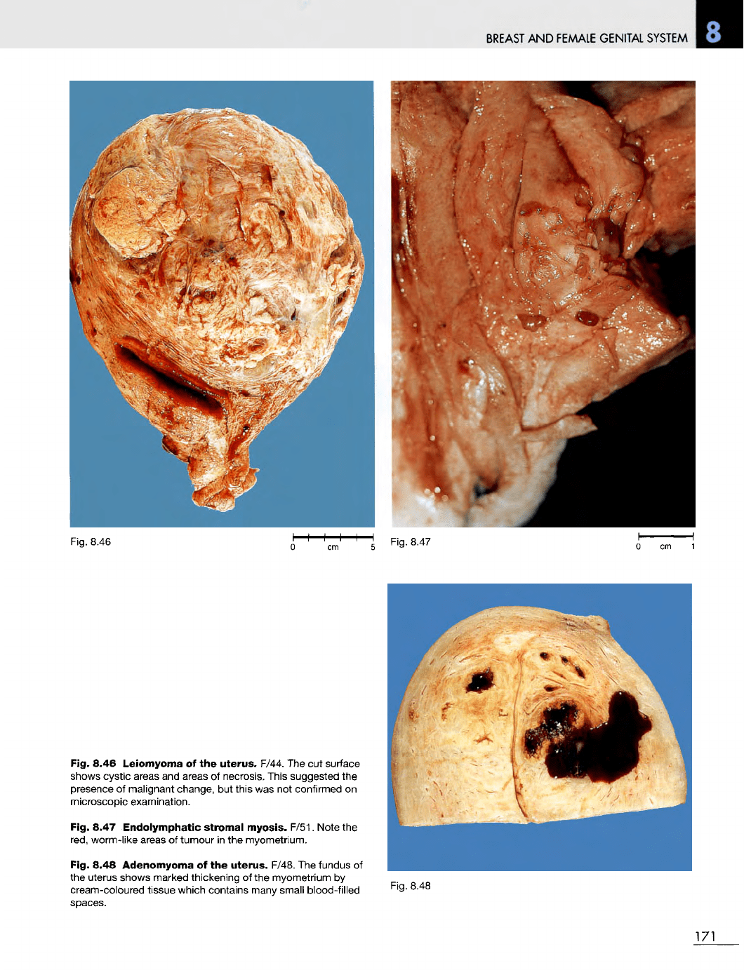
Fig. 8.46
Leiomyoma
of the
uterus.
F/44.
The cut
surface
shows cystic areas
and
areas
of
necrosis. This suggested
the
presence
of
malignant change,
but
this
was not
confirmed
on
microscopic examination.
Fig. 8.47
Endolymphatic
stromal myosis. F/51. Note
the
red,
worm-like areas
of
tumour
in the
myometrium.
Fig. 8.48 Adenomyoma
of the
uterus.
F/48.
The
fundus
of
the
uterus shows marked thickening
of the
myometrium
by
cream-coloured tissue which contains many small blood-filled
spaces.
Fig.
8.48
171
Fig.
8.47
BREAST
AND
FEMALE GENITAL SYSTEM
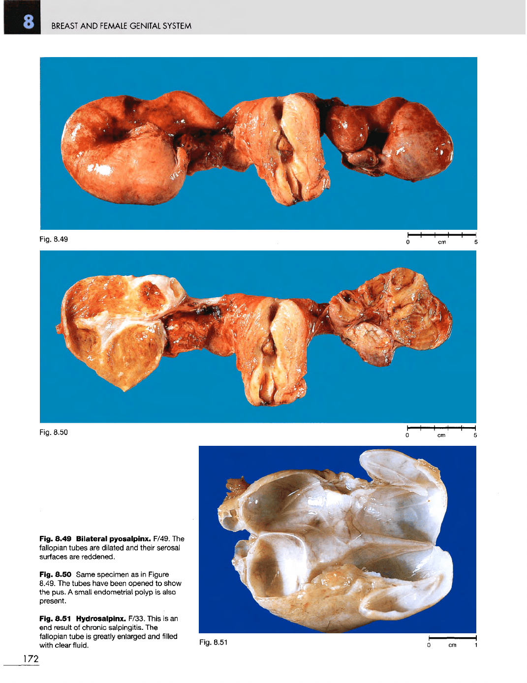
Fig. 8.49
Bilateral
pyosalpinx. F/49.
The
fallopian tubes
are
dilated
and
their serosal
surfaces
are
reddened.
Fig. 8.50 Same specimen
as in
Figure
8.49.
The
tubes have been opened
to
show
the
pus.
A
small
endometrial
polyp
is
also
present.
Fig. 8.51
Hydrosalpinx.
F/33. This
is an
end
result
of
chronic
salpingitis.
The
fallopian
tube
is
greatly enlarged
and
filled
with clear fluid.
Fig. 8.51
172
Fig. 8.49
Fig. 8.50
BREAST
AND
FEMALE
GENITAL
SYSTEM
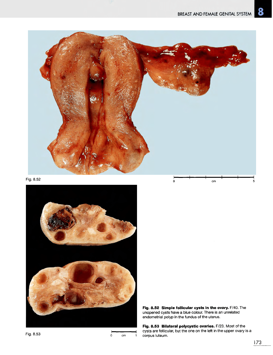
Fig. 8.52
Fig. 8.53
Fig. 8.52
Simple
follicular
cysts
in the
ovary. F/40.
The
unopened cysts have
a
blue colour. There
is an
unrelated
endometrial
polyp
in the
fundus
of the
uterus.
Fig. 8.53
Bilateral
polycystic ovaries. F/23. Most
of the
cysts
are
follicular,
but the one on the
left
in the
upper
ovary
is a
1
corpus luteum.
173
BREAST
AND
FEMALE
GENITAL
SYSTEM
Fig.
8.56
0 cm 1
174
Fig.
8.54
Fig.
8.55
Fig.
8.57
BREAST
AND
FEMALE
GENITAL
SYSTEM
Fig.
8.58
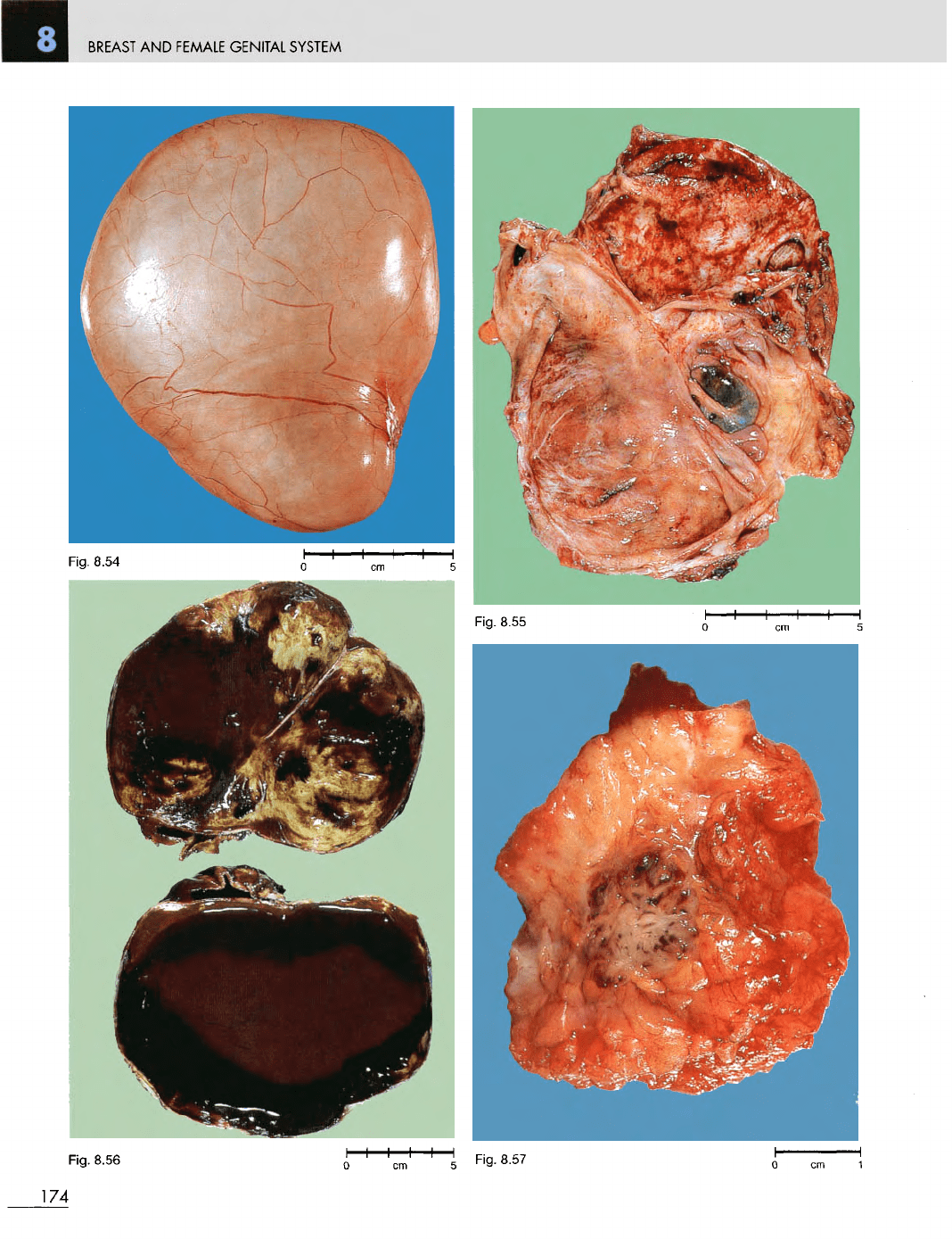
Fig. 8.54
Benign
serous
cystadenoma
of
the
ovary. F/19. This
is a
unilocular,
thin-walled cyst containing clear fluid.
Fig. 8.55
Endometriosis
of the
ovary.
F/40.
The
ovarian cyst
is
multiloculated.
The
blood
has
been removed from most
of
the
loculi.
Fig. 8.56
Endometriosis
of the
ovary.
F/31. Blood
has not
been removed
so as to
demonstrate
the
so-called 'chocolate cyst'.
Fig. 8.57
Extrapelvic
endometriosis.
F/43.
A
subcutaneous lump appeared
at
the
site
of an
abdominal scar,
the
result
of
Caesarian
section
5
years
previously. This
was
excised,
and on
cross-section
the
blood-filled cysts
can be
seen
in the
centre
of the
specimen.
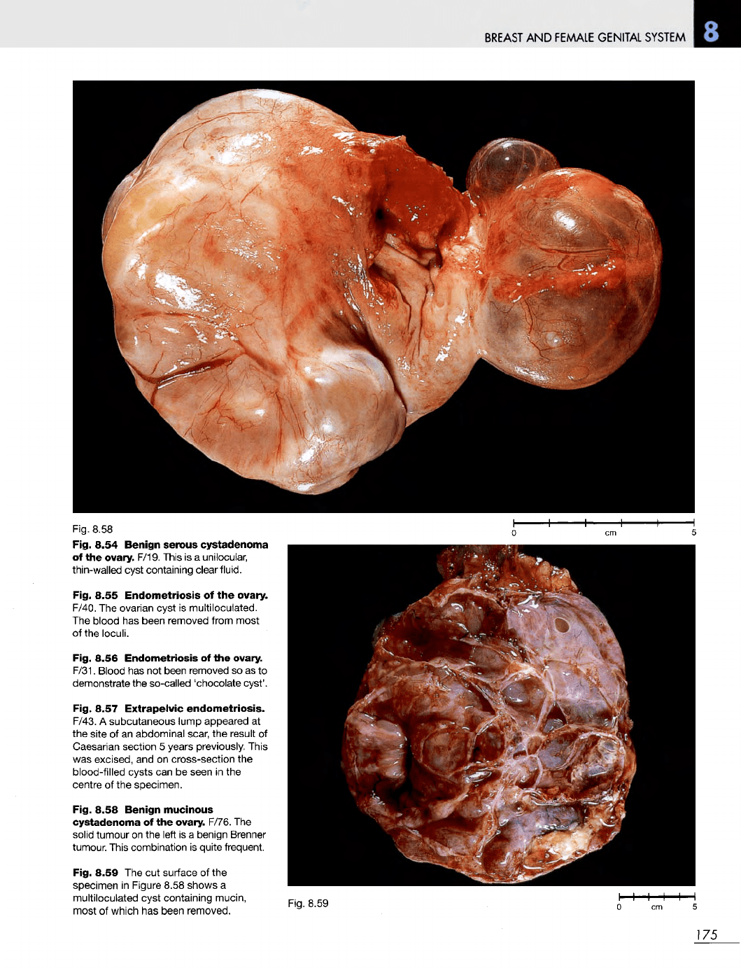
Fig. 8.58
Benign
mucinous
cystadenoma
of the
ovary. F/76.
The
solid
tumour
on the
left
is a
benign Brenner
tumour. This combination
is
quite frequent.
Fig. 8.59
The cut
surface
of the
specimen
in
Figure 8.58 shows
a
multiloculated cyst containing mucin,
most
of
which
has
been removed.
Fig.
8.59
175
BREAST
AND
FEMALE
GENITAL
SYSTEM
176
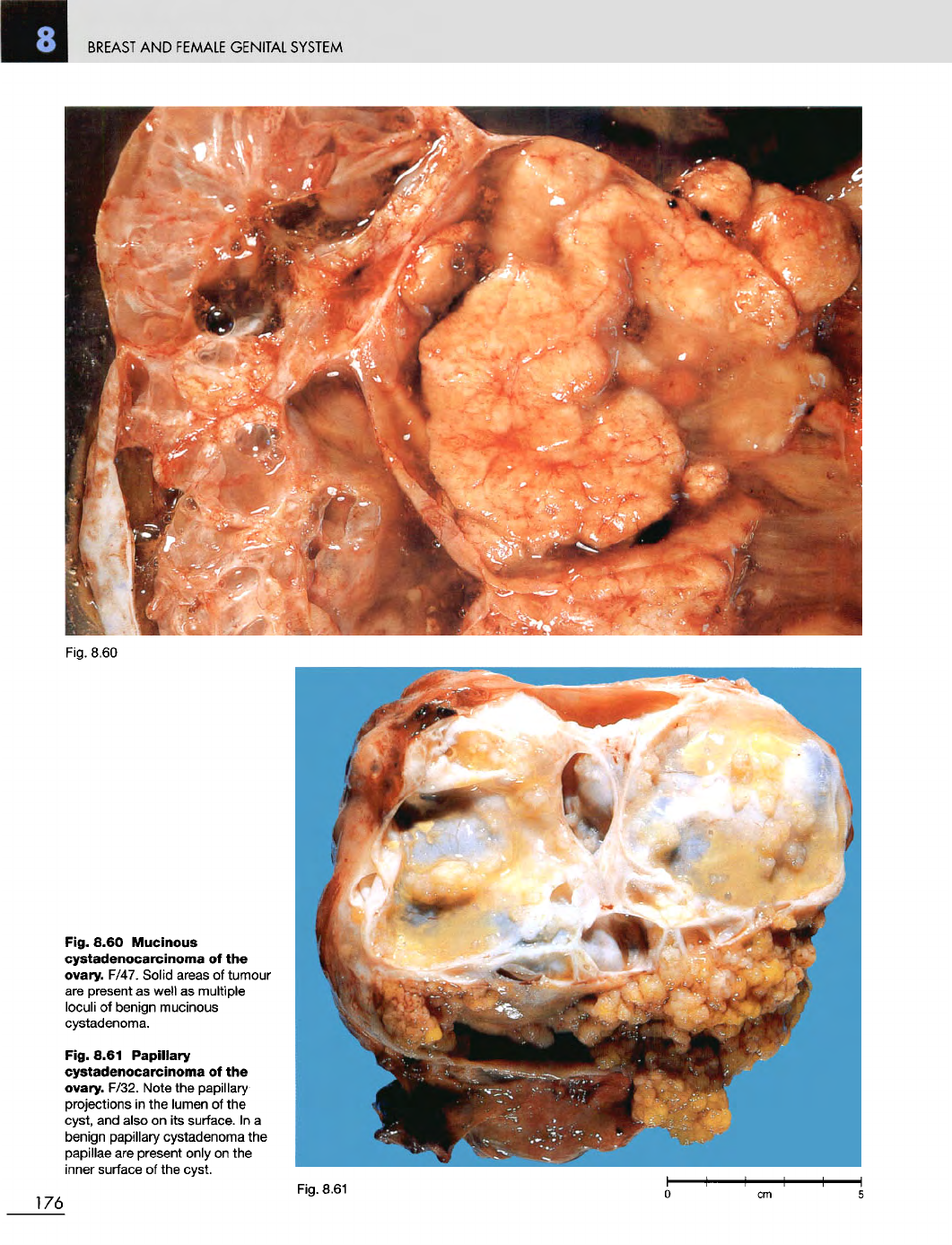
Fig. 8.60
Mucinous
cystadenocarcinoma
of the
ovary.
F/47. Solid areas
of
tumour
are
present
as
well
as
multiple
loculi
of
benign mucinous
cystadenoma.
Fig. 8.61
Papillary
cystadenocarcinoma
of the
ovary. F/32. Note
the
papillary
projections
in the
lumen
of the
cyst,
and
also
on its
surface.
In a
benign papillary cystadenoma
the
papillae
are
present only
on the
inner
surface
of the
cyst.
Fig. 8.61
Fig.
8.60
BREAST
AND
FEMALE GENITAL SYSTEM
Fig. 8.62
Fig. 8.63
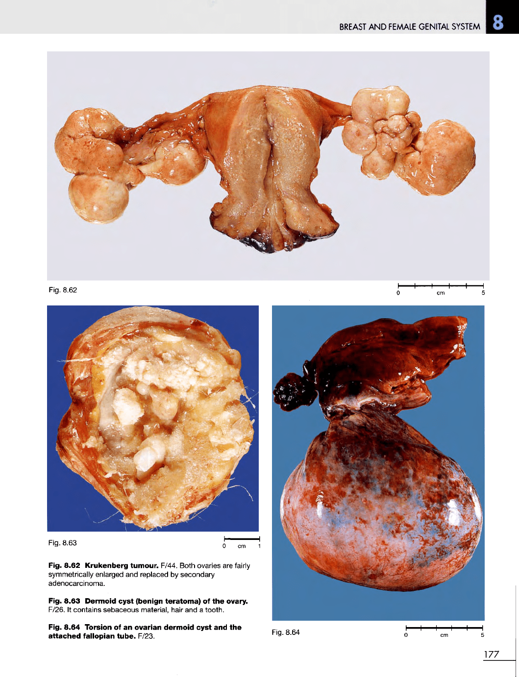
Fig. 8.62
Krukenberg
tumour.
F/44. Both ovaries
are
fairly
symmetrically enlarged
and
replaced
by
secondary
adenocarcinoma.
Fig. 8.63
Dermoid
cyst
(benign
teratoma)
of the
ovary.
F/26.
It
contains sebaceous material, hair
and a
tooth.
Fig. 8.64 Torsion
of an
ovarian
dermoid
cyst
and the
attached
fallopian
tube.
F/23.
Fig. 8.64
177
BREAST
AND
FEMALE
GENITAL
SYSTEM
Fig.
8.67
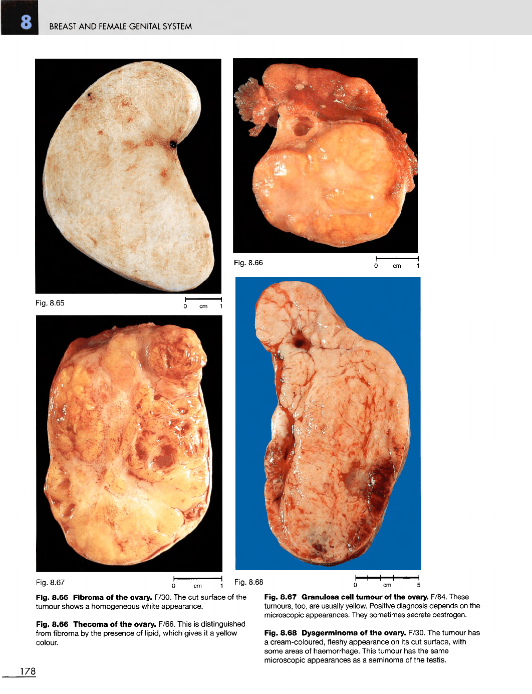
Fig. 8.65
Fibroma
of the
ovary. F/30.
The cut
surface
of the
Fig. 8.67
Granulosa
cell
tumour
of the
ovary. F/84. These
tumour shows
a
homogeneous white appearance.
Fig. 8.66
Thecoma
of the
ovary. F/66. This
is
distinguished
from
fibroma
by the
presence
of
lipid,
which gives
it a
yellow
colour.
tumours, too,
are
usually yellow. Positive diagnosis depends
on the
microscopic appearances.
They
sometimes secrete oestrogen.
Fig. 8.68
Dysgerminoma
of the
ovary. F/30.
The
tumour
has
a
cream-coloured, fleshy appearance
on its cut
surface, with
some areas
of
haemorrhage. This tumour
has the
same
microscopic appearances
as a
seminoma
of the
testis.
178
Fig.
8.65
Fig.
8.66
Fig.
8.68
BREAST
AND
FEMALE
GENITAL
SYSTEM
Fig.
8.69
Fig. 8.70
0 cm
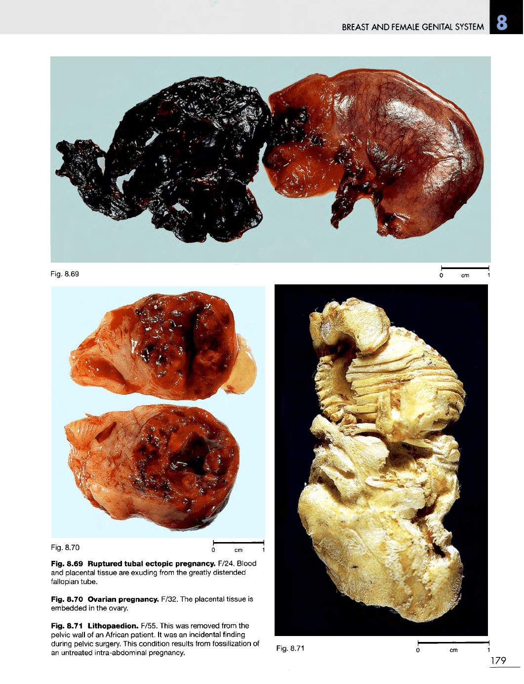
Fig. 8.69
Ruptured
tubal
ectopic pregnancy. F/24. Blood
and
placental tissue
are
exuding from
the
greatly distended
fallopian tube.
Fig. 8.70 Ovarian
pregnancy.
F/32.
The
placental tissue
is
embedded
in the
ovary.
Fig. 8.71
Lithopaedion.
F/55. This
was
removed from
the
pelvic
wall
of an
African patient.
It was an
incidental finding
during pelvic surgery. This condition results from fossilization
of
an
untreated intra-abdominal pregnancy.
Fig.
8.71
179
BREAST
AND
FEMALE
GENITAL
SYSTEM
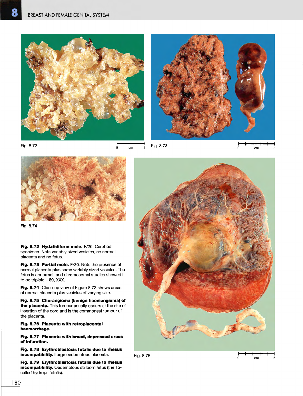
Fig. 8.72
Hydatidiform
mole.
F/26. Curetted
specimen. Note variably sized vesicles,
no
normal
placenta
and no
fetus.
Fig. 8.73
Partial
mole.
F/30. Note
the
presence
of
normal placenta plus some variably sized vesicles.
The
fetus
is
abnormal,
and
chromosomal studies showed
it
to
be
triploid
- 69,
XXX.
Fig. 8.74 Close-up view
of
Figure 8.73 shows areas
of
normal placenta plus vesicles
of
varying size.
Fig. 8.75 Chorangioma
(benign
haemangioma)
of
the
placenta.
This tumour usually occurs
at the
site
of
insertion
of the
cord
and is the
commonest tumour
of
the
placenta.
Fig. 8.76
Placenta
with
retroplacental
haemorrhage.
Fig. 8.77
Placenta
with
broad,
depressed
areas
of
infarction.
Fig. 8.78
Erythroblastosis
fetalis
due to
rhesus
incompatibility.
Large oedematous placenta.
Fig. 8.79
Erythroblastosis
fetalis
due to
rhesus
incompatibility.
Oedematous stillborn fetus (the
so-
called hydrops fetalis).
180
Fig.
8.72
Fig.
8.73
Fig.
8.74
Fig 8.75