Cui Dongmei. Atlas of Histology: with functional and clinical correlations. 1st ed
Подождите немного. Документ загружается.


CHAPTER 14
■
Oral Cavity
265
Teeth
Introduction and Key Concepts for Teeth
Teeth are prominent structures in the oral cavity. They can be
divided into maxillary (upper) and mandibular (lower) teeth.
The root of the tooth is surrounded by the alveolar bone (alveo-
lar process or alveolar arch), which forms a socket to hold and
support each tooth. The alveolar bone of the maxillary teeth
is a part of the maxilla (upper jaw); the alveolar bone of the
mandibular teeth is a part of the mandible (lower jaw). There
are two sets of teeth: primary (baby) and permanent teeth. The
primary teeth are eventually replaced by permanent teeth. In
adults, there are 32 permanent teeth including two incisors, one
canine, two premolars, and three molars on each of the four
quadrants in the maxillary and mandibular arches (Fig. 14-7).
Each tooth can be divided into three parts: the crown, the
cervix (neck), and the root. The crown of the tooth is covered
by enamel and extends above the gingiva (gum); the shape of
the crown is unique in different types of teeth and is adapted to
their functions. The cervix is also called the neck of the tooth
and is the junction between the crown and root. The region
where the enamel and cementum meet is called the cemen-
toenamel junction (CEJ). The root of the tooth is covered by
cementum and surrounded by the alveolar bone. A tooth has
one or more roots; the apex is the end of the root. The api-
cal foramen is a small opening where nerves and blood vessels
enter and exit the dental pulp (Figs. 14-1 and 14-7).
TOOTH DEVELOPMENT (ODONTOGENESIS) stems from
two different origins: ectodermal and neural crest–derived mes-
enchymal tissue. The oral epithelium derives from ectodermal
tissue, which gives rise to the dental lamina, and later becomes
the enamel organ. The dental papilla derives from mesenchymal
tissue and gives rise to the future dental pulp. The enamel organ,
dental papilla, and dental sac (follicle) as a whole are described
as a tooth germ, which eventually forms a tooth. The initiation
of tooth germs occurs along the oral epithelium of the maxillary
and mandibular prominences (processes). Tooth germs undergo
initiation phases (development of the dental lamina and initia-
tion of tooth germs), morphogenesis phases (cell movement and
formation of the shape of the tooth), and histogenesis phases
(formation of the hard tissue and development of the tooth
root). Although tooth development is a continuous process,
odontogenesis can be divided into several stages based on the
shape of the tooth germ and formation of the tooth structure.
These stages include initiation, bud, cap, bell, and apposition
(crown) stages. When the primitive oral cavity is forming, the
fi rst pharyngeal arch gives rise to the maxillary and mandibu-
lar prominences or processes (developing dental arches), which
lead to the future upper and lower jaws (Fig. 14-9A).
1. Initiation stage: At about 6 to 7 weeks into development,
some oral epithelial cells on the surface of the maxillary
and mandibular prominences increase proliferation activ-
ity, become thicker, and invaginate into the underlying mes-
enchymal tissue to form the primary epithelial band (Fig.
14-9A).
2. Bud stage: At about 8 to 9 weeks, the primary epithelial
band gives rise to the vestibular lamina
and dental lamina.
The vestibular lamina forms a cleft that becomes the vesti-
bule between the cheek and tooth; the dental lamina forms
a U-shaped structure and develops into tooth buds (enamel
organs and mesenchymal tissue), which become primary
deciduous teeth (Fig. 14-9A). The development of perma-
nent teeth comes from the secondary tooth buds, which
sprout from the primary tooth buds.
3. Cap stage: At about 10 to 11 weeks, the enamel organ
appears as a cap shape and the condensed mesenchymal
cells beneath the enamel organ form the dental papilla (Fig.
14-9B).
4. Bell stage: At about 12 to 14 weeks, the enamel organ con-
tinues to grow into a bell shape, and cells of the enamel
organ differentiate into four distinguishable cell layers: the
outer enamel epithelium, inner enamel epithelium, stellate
reticulum, and stratum intermedium. At the same time, the
dental papilla grows and helps to form the shape of the
tooth crown. Cells in the dental papilla are differentiated
into outer and inner cell groups. The outer cells of the dental
papilla will develop into odontoblasts that produce future
dentin; the inner cells will develop into future dental pulp
tissues (Fig. 14-10A,B).
5. Apposition (crown) stage: At about 18 to 19 weeks, cells of
the tooth germ continue to differentiate; the inner enamel
epithelial cells have become preameloblasts which induce
the outer cells of the dental papilla to become odontoblasts
and begin to produce the dentinal matrix at the tooth crown
region. This material is called predentin, and after undergo-
ing calcifi cation will become dentin. When dentin is formed,
it induces preameloblasts to differentiate and become active
ameloblasts, which produce enamel. Thus, the production of
both the enamel matrix and the dentin for the tooth crown
has begun (Fig. 14-11A,B). When the two hard tissues, den-
tin and enamel, have formed and the shape of the crown is
completed, the root of the tooth begins to develop. The tooth
then starts to erupt into the oral cavity in order to make
room for the tooth root to grow (Fig. 14-12A,B).
ENAMEL, DENTIN, AND DENT
AL PULP are components
of teeth. Enamel is the hardest tissue in the body. The basic
morphological unit of enamel is the rod, also called the prism,
which is composed of a head and a tail. The rods are arranged
in a three-dimensional complex, perpendicular to the dentinoe-
namel junction (DEJ). They extend from the DEJ to the surface
of the enamel. Enamel provides a seal for the dentin and makes
a strong surface for chewing (Fig. 14-14A,B). The crown of the
tooth is covered by enamel and the root of the tooth is covered
by cementum. Dentin surrounds and forms the walls of the den-
tal pulp. It is composed of numerous dentinal tubules, odon-
toblastic processes, and the dentinal matrix and is the second
hardest tissue of the body (Fig. 14-15A,B). The central core of
the tooth is occupied by dental pulp, which is made up of loose
mucous connective tissue containing blood vessels and nerve
fi bers (Fig. 14-17A,B).
THE PERIODONTIUM refers to the structures surrounding
and supporting the tooth root and includes the cementum, the
PDL, and the alveolar bone. The cementum is a thin layer of hard
tissue that covers the root dentin (Fig. 14-18A,B). The PDL is
the dense fi brous connective tissue that attaches the cementum to
the alveolar bone (Figs. 14-18C and 14-19A). The alveolar bone,
also called the alveolar process, is part of the maxilla and man-
dible. The alveolar bone supports and protects the tooth root. It
includes the alveolar crest, the alveolar bone proper, and support-
ing bone
(Figs. 14-18C and 14-19A).
CUI_Chap14.indd 265 6/2/2010 8:23:14 AM
266
UNIT 3
■
Organ Systems
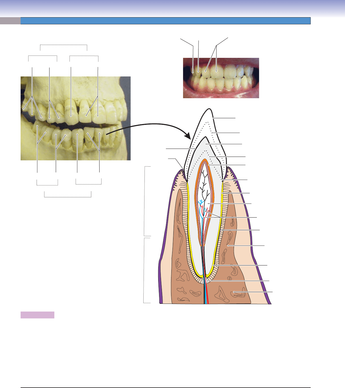
Figure 14-7. Overview of the teeth.
There are two sets of teeth that form during a person’s lifetime: the primary (baby) teeth and the secondary (adult) teeth. The
secondary teeth will eventually replace the primary teeth. The secondary teeth are more commonly called permanent teeth by
dentists. There are 32 permanent teeth in an adult. Each of the four quadrants includes two incisors, one canine, two premolars,
and three molars in the mandibular and maxillary dental arches (left). The tooth is composed of several types of hard tissues: dentin,
enamel, cementum, and the alveolar bone. The central core of each tooth is a chamber containing dental pulp made up of mucous
connective tissue with a rich supply of nerve fi bers and blood vessels. Each tooth has a crown, cervix, and root (see Fig. 14-1). The
crown of the tooth is covered by enamel, the hardest tissue found in the body; the cervix is the junction between the crown and
root; and the surface of the root is covered by cementum, which connects to the alveolar bone by the PDL. The junction between
the enamel and cementum is the CEJ, and the border between the dentin and enamel is the DEJ.
T. Yang
Incisors
Incisors
Incisors
Canine
Canine
Canine
Canine
Canine
Molars
Molars
Premolars
Premolar
Premolars
Maxillary teeth
Mandibular teeth
Anterior teeth
Anterior teeth
Posterior teeth
Posterior teeth
Enamel
Dentin
DEJ
Neonatal line of enamel
Neonatal line of dentin
Cementoenamel junction
Acellular
cementum
Cellular
cementum
Alveolar bone
(alveolar process)
Apical
foramen
Gingiva
Alveolar
mucosa
Gingival
sulcus
Dental pulp
Dental pulp
Dental pulp
Nerve fibers and
blood vessels
Predentin
Predentin
Predentin
Alveolar
Alveolar
bone
bone
Alveolar
bone
Periodontal
ligament (PDL)
CUI_Chap14.indd 266 6/2/2010 8:23:14 AM
CHAPTER 14
■
Oral Cavity
267
Tooth Development (Odontogenesis)
5. Apposition stage
(dentinogenesis)
6. Apposition stage
(amelogenesis)
7. Root formation
and eruption
8. Function
2. Bud stage
(weeks 8–9)
1. Initiation stage
(weeks 6–7)
3. Cap stage
(weeks 10–11)
4. Bell stage
(weeks 12–14)
Dental
lamina
Dental
Dental
lamina
lamina
Dental
lamina
Dental
Dental
Dental
papilla
Dental
Dental
papilla
papilla
Dental
Dental
papilla
papilla
Dental
papilla
Dental
Dental
papilla
papilla
Dental
papilla
Dental
Dental
pulp
pulp
Dental
papilla
Dental
pulp
Dentin
Dentin
Dentin
Enamel
Enamel
Enamel
E
E
n
n
a
a
m
m
e
e
l
l
o
o
r
r
g
g
a
a
n
n
Enamel
organ
Enamel
Enamel
organ
organ
Enamel
organ
Primary
epithelial band
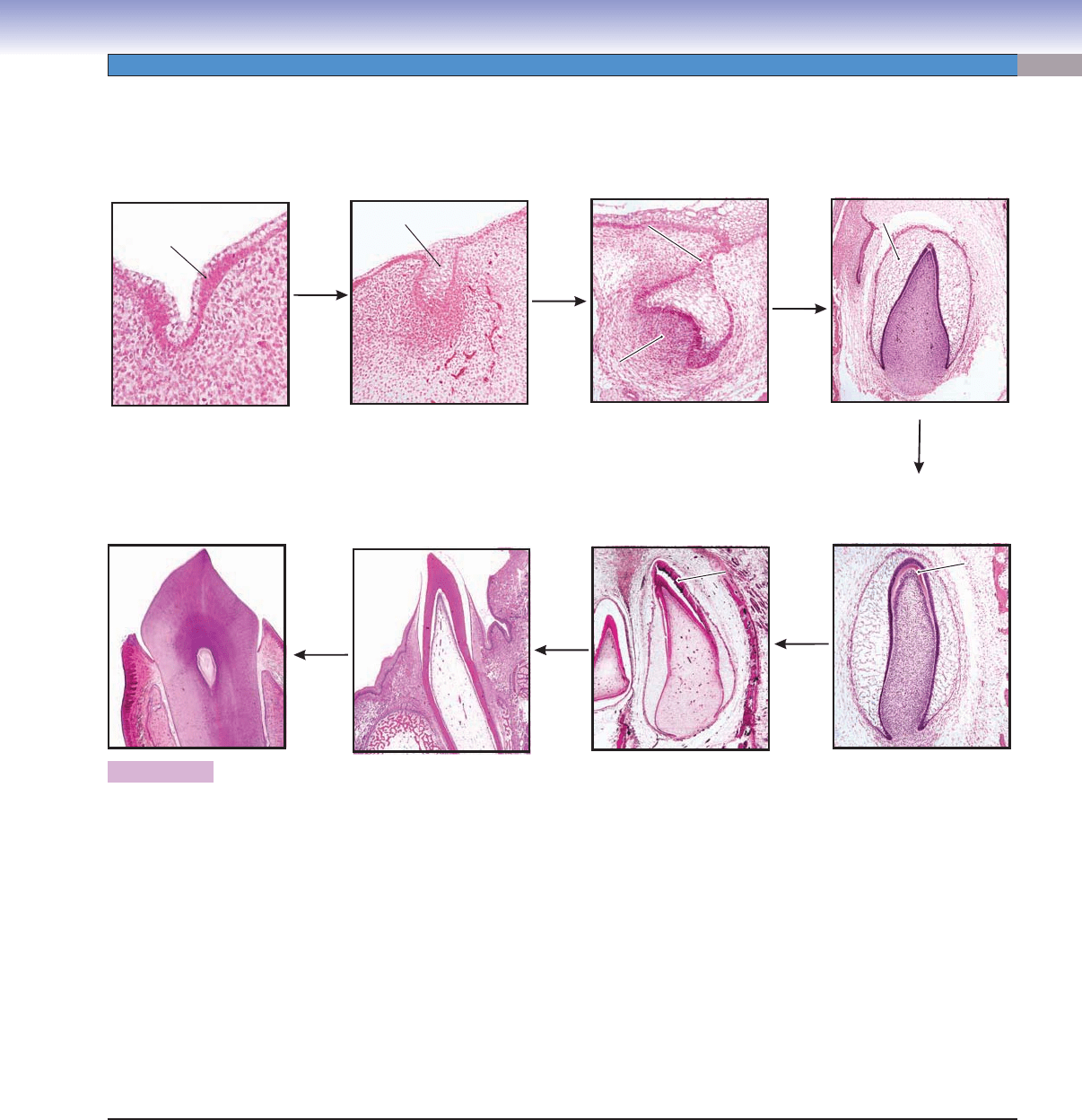
Figure 14-8. Overview of tooth development (odontogenesis). H&E, 5 to 128
Tooth development is also called odontogenesis. The tooth derives from two types of tissues: ectoderm (oral epithelium) and
mesenchymal tissues that originate from the neural crest. The oral epithelium forms a U-shaped structure called the dental lamina,
which develops into an enamel organ. The mesenchymal tissue develops into a dental papilla and also forms a dental sac (follicle),
which surrounds the dental papilla and enamel organ. The enamel organ, dental papilla, and dental sac (follicle) collectively are
described as a tooth germ. Tooth development is a continuous process and can be described in several stages (like snapshots) based on
the shape of the tooth germs: initiation, bud, cap, bell, and apposition stages. The illustrations represent the process of tooth develop-
ment. (1) Initiation stage: Proliferating oral epithelial cells become thickened and form a primary epithelial band. (2) Bud stage: The
dental lamina (derived from the primary epithelial band) forms a U-shaped structure (tooth bud). (3) Cap stage: The enamel organ
develops into a cap shaped structure. At the same time mesenchymal tissue becomes condensed and forms the dental papilla. (4) Bell
stage: The enamel organ continues to grow into a bell shape that has fi ve distinct cell layers (Fig. 14-10A,B). (5) Apposition stage
(dentinogenesis): Odontoblasts begin to form dentin on the crown region. (6) Apposition stage (amelogenesis): After a layer of dentin
is formed at the crown of the tooth, the formation of enamel begins (produced by ameloblasts). (7) Root formation and eruption:
When the shape of the crown and the dentin and enamel of the crown have completed, the tooth begins to develop its root structure
(cementum, PDL). During this process, the tooth gradually erupts into the oral cavity. (8) Function: Development of the tooth root
and its associated structures continues until the tooth is functional.
CUI_Chap14.indd 267 6/2/2010 8:23:16 AM
268
UNIT 3
■
Organ Systems
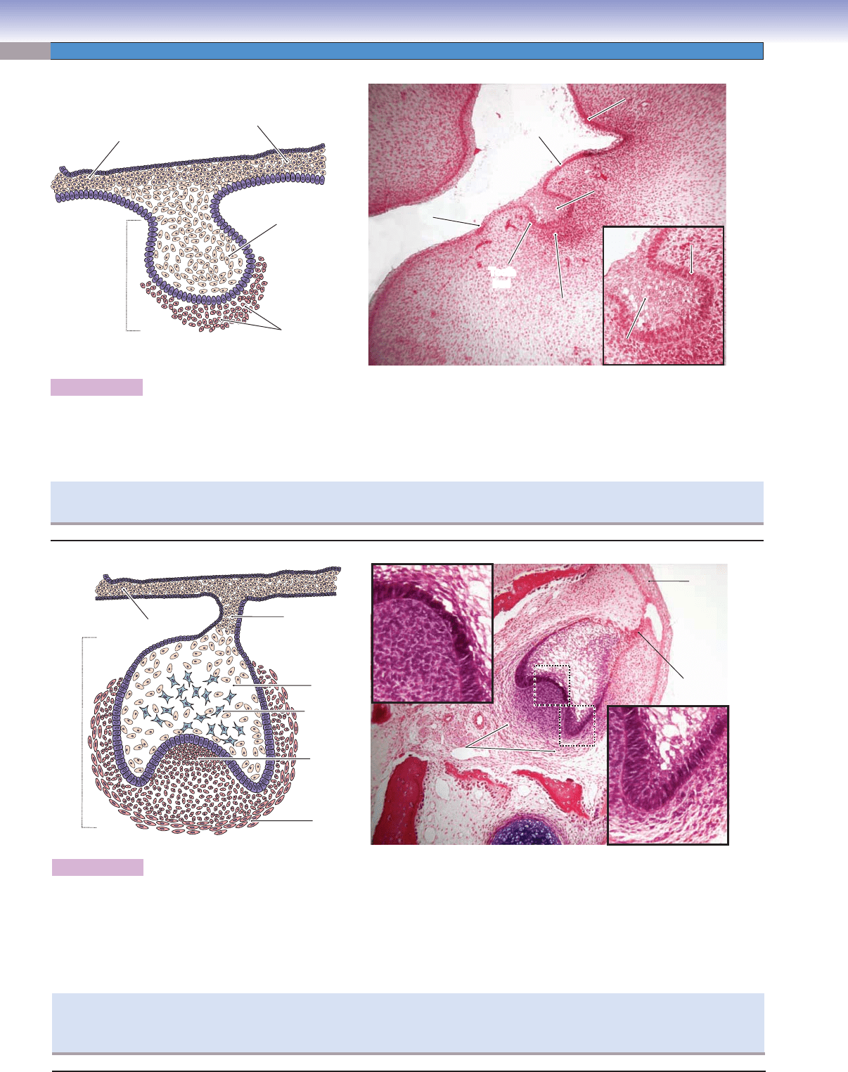
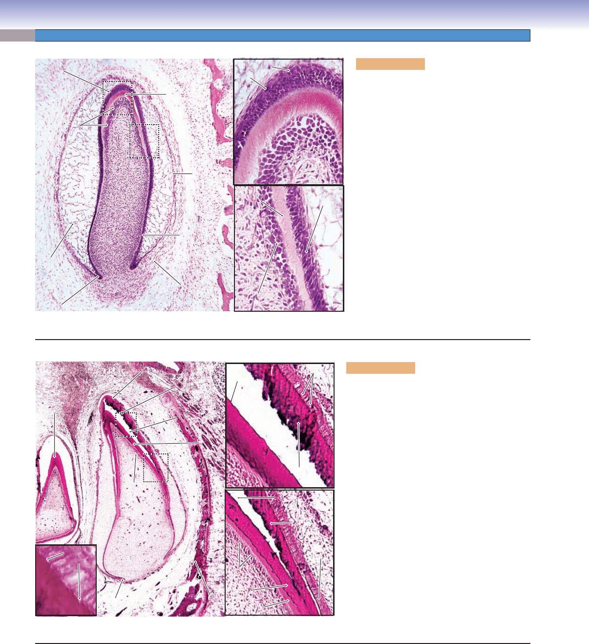
Figure 14-9A. Bud stage, weeks 8–9. H&E, 72; inset 140
The bud stage is a continuation of the initiation stage in which the proliferating and thickening oral epithelium forms the primary
epithelial band. In the bud stage, the tooth germ forms a budlike structure that is surrounded by proliferating and accumulating
mesenchymal cells. The combination of the dental lamina and condensed mesenchymal tissues is called the tooth bud and develops
into a primary tooth. The maxillary and mandibular prominences lead to the future upper and lower jaws.
T. Yang
Dental
lamina
Oral
epithelium
Oral
epithelium
Mesenchymal
cells
Tooth
bud
Tooth
bud
Tooth
bud
Tooth bud
Tooth bud
Tooth bud
Primary
Primary
epithelial band
epithelial band
Primary
epithelial band
Dental
Dental
lamina
lamina
Dental
lamina
Dental
Dental
lamina
lamina
Dental
lamina
cells
cells
Mesenchymal
Mesenchymal
Mesenchymal
cells
Mandibular
Mandibular
prominence
prominence
(process)
(process)
Mandibular
prominence
(process)
Oral
epithelium
Maxillary
Maxillary
prominence
prominence
(process)
(process)
Maxillary
prominence
(process)
Oral
epithelium
A
During the bud stage, any disturbance can cause the formation of abnormal teeth, such as microdontia (abnormally small teeth)
and macrodontia (abnormally large teeth).
T. Yang
Dental
papilla
Enamel
organ
Dental
lamina
Oral
epithelium
Dental
sac
Tooth
germ
Dental
Dental
papilla
papilla
Dental
papilla
Dental
Dental
sac (follicle)
sac (follicle)
Dental
sac (follicle)
Dental
Dental
papilla
papilla
Dental
papilla
Enamel
Enamel
organ
organ
Enamel
organ
Dental
lamina
Oral
epithelium
Dental
Dental
sac
sac
Dental
sac
Stellate
reticulum
B
Figure 14-9B. Cap stage, weeks 10–11. H&E, 72; inset 204
The cap stage is a continuation of the bud stage and results from the enlargement and unequal growth of the tooth bud. At this stage,
the enamel organ is formed and attaches to the remaining dental lamina. The base of the enamel organ becomes concave and appears
as a cap-shaped structure with mesenchymal cells beneath it. These mesenchymal cells are condensed, forming a dental papilla that
will form the future dental pulp. Other mesenchymal cell layers surrounding the enamel organ and dental papilla are called the
dental sac (dental follicle). At this stage, the morphogenesis and formation of the tooth germ are ongoing. The cell proliferation and
movement determine the shape of the tooth.
A disturbance during the cap stage of tooth development can cause clinical tooth fusion (two adjacent tooth germs fuse and
develop into a large macrodontic tooth), or gemination (one tooth germ develops into two teeth which share a single pulp but have
two crowns [Fig. 14-13A]) and dens invaginatus (or dens in dente, meaning, “tooth within a tooth”).
CUI_Chap14.indd 268 6/2/2010 8:23:19 AM
CHAPTER 14
■
Oral Cavity
269
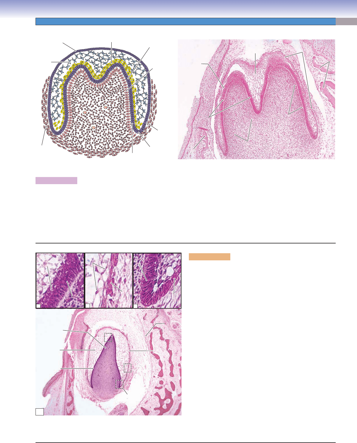
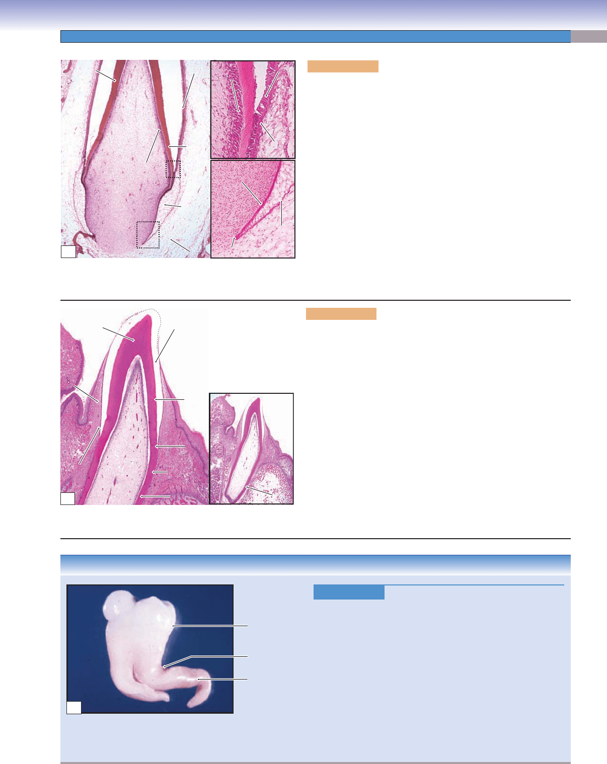
Figure 14-10A. Bell stage, weeks 12–14. H&E, 72
The bell stage is a continuation of the cap stage. The tooth germ undergoes further morphogenesis, proliferation, and differentiation.
At this stage, the enamel organ detaches from the dental lamina and loses connection with the oral epithelium. The dental lamina
gradually degenerates. However, the enamel organ continues to differentiate and forms four cell layers: the inner enamel epithelium,
stratum intermedium, stellate reticulum, and outer enamel epithelium (Fig. 14-10B). Some cell types may also be found in the later
cap stage. The dental papilla differentiates and forms the outer cells of the dental papilla and the inner cells of the dental papilla near
the base of the enamel organ. As the tooth germ continues to grow, the outer cells of the dental papilla will differentiate into future
odontoblasts; the inner cells of the dental papilla will differentiate into future dental pulp tissues.
T. Yang
Outer enamel
epithelium
Outer
enamel
epithelium
Stellate
reticulum
Dental
sac
Inner enamel
epithelium
Inner enamel
epithelium
Stratum intermedium
Outer cells
of dental
papilla
Cervical
loop
Bone
Bone
Bone
Outer enamel
Outer enamel
epithelium
epithelium
Outer enamel
epithelium
Stellate
Stellate
reticulum
reticulum
Stellate
reticulum
Stratum
intermedium
Enamel
Enamel
organ
organ
Enamel
organ
Outer cells of
Outer cells of
dental papilla
dental papilla
Outer cells of
dental papilla
Dental
Dental
papilla
papilla
Dental
papilla
Inner cells of
Inner cells of
dental papilla
dental papilla
Inner cells of
dental papilla
Bone
Bone
Bone
Inner enamel
Inner enamel
epithelium
epithelium
Inner enamel
epithelium
A
1
2
3
Stratum
Stratum
intermedium
intermedium
Stratum
intermedium
Inner enamel
Inner enamel
epithelium
epithelium
Inner enamel
Inner enamel
epithelium
epithelium
Inner enamel
epithelium
Inner enamel
epithelium
Stellate
Stellate
reticulum
reticulum
Stellate
reticulum
Outer
Outer
enamel
enamel
epithelium
epithelium
Outer
enamel
epithelium
Outer
Outer
enamel
enamel
epithelium
epithelium
Outer
enamel
epithelium
Outer enamel
Outer enamel
epithelium
epithelium
Outer enamel
epithelium
Dental
sac
Bone
Bone
Bone
Cervical
loop
Dental
papilla
Enamel
organ
Mesenchymal
tissue
Inner enamel
epithelium
Outer cells of
dental papilla
Stellate
reticulum
1
2
3
3
3
B
Figure 14-10B. Bell stage, cell layers. H&E, 37; insets 472
During the differentiation process of the enamel organ at the bell
stage, four cell layers are distinguishable. (1) The inner enamel
epithelium is a row of columnar cells that contain rich RNA and
are much taller in the tip region where initial enamel formation
will take place at the apposition stage. The inner enamel epithe-
lium differentiates and later becomes ameloblasts. (2) The stra-
tum intermedium is composed of two to three layers of squamous
or cuboidal cells and is located between the stellate reticulum and
inner enamel epithelium. Cells in this layer are rich in alkaline
phosphatase, which assists in the inner enamel epithelium pro-
duction of enamel. (3) The stellate reticulum is located within
the enamel organ. It may also be present in the cap stage, but is
more developed in the bell stage. The cells of the stellate reticu-
lum are star shaped and have many cellular processes intercon-
nected with one another to form a network within the enamel
organ. The extracellular spaces of the stellate reticulum are large
and fi lled with a fl uidlike, fl uffy material. These cells contain
glycosaminoglycans and alkaline phosphatase and have desmo-
somes and gap junctions between the cells. The stellate reticulum
plays a role in maintaining tooth shape and protecting underly-
ing dental tissue. (4) The outer enamel epithelium is composed of
cuboidal cells and is the outermost layer of the enamel organ. This
cell layer separates the enamel organ from the nearby mesenchy-
mal tissues. It serves as a protective barrier and also helps main-
tain the shape of the enamel organ. The inner enamel epithelium
and outer enamel epithelium meet to form the cervical loop.
CUI_Chap14.indd 269 6/2/2010 8:23:25 AM
270
UNIT 3
■
Organ Systems
Dental
Dental
papilla
papilla
Dental
Dental
papilla
papilla
Dental
Dental
papilla
papilla
Dental
papilla
Preameloblasts
Preameloblasts
Preameloblasts
Dentinal
Dentinal
matrix
matrix
Dentinal
matrix
Odontoblasts
Odontoblasts
Odontoblasts
Stratum
Stratum
intermedium
intermedium
Stratum
intermedium
Preameloblasts
Preameloblasts
Preameloblasts
Dentinal
Dentinal
matrix
matrix
Dentinal
matrix
Odontoblasts
Odontoblasts
Odontoblasts
Cervical
Cervical
loop
loop
Cervical
loop
Stellate
Stellate
reticulum
reticulum
Stellate
reticulum
Enamel
Enamel
organ
organ
Enamel
organ
Odontoblasts
Odontoblasts
Odontoblasts
Preameloblasts
Preameloblasts
Preameloblasts
Dentinal
Dentinal
matrix
matrix
Dentinal
matrix
Outer enamel
Outer enamel
epithelium
epithelium
Outer enamel
epithelium
Inner enamel
Inner enamel
epithelium
epithelium
Inner enamel
epithelium
Dental
Dental
sac
sac
Dental
sac
A
Figure 14-11A. Apposition (crown) stage,
dentinogenesis. H&E, 76; insets 315
During the apposition (crown) stage, induction
occurs between the ectodermal (enamel organ)
and mesenchymal (dental papilla) tissues. The
inner enamel epithelium from the enamel organ
has become preameloblasts, which induce
outer cells of the dental papilla (Fig. 14-11B)
to differentiate into odontoblasts. When
odontoblasts become mature and active, they
appear columnar in shape and begin to secrete
dentinal matrix (predentin), which is the fi rst
hard tissue formed during tooth development.
The formation of dentin is called dentinogen-
esis. The crown of the tooth develops much
earlier than the root of the tooth. The crown
dentin is fi rst produced as predentin by odon-
toblasts. The predentin soon becomes calcifi ed
and is called dentin (Fig. 14-11B). The cervical
loop is formed by the inner enamel epithelium
and the outer enamel epithelium. Along with
the dental sac, the cervical loop will contribute
to develop future root structures.
Ameloblasts
Ameloblasts
Ameloblasts
E
E
n
n
a
a
m
m
e
e
l s
l
s
p
p
a
a
c
c
e
e
Ename
l space
Dentin
Dentin
Dentin
DEJ
Enamel matrix
Enamel matrix
(uncalcified)
(uncalcified)
Enamel matrix
(uncalcified)
Enamel matrix
Enamel matrix
(uncalcified)
(uncalcified)
Enamel matrix
(uncalcified)
Ehe en space
Ehe en space
Enamel space
Ameloblasts
Ameloblasts
Ameloblasts
Odontoblasts
Odontoblasts
Odontoblasts
Predentin
Predentin
Predentin
Dentin
Dentin
Dentin
Alveolar
Alveolar
bone
bone
Alveolar
bone
Epithelial
Epithelial
diaphragm
diaphragm
Epithelial
diaphragm
Tomes
Tomes
processes
processes
Tomes
processes
Dental
Dental
papilla
papilla
Dental
papilla
Dental
Dental
papilla
papilla
Dental
papilla
Odontoblasts
Odontoblasts
Odontoblasts
Enamel matrix
Enamel matrix
(uncalcified)
(uncalcified)
Enamel matrix
(uncalcified)
Enamel space
Enamel space
Enamel space
Dentin
Dentin
Dentin
Ameloblasts
Ameloblasts
Ameloblasts
Dentin
Dentin
Dentin
E
E
n
n
a
a
m
m
e
e
l
l
m
m
a
a
trix
trix
Enamel
matrix
B
Figure 14-11B. Apposition (crown) stage,
amelogenesis. H&E, 19; insets (right) 104;
inset (left) 354
The formation of crown dentin induces preamelo-
blasts to differentiate and become ameloblasts.
Active ameloblasts are columnar cells, and their
nuclei are located toward the stratum interme-
dium. They actively secrete enamel matrix with
assistance from the stratum intermedium. The
newly secreted enamel matrix, in contact with the
dentin matrix, forms the DEJ. At the same time,
the ameloblasts and the odontoblasts retreat
from the DEJ as deposition of the matrix pro-
ceeds. Enamel formation moves outward (toward
the enamel organ), and dentin formation moves
inward (toward the dental pulp). The process of
enamel formation is called amelogenesis. Dur-
ing amelogenesis, conical processes, known as
Tomes processes, are developed at the secretory
(apical) surface of the ameloblasts. In the apposi-
tion stage, the two types of hard tissues (dentin
and enamel) begin to form at the tooth crown.
These two types of hard tissues are formed in a
regular rhythm.
CUI_Chap14.indd 270 6/2/2010 8:23:29 AM
CHAPTER 14
■
Oral Cavity
271
Stratum
Stratum
intermedium
intermedium
Stratum
intermedium
Odontoblasts
Odontoblasts
Odontoblasts
Ameloblasts
Ameloblasts
Ameloblasts
Ameloblasts
Ameloblasts
Ameloblasts
Enamel space
DEJ
Reduced
enamel
organ
Dental
Dental
sac
sac
Dental
sac
Primary
apical
foramen
Odontoblasts
Dental
papilla
Crown dentin
Crown dentin
Crown dentin
Inner enamel
epithelium
Outer enamel
epithelium
Begining of root sheath
Begining of root sheath
Begining of root sheath
A
Figure 14-12A. Tooth root development. H&E, 29; (upper)
inset 160; (lower) inset 90
Root development begins after the formation of the crown has been
completed. It involves three structures: the epithelial root sheath,
the dental sac (follicle), and the dental papilla. Dentin formation
proceeds from the crown to the root. The formation of the root
begins at the epithelial root sheath (Hertwig epithelial root sheath),
which develops from the cervical loop (Fig. 14-11A). The inner and
outer enamel epithelia of the cervical loop extend to form the root
sheath. The epithelial root sheath grows around the dental papilla.
It bends at a 45-degree angle and is called the epithelial diaphragm
(Fig. 14-11B). The epithelial diaphragm gradually encloses the dental
papilla with the exception of the apical foramen. The root sheath is
important in forming the shape of the root. It induces the outer cell
layer of the dental papilla to differentiate into odontoblasts, which
produce the root dentin. When the root dentin has formed, the mes-
enchymal cells from the dental sac come in contact with the surface
of the root dentin and induce these cells to differentiate into cemen-
toblasts, which produce cementum. Fibroblasts, which differentiate
from the dental sac, begin to form the PDL.
Enamel
space
Root
Root
dentin
dentin
Root
dentin
Tooth
Tooth
root
root
Tooth
root
Cementum
Cementum
Cementum
DEJ
CEJ
CEJ
CEJ
Crown
dentin
Initial
Initial
gingiva
gingiva
Initial
Initial
junction
junction
epithelium
epithelium
Initial
junction
epithelium
Dental
Dental
Pulp
Pulp
Dental
Pulp
Initial
gingiva
B
Figure 14-12B. Tooth eruption. H&E, 14; inset 5
As root length increases, additional space is needed for root growth, and
the tooth gradually moves and erupts into the oral cavity. Tooth erup-
tion and root growth, therefore, occur at the same rate. The formation
of the tooth root includes root dentinogenesis (formation of root den-
tin), cementogenesis (formation of cementum), pulp formation, forma-
tion of the PDL, and alveolar bone development. The crown of the tooth
passes through the bony crypt, dental epithelium, and oral epithelium to
emerge into the oral cavity. As the tooth moves vertically, the overlying
bone gradually becomes absorbed by osteoclasts. The dental epithelium
(outer enamel epithelium, stellate reticulum, stratum intermedium, and
ameloblasts) is compressed into a thin layer called the reduced enamel
epithelium (REE). The oral epithelium fuses with the REE and gradu-
ally degenerates, enabling the tooth crown to erupt into the oral cavity.
The initial junction epithelium is created during tooth movement and
eruption; later it becomes the junction epithelium (Fig. 14-1), forming
a seal between the gingiva and enamel. In a regular H&E stain, highly
calcifi ed enamel disappears because of the decalcifi cation process during
tissue preparation.
CLINICAL CORRELATION
Figure 14-12C.
Dilaceration.
Dilaceration is a developmental anomaly of the tooth in
which there is an abrupt bend between the crown and the
root of a tooth. T
rauma to the primary predecessor tooth and
developmental disturbances to the primary tooth germ are the
possible causes of this condition. Dilaceration can happen in
any tooth, but is more common in mandibular third molars,
maxillary second premolars, and mandibular second molars.
Visual examination can fi nd a crown dilaceration, but the x-ray
is the most appropriate diagnostic tool to diagnose a root dilac-
eration. Dilacerations may be associated with some syndromes,
such as Smith-Magenis syndrome, a chromosomal disorder
that produces a set of characteristic physical, behavioral, and
developmental features. Dilacerations can complicate cavity
preparation, root canal preparation, and other treatments.
Abrupt bend
Tooth crown
Tooth root
C
CUI_Chap14.indd 271 6/2/2010 8:23:35 AM
272
UNIT 3
■
Organ Systems
CLINICAL CORRELATIONS
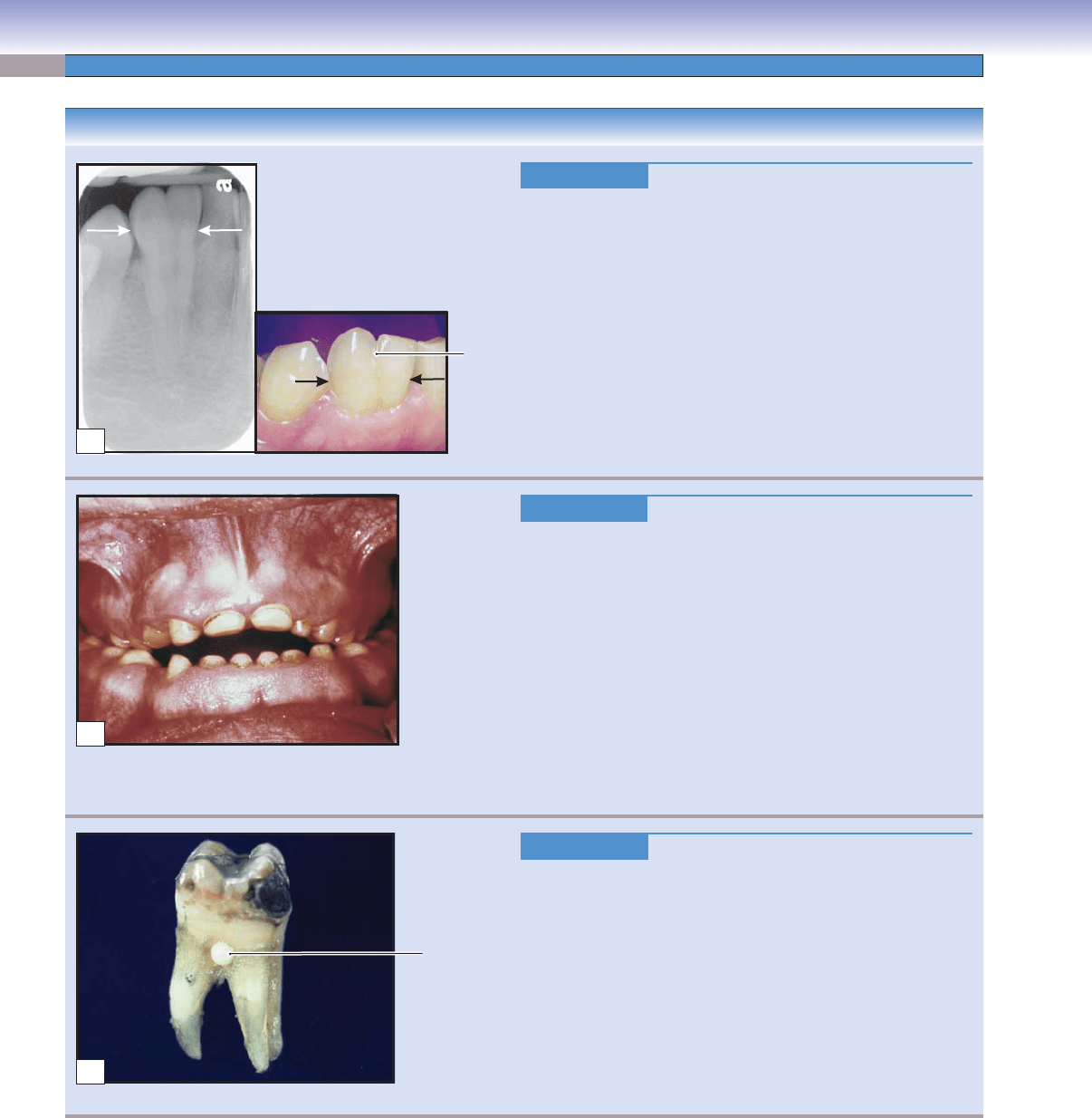
Figure 14-13A.
Gemination, Incisor.
Gemination is a variation in tooth structure that occurs when
two teeth develop from the same tooth bud, resulting in a larger
than normal tooth crown, but with a normal number of teeth.
The gemination results from a disturbance that occurs at the
cap stage. During the cap stage, the tooth germ is undergo-
ing early morphogenesis and beginning to form the shape of
the tooth. Gemination represents the unsuccessful division of a
single tooth germ into two tooth germs. There is a deep cleft on
the surface of the tooth, giving the appearance of two crowns.
The radiograph at left shows two crowns (arrows) sharing one
root. Occasionally
, x-ray images may show two independent
pulp chambers and root canals. The cause of gemination is
unknown.
B
Figure 14-13B.
Amelogenesis Imperfecta.
Amelogenesis imperfecta is a group of hereditary disorders
that affect the dental enamel formation at the apposition and
maturation stages. The teeth are covered with a thin, defec-
tive enamel or lack enamel covering. Mutations in different
genes may affect enamel proteins, such as amelogenin, which
is involved in enamel mineralization. The most common forms
of amelogenesis imperfecta include hypoplastic and hypocal-
cifi ed types. In the hypoplastic form, the teeth do not have a
suffi
cient amount of enamel, whereas in the hypocalcifi ed form,
the quantity of enamel is normal, but it is soft and easily worn.
Affected teeth are most often yellow-brown and blue-gray in
color, and are vulnerable to dental caries, damage, and loss.
Full crowns will improve the appearance of the teeth and pro-
tect the teeth from damage.
Enamel
pearl
C
Figure 14-13C.
Enamel Pearl.
Enamel pearl is an ectopic formation of enamel that is usually
found close to the CEJ between tooth roots. Localized failure of
Hertwig epithelial root sheath to separate from the dentin may
be the cause of this anomaly
. The enamel pearl results from a
disturbance during the apposition or maturation stages during
the tooth development. An enamel pearl may lead to periodon-
tal disease because of the extension of a periodontal pocket.
Histologically, enamel pearls have reduced enamel epithelium
and are surrounded by normal cementum. X-rays can reveal
enamel pearls. Enamel pearls cannot be removed by scaling,
but they can be ground away to restore the normal shape of the
tooth. An enamel pearl on a molar is shown here.
Cleft
A
CUI_Chap14.indd 272 6/2/2010 8:23:38 AM
CHAPTER 14
■
Oral Cavity
273
CLINICAL CORRELATION
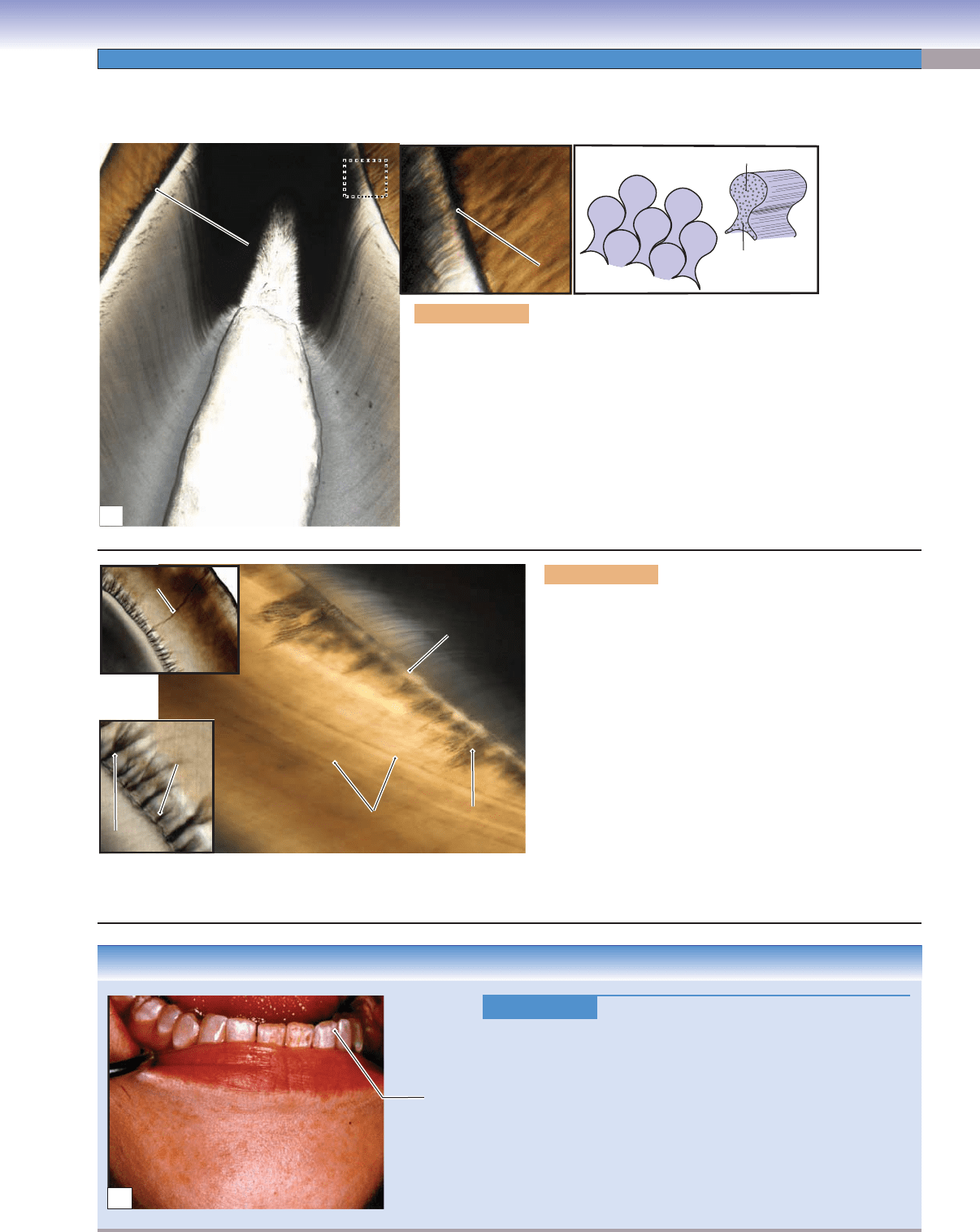
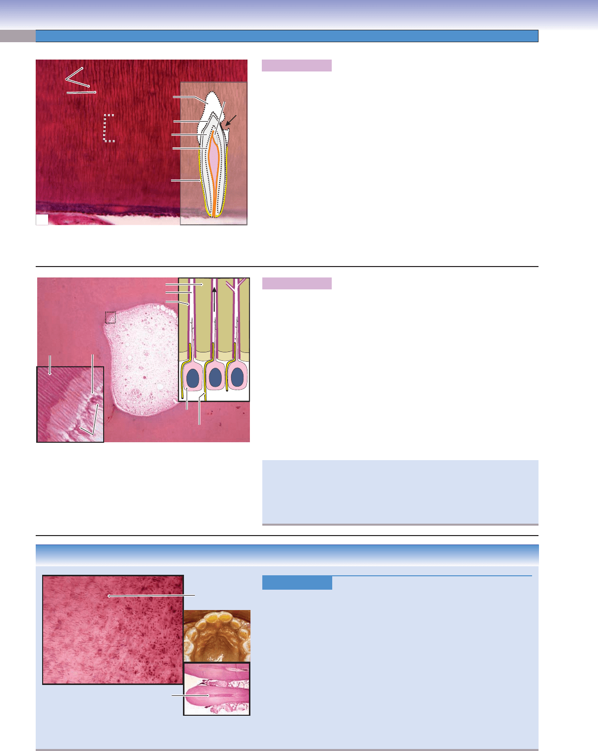
Figure 14-14C.
Enamel Fluorosis.
Enamel fl uorosis is the general term for enamel changes caused
by excessive fl uoride intake during tooth development (before age
8 years). Fluoride helps to prevent and control tooth caries, but
too much fl
uoride intake during tooth development can result in
hypomineralization of the enamel surface. These changes are char-
acterized by diffuse, opaque, and white streaks that run horizontally
across the enamel. Stains with rough, irregular enamel surfaces are
common. A fl uoride-induced toxic effect on ameloblasts during
enamel formation is believed to be the mechanism of the condition.
Prevention is through controlling the fl uoride intake, and treat-
ments include bleaching and enamel microabrasion.
Enamel, Dentin, and Dental Pulp
D. Cui
Tail
Head
Rods (prisms)
Dental
pulp
Dental
pulp
Dentin
Dentin
Dentin
Dentin
Dentin
Dentin
DEJ
DEJ
DEJ
DEJ
DEJ
DEJ
Dentin
Dentin
Dentin
Enamel
Enamel
Enamel
Enamel
Enamel
Enamel
A
Figure 14-14A. Enamel, tooth. Ground specimen, 17; inset 46
Enamel is the hardest tissue in the body and is highly mineralized. It is composed
of 96% inorganic material in the form of hydroxyapatite (crystalline calcium
phosphate), 1% organic material, and 3% water. Enamel covers the dentin of the
crown to make the tooth surface strong and suitable for chewing and to seal and
protect the dentin. Enamel cannot be renewed after enamel formation has been
completed, because enamel is produced by ameloblasts, which disappear after
tooth eruption. However, enamel can be strengthened by fl uoride. This ground
specimen shows the enamel, dentin, and DEJ. The basic morphological unit of
enamel is the rod (prism). Each rod has a head and tail and is tightly packed with
hydroxyapatite crystals. The rods are arranged in a three- dimensional complex
and are oriented generally perpendicular to the DEJ.
DEJ
DEJ
DEJ
Dentin
Dentin
Dentin
Enamel
Enamel
lamella
lamella
Enamel
lamella
Enamel
Enamel
Enamel
Enamel
Enamel
spindle
spindle
Enamel
spindle
Enamel tuft
Enamel tuft
Enamel tuft
Incremental lines
Incremental lines
(striae of Retzius)
(striae of Retzius)
Incremental lines
(striae of Retzius)
Enamel tufts
Enamel tufts
Enamel tufts
B
Figure 14-14B. Enamel structures, tooth. Ground speci-
men, 68; inset (upper) 13; inset (lower) 62
The incremental growth lines of the enamel, also called
the striae of Retzius, can be found in either cross (bands)
or longitudinal sections (arcs) of the mature enamel. These
patterns refl ect the changes in enamel secretory rhythm.
The neonatal line (see Fig. 14-7) is much more prominent
than the striae. It is a landmark that indicates the transition
from enamel produced before birth and enamel produced
after birth. This line is produced by a metabolic change that
occurs at birth. There are three defects of enamel: (1) Enamel
tufts are hypomineralized areas fi lled with organic material
at the DEJ and toward the surface; (2) enamel lamellae are
hypomineralized, thin, sheetlike defects that can run through
the entire enamel and are commonly caused by cracks; and
(3) enamel spindles are thin, needlelike lines extending from
the DEJ to the enamel and are because of odontoblast pro-
cesses trapped in the enamel during early amelogenesis.
White
streaks
C
CUI_Chap14.indd 273 6/2/2010 8:23:41 AM
274
UNIT 3
■
Organ Systems
T. Yang / D. Cui
Mantle dentin
Mantle dentin
Mantle dentin
Circumpulpal dentin
Circumpulpal dentin
Circumpulpal dentin
Secondary dentin
Secondary dentin
Secondary dentin
Cementum
Cementum
Cementum
Enamel
Enamel
Enamel
Tertiary
Tertiary
dentin
dentin
Tertiary
dentin
Primary dentin
Primary dentin
Primary dentin
Dentinal
Dentinal
tubules
tubules
Dentinal
tubules
Dentinal
Dentinal
Matrix
Matrix
Dentinal
Matrix
Dental pulp
A
Figure 14-15A. Dentin, tooth. H&E, 284
Dentin, which forms the bulk of the tooth, is covered by enamel on the
crown and cementum at the root. It is about 70% inorganic materials
(hydroxyapatite), 20% organic components (type I collagen and proteo-
glycans), and 10% water. Dentin is harder than bone and cementum but
weaker than enamel. Dentin can be classifi ed into three types: (1) Primary
dentin, deposited before the formation of the tooth root and tooth eruption
have been completed, includes mantle dentin (at the DEJ) and circumpulpal
dentin; (2) secondary dentin, produced after tooth eruption and root
formation have been completed, is deposited very slowly and is located
beneath the primary dentin; and (3) tertiary (reparative) dentin is produced
in response to injures (caries [arrow], drilling, or attrition). Tertiary dentin
is produced only by the odontoblasts that are directly stimulated when
the tooth is injured. This type of dentin has few, mostly irregular, dentin
tubules, which provide a seal to prevent bacteria and harmful molecules
from invading the dental pulp. Dentin has three basic components: dentinal
tubules, dentinal matrix, and odontoblastic processes (Fig. 14-15B).
T. Yang / J. Naftel
Fluid
Dentinal tubule
Intertubular dentin
Peritubular dentin
Dentin
Dentinal
tubule
Odontoblastic
process
Dental
pulp
Nerve
ending
Odontoblast
Dentin
D
D
e
e
n
n
tin
tin
Dentin
P
P
re
re
d
d
e
e
n
n
tin
tin
Predentin
Odontoblasts
Odontoblasts
Odontoblasts
B
Figure 14-15B. Dentin and dentinal tubules, tooth. H&E, 35; inset
359
Dentin is produced by odontoblasts, and the formation of dentin is
continuous throughout life. During the dentinogenesis process, the cell
bodies of the odontoblasts retreat, but their cytoplasmic processes remain
and are embedded in the mineralized dentinal matrix. Each odontoblastic
process resides in a narrow channel, the dentinal tubule, which is lined by
highly calcifi ed peritubular dentin and traverses the entire thickness of the
dentin layers. The dentinal matrix, between the dentinal tubules, is called
intertubular dentin and is less mineralized than the peritubular dentin.
The dentinal tubules run from the dental pulp surface to the DEJ. These
tubules are larger close to the pulp and more branched near the DEJ. Each
dentinal tubule contains an odontoblastic process about halfway toward
the DEJ; the rest of the space is fi lled with fl uid. Nerve fi bers for pain
extend from the dental pulp to the inner part of the dentinal tubule.
CLINICAL CORRELATION
Figure 14-15C.
Dentinogenesis Imperfecta. H&E, 329; inset
(lower) ×2
Dentinogenesis imperfecta is an autosomal dominant genetic disorder of
tooth development caused by mutations in the dentin sialophosphoprotein
gene. Affected teeth are often blue-gray or yellow-brown in color and
translucent. Dentinogenesis imperfecta is classifi ed as types I–III. Type I
is associated with osteogenesis imperfecta, in which bones are brittle
and easily fractured and pulp chambers are diminished. Type I collagen
defects are also associated with type I dentinogenesis imperfecta. Type
II is characterized by diminished pulp chambers and may be associated
with progressive hearing loss. Type III has large pulp chambers. Patients
suffer frequent enamel fractures and enamel attrition. Full crowns will
improve the appearance of the teeth and protect the teeth from damage.
Histologically, dentinal tubules are irregular and are larger than normal
in diameter, and uncalcifi ed matrix may be present.
Irregular
dentinal
tubules
Diminished
pulp chamber
C
Under certain circumstances, such as in sensitive teeth, the exposure of
dentin to air blasts or drinking or eating cold, hot, or sweet substances
will cause movement of the fl uid in the dentinal tubules. This move-
ment produces a mechanical disturbance that activates the nerve end-
ings of pain fi bers, inducing intense pain (hydrodynamic theory).
CUI_Chap14.indd 274 6/2/2010 8:23:48 AM