Hatano Y., Katsumura Y., Mozumder A. (Eds.) Charged Particle and Photon Interactions with Matter - Recent Advances, Applications, and Interfaces
Подождите немного. Документ загружается.

520 Charged Particle and Photon Interactions with Matter
In the core, energy is deposited by direct interactions with the ion (Section 19.1.1), and the density
of energy deposition is much higher than in the penumbra. Anionic and cationic base radicals form
very close to each other and columbic forces drive substantial recombination of these radicals before
they are trapped at 77K. Neutral sugar radicals, however, are able to survive core recombinations
more easily and as a consequence, most of the trapped neutral sugar radicals are produced in the core.
With the presumption that most of the base radicals are produced in the penumbra, it is pos-
sible to use their yields to determine the partition of energy between the core and the penumbra.
For example, in the sample illustrated in Figure 19.3 and Table 19.5, one can conclude that 53% of
the ion energy (0.10/0.19) is deposited in the penumbra, and the remainder in the core. For a series
of experiments using argon ion irradiation, it was concluded that for ve samples with hydration
9≥Γ≥ 16 D
2
O/nucleotide and LET ≤ 650keV/μm, the percentage energy deposited in the penumbra
ranged
from 49% to 56%, or roughly 50%.
The
large absolute Gs for neutral sugar radical production with ion-beams are not simply
explained by the partition of energy. A further hypothesis regarding the effects of track structure
on yields is that excited state reactions in the core are partially responsible for the relatively high
yield of sugar radicals (Becker et al., 2003). The high density of energy deposition in the core could
clearly lead to excited states. For those excited states that are created on ion radicals, excited state
chemical reaction channels likely exist. A striking conrmation of this hypothesis was obtained in
a series of papers in which the direct excitation of G
•+
in DNA and in model compounds and of A
•+
in
model compounds leads to sugar radicals (Becker et al., 2007; Becker and Sevilla, 2008; Kumar
and
Sevilla, 2008a). This work is described in detail in Section 19.5.
Further
LET effects with regard to the irradiation of hydrated DNA are observed in the yields of
the prompt strand-break radicals
ROPO
2
i−
and
C
dephos
′
3
i
. The yield of these radicals with argon and
oxygen ion-beams is ca. 10 times higher than that found with γ-irradiation (Tables 19.1 and 19.5).
The pathway for formation of these radicals originates with LEE, so a question arises regarding what
factor is causing a approximate 10-fold increase in the number of such radicals. The likely answer
is that an electronic and/or vibrational excited state is involved in strand-break formation. Thus,
LEE capture induces excited anionic states that are one source of
ROPO
2
i−
and
C
dephos
′
3
i
. A second
source can arise from excited states of existing anionic radicals. For example, the higher density of
energy deposition in the core facilitates formation of excited states in proximity to existing radicals.
DFT calculations also indicate that LEE capture by DNA can be modeled by calculations of excited
states of the anion radicals. Thus excited states are very much involved in LEE capture by DNA and
clearly lead to subsequent strand breaks (Kumar and Sevilla, 2007, 2008b, 2009b).
Because a strand break that is part of a multiply damaged site (damage cluster) is likely to be
less repairable than an isolated strand break (vide infra), the spatial clustering of radicals is highly
S
S
δ-ray
Ar
18+
ca. 50% of energy
in core
ca. 50% of energy
in penumbra
+
+
–
–
+
+
–
–
–
+
–
+
–
+
r
core
= ca. 6.8 nm
S
S
S
S
S
S
S
S
S = sugar radicals, ROPO
2
•
–
G
•
+
, C(N3)H
•
, T
•
–
•
•
•
•
•
•
•
•
•
•
•
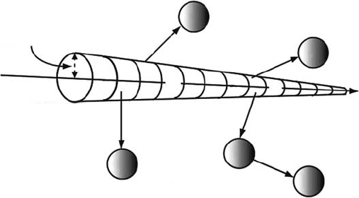
Figure 19.4 Schematic depiction of chemical track structure from Ar ion beam irradiation of hydrated DNA.

PhysicochemicalMechanisms of Radiation-Induced DNA Damage 521
relevant to understanding damage pathways. A pulsed electron double resonance (PELDOR) study
of argon ion irradiated hydrated DNA found that clusters of radicals with a local radical concen-
tration higher than the overall radical concentration do exist (Bowman et al., 2005). In this study,
the observing magnetic eld was set outside the range of the base radical ESR spectra, but on the
neutral sugar radical spectrum, and a strong pulse was applied. The pulse causes transitions in most
of the radicals present. The effect of the pulse on the neutral sugar radicals is then observed. It was
concluded
that clusters of radicals do exist, as shown in Table 19.6.
What
is most remarkable is that in the 1.7 kGy dose sample, although the average radical concen-
tration is 0.20μmol/cm
3
, the cluster radical concentration is 18.8μmol/cm
3
. This is conrmation of
the existence of signicant multiply damaged sites in Ar-irradiated DNA. Since the observing mag-
netic eld detects effects on the neutral sugar radicals, and the experimental technique used detects
mainly a local concentration of radicals, the spherical cluster radius observed is actually a measure
of the core radius (Figure 19.4). In addition, because the core is largely populated with neutral sugar
radicals, it is likely that more than one of the radicals in a cluster will lead to a strand break. Since
two strand breaks within 10–20 base pairs (3.4–6.8nm) on opposite strands is generally regarded to
constitute a double strand break, if two breaks on opposite strands occur within a cluster, a double
strand
break will almost certainly result.
The
track parameters reported, that is, the core radius, the radical concentration in the core, and
the energy partition between the core and penumbra all agreed well with track-structure calcula-
tions
(Magee and Chatterjee, 1987; Chatterjee and Holley, 1993; Chatterjee, 2006).
19.4.3 Strand breakS and daMage cluSterS froM heavy-ion direct effectS
Very little work has been published (also see Chapter 21 for further information) regarding strand
breaks from ion-beam irradiation using experimental protocols that assure direct-type effects
are being measured. Using hydrated (Γ = 35 H
2
O/nucleotide) pUC18 prepared in a tris buffer,
Urushibara et al. (2008) determined the yield of ssb, dsb, and damage clusters in pUC18 DNA using
α-radiations of varying LET at 5.6°C. Using α-radiation, it was found that the yield of frank ssb
decreased as LET increased (Table 19.7) and that heat treatment did not change the yield of ssb.
Contrary to the behavior of ssb, the yield of dsb increased to a maximum at an LET of 141keV/μm,
and then dropped at 148keV/μm.
Determination of the yield of newly formed ssb after enzymatic treatment with Nth and Fpg to
reveal isolated base damage indicated the presence of such signicant damage at the lower LETs
used, but the yield of these sites decreased markedly as the LET increased. In addition, measure-
ment of newly formed dsb after treatment with Nth and Fpg, to reveal damage clusters, indicated the
presence of signicant yields of such clusters, which also appeared to decrease as LET increased.
This latter decrease was interpreted as possibly being an artifact caused by the failure of the
BER glycolases to function properly, rather than as an actual decrease in base lesions. However, as
indicated earlier (vide infra), there actually is a substantial decrease in initial trapped base radical
yields as LET increases, caused by the partition of energy in a core and a penumbra, and the effects
table 19.6
multiply
d
amaged
s
ites
in a
r-irradiated
dna
track parameter
value for doses
1.7–50 kgy
Radicals
in cluster 17.7 ± 0.7
Cluster
radius (spherical cluster assumed) 6.8
nm
Radical
concentration in cluster
18.8
μmol/cm
3
Radical concentration for whole sample (dose dependent)
0.20μmol/cm
3

522 Charged Particle and Photon Interactions with Matter
thereof. It may be the case that the loss of enzyme sensitive sites is real and due to track-structure
effects. Earlier experimental evidence from the same group using γ-irradiation showed that the
yields of ssb and dsb do not increase for Γ ≥ 15, suggesting that the indirect effect is suppressed in
the
samples (Yokoya et al., 2002).
In
earlier experiments by the same team, α-particle irradiated DNA was investigated at three
hydration levels, from dry DNA to Γ = 35 H
2
O/nucleotide (Yokoya et al., 2003). The dsb yield
increased from the rst to the second hydration level, and then appeared to plateau for fully hydrated
DNA. At all three hydration levels used, the yield of dsb was two to three times higher than that
found for γ-irradiated samples, indicating clusters of damage originated in the ion track core. In
a manner consistent with the work cited above, fewer Nth and Fpg enzyme sensitive sites were
detected than one might expect. As discussed earlier, it may be the case that the lack of enzyme
sensitive sites is actually due to fewer base lesions being formed rather than the inability of the BER
enzymes
to properly catalyze strand cleavage.
19.4.4 daMage by Soft and ultraSoft x-rayS
Extensive literature exists regarding the damage caused by soft and ultrasoft x-rays. For these radia-
tions, the photoelectric effect induces inner shell ionizations of light atoms, and damage is caused
both by the original ionization of the target atom as well as by the resulting auger and secondary
electrons as well. These beams have LETs higher than that of γ-rays or hard x-rays. A discussion
of these effects is outside the scope of this chapter, but recent reviews (Yokoya et al., 2006) as well
Chapter 21 in this book can be consulted.
19.5 exCited states oF ion-radiCals as dna damage preCursors
19.5.1 Sugar–phoSphate daMage via excited StateS of dna baSe ion-radicalS
Early ESR studies using γ-irradiated hydrated salmon sperm DNA (Wang et al., 1994) showed
that the yield of neutral sugar radicals increased with radiation dose, in apparent direct cor-
relation with the loss of existing one-electron oxidized guanine radicals (Wang et al., 1994).
This observation suggested that, in these γ-irradiated hydrated DNA samples, a portion of the
table 19.7
yields
of s
trand
b
reaks
in
- 2008 — 2026 «СтудМед»