Kasper C., van Griensven M., P?rtner R. (Eds.) Bioreactor Systems for Tissue Engineering II: Strategies for the Expansion and Directed Differentiation of Stem Cells
Подождите немного. Документ загружается.


Table 2 Occurrence and actions of growth factors in developing and mature cartilage, diseased cartilage tissue, and applications in cartilage cultivation
in vitro
Growth factor/
receptor
Role in the development of
cartilage tissue in vivo
Role in adult cartilage tissue Role in cartilage lesions or in
articular cartilage disease
In vitro applications and functions
FGF family
FGF-2 Present in the AER Bound to heparan sulfate in
the ECM, possible role in
mechanotransduction
[97–99]
Recruitment of MSCs to
cartilage defects
Stimulation of proliferation of
MSC in vitro [146]
Stimulation of Tenascin synthesis
in mesenchymal
condensations [147]
– Induction of MMP-13 [101] and
noggin [104]
Maintenance of differentiation
potential of MSCs [126, 127]
Stimulation of tenascin and
syndecan synthesis in
condensations limiting size of
condensations [64]
– Increased production in RA
[148]
Increased integrin alpha10-
expression on cultured MSCs
in vitro [128]
Activation by endothelial
proteases in endochondral
ossification [149]
– – Maintenance of differentiation
potential of mammalian
chondrocytes in vitro [123]
– – – “Dedifferentiation” of
chondrocytes [33]
FGF-4 Occurrence in the AER,
chemoattractant for limb bud
mesenchymal cells [46]
–– –
Occurrence in the ZPA, role in
AP axis formation, controls
the formation of gap junctions
[150, 151]
–– –
FGF-8 Expression in the AER,
maintenance of fgf-10
expression in the underlying
mesenchyme
[47, 75, 77]
–– –
(continued)
Cartilage Engineering from Mesenchymal Stem Cells 169

Table 2 (continued)
Growth factor/
receptor
Role in the development of
cartilage tissue in vivo
Role in adult cartilage tissue Role in cartilage lesions or in
articular cartilage disease
In vitro applications and functions
FGF-10 Lateral plate mesenchyme: – – –
Proliferation, migration [73, 77]– – –
Expression in blastemas of
regenerating limbs of
amphibia [78]
–– –
FGF-18 Role in endochondral ossification,
coordination of
chondrogenesis and
osteogenesis [152, 153]
–– –
Signaling via FGFR3, promotion
of chondrogenesis [154]
–– –
FGF receptors
FGFRI Mesenchymal cells of the limb
bud prior to condensation
[155, 156]
– – Expression in proliferating cells
of mesenchymal lineage
in vitro [157]
Absence of FGFR1 causes
apoptosis in limb buds [158]
– – Expression prior to differentiation
[130, 131]
– – – Expression in hypertrophic
cultures of MSCs [130, 131]
FGFRII Mesenchymal condensations
[156], essential for limb
outgrowth [74]
Adult articular cartilage
[130]
– Expression in condensations of
mesenchymal micromass
cultures [157]
Expression in limb bud
ectodermal epithelium [155]
– – Expression during condensation
in vitro [131]
Ectodermal FGF-2 and
FGF-8 inhibit chondrogenesis
through FGFRII signaling
[159]
– – Down regulation during
differentiation in vitro [130]
In prechondrocytic cells prior to
condensation [155]
–– –
170 C. Goepfert et al.

FGFRIII Growth plate chondrocytes [155,
160]
– – Prior to calcification of
mesenchymal micromass
cultures [157]
TGF-b superfamily
TGF-b1 Expression in condensations,
stimulates synthesis of
fibronectin Tenascin, N-
CAM, N-cadherin [50, 64, 65]
Present in adult cartilage,
bound to cartilage matrix
[107]
Induction of OA-like changes
upon overexpression [111]
Induction of chondrogenesis of
bone marrow MSC [133] and
synovium derived MSC [137]
Fn positive areas precede
chondrogenic differentiation
[86]
Stimulation of fibronectin
synthesis in cartilage
explants [161]
Stimulation of proteoglycan
synthesis by OA
chondrocytes [ 162, 163]
Inhibitory effect on primary
chondrocytes [164, 165]
Inhibition of terminal
differentiation of epiphyseal
chondrocytes [166]
TGF binding domain in
Procollagen type IIA [167]
– Upregulation of TGF-b
expression in OA [168]
–
Storage in growth plate matrix
bound to LTBP, activation by
matrix vesicles [107, 108]
–– –
Stimulation of PTHrP synthesis in
epiphyseal chondrocytes
[169]
–– –
Proliferation of undifferentiated
mesenchymal cells [79]
–– –
TGF-b2 Condensations Present in adult cartilage Proliferation Induction of matrix synthesis,
Endogenous synthesis by
ATDC-5 aggregate cultures
[170]
– – – Inhibitory effect on proliferation
of MSCs [
146]
(continued)
Cartilage Engineering from Mesenchymal Stem Cells 171

Table 2 (continued)
Growth factor/
receptor
Role in the development of
cartilage tissue in vivo
Role in adult cartilage tissue Role in cartilage lesions or in
articular cartilage disease
In vitro applications and functions
TGF-b3 – – Proliferation, stimulation of
matrix synthesis
Chondrogenic differentiation of
MSCs [ 10, 36]
– – Induction by IL-1 [112]–
BMP (general) Negative regulators of the AER
[81]
– – ATDC5 cell line: stage specific
expression of BMP-2, -4, -6,
-7 [171]
Feedback inhibition by Noggin
[172]
–– –
BMP signaling required for
maintenance of differentiated
phenotype, cell proliferation
and hypertrophy [173]
–– –
Inhibition of BMP signaling by
noggin inhibits condensation
and differentiation [91]
–– –
BMPs act as heterodimers [82]– – –
Co-localization of BMPs [174]– – –
Feedback inhibition of BMP
signaling by noggin [175]
–– –
BMP-2 Expression in the AER and ZPA – – Stimulation of N-cadherin
synthesis and chondrogenesis
in C3H10T1/2 cells [176]
Induction of collagen type X
synthesis and alkaline
phosphatase expression in
growth plate chondrocytes
[177]
– – Induction of chondrogenesis of
bone marrow MSC [178] and
synovium derived MSC [15]
and synovial explants [137]
172 C. Goepfert et al.

BMP-4 Induction of collagen type X
synthesis and alkaline
phosphatase expression in
growth plate chondrocytes
[177]
–– –
BMP-6 Transient expression in
prehypertrophic chondrocytes
[179, 180]
– – Chondrogenic differentiation of
MSCs, synthesis by
prehypertrophic cells in vitro
[171]
BMP-7
(OP-1)
Expression during
chondrogenesis
Synovial fluid, cartilage
tissue
Reduced expression in aged
cartilage tissue [113]
Synthesis of collagen type II,
aggrecan, GAG
Induction of collagen type X
synthesis and alkaline
phosphatase expression in
growth plate chondrocytes
[177]
– – Induction of chondrogenesis in
ATDC5 cell line [181]
– – – Induction of chondrogenesis of
synovial explants [137]
– – – Redifferentiation of articular
chondrocytes [182, 183]
BMP receptors BMPRIB expression prefigures
the cartilage primordia [184]
–– –
BMPRIB present on
chondrogenic cells,
stimulation of BMPRIB
expression by TGF-b [65,
185]
–– –
Inhibition of BMPRIB expression
by FGF causes apoptosis [80]
–– –
GDFs Formation of joints and joint
spaces [186, 187]
–– –
(continued)
Cartilage Engineering from Mesenchymal Stem Cells 173

Table 2 (continued)
Growth factor/
receptor
Role in the development of
cartilage tissue in vivo
Role in adult cartilage tissue Role in cartilage lesions or in
articular cartilage disease
In vitro applications and functions
IGF family
IGF-I IGF promotes limb bud
outgrowth [188–190]
IGF-I is detected in articular
cartilage [115] and in
synovial fluid [191]
Increased synthesis of IGF-I and
IGFBP-3 in OA [192–194]
and RA [194]
Chondrogenic effect on MSCs in
the absence of insulin [195]
Co-expression of IGF-I and
IGFBP-2 in condensing
mesenchyme [196]
Autocrine effect [197]– –
Stimulation of IGF synthesis in
growth plate by growth
hormone [116]
–– –
Expression mainly in
proliferating chondrocytes of
the growth plate [121]
–– –
IGF-II Expression in chondrocyte
precursors, co-expression
with both types of IGF
receptors [ 95]
Autocrine effect [197]– –
IGF binding proteins
IGFBP-3 Perichondrium [94] Transport of IGF-I Inhibits of IGF-I induced matrix
synthesis
Antiproliferative effect of
IGFBP-3 on RCJ3.1C5.18 cell
line [198]
– – Increased in synovial fluid of
OA and RA [194]
–
– – Increased synthesis in OA [192,
193]
–
IGFBP-4 Developing cartilage [95]– – –
IGFBP-5 Mesenchymal condensations [95]– – –
IGFBP-6 Mesenchymal condensations [95]– – –
IGF receptors – – Increased number of binding
sites in OA chondrocytes,
decreased responsiveness to
IGF [192]
–
174 C. Goepfert et al.

PTHrP
PTHrP Synthesis in epiphyseal
chondrocytes [ 67, 169]
– Prevention of collagen type X
synthesis by MSC of OA
donors [144]
Prevention of collagen type X
synthesis by bone marrow
MSC [143, 144] and by
adipose tissue derived MSC
[143]
Hedgehog proteins
Ihh Production by prehypertrophic
chondrocytes, induction of
PTHrP in epiphyseal cartilage
[68, 69]
–– –
Shh Expression in the ZPA, role in AP
axis formation [89, 199]
–– –
Growth factor interactions
FGF/TGF Stimulation of TGF synthesis by
FGF in progress zone =>
promotes transition of
precursor cells towards
differentiation [46]
– – FGF suppresses senescence
induced by TGF-b2[129]
– – – “Secondary progenitor cells”
from human articular
chondrocytes [35]
– – – Stimulation of chondrocyte
growth in vitro [203]
– – – Synergistic effect on chondrocyte
proliferation together with
PDGF-BB [124]
FGF/BMP FGF and BMP have opposite
effects on limb outgrowth
[208]
–– –
FGF and BMP interact to induce
apoptosis of the interdigital
mesenchyme [80]
–– –
IGF/TGF – – – Additive effect on chondrogenesis
of MSC [138]andon
synovium derived MSC [200]
(continued)
Cartilage Engineering from Mesenchymal Stem Cells 175

Table 2 (continued)
Growth factor/
receptor
Role in the development of
cartilage tissue in vivo
Role in adult cartilage tissue Role in cartilage lesions or in
articular cartilage disease
In vitro applications and functions
– – – Enhanced chondrogenesis of
periosteal explants [201]
– – – Synergistic effect of IGF-I and
TGF-b on redifferentiation of
human articular chondrocytes
[202]
IGF/BMP – – Synergistic effect on matrix
synthesis in OA
chondrocytes [ 204]
Synergistic effect on matrix
synthesis [204, 206]
– – Inhibitory effect on MMP-13
expression [205]
IGF/TGF/FGF – – – Enhance proliferation of
synovium derived stem cells
[200]
IGF/BMP/FGF – – – Inhibition of anabolic effects of
IGF-I and BMP-7 on
chondrocytes by FGF [105]
TGF/BMP – – – Induction of chondrogenesis of
adipose-derived stem cells
by TGF-b3 combined with
BMP-6 [135]
– – – Induction of chondrogenesis of
bone marrow and adipose
tissue derived MSC by
TGF-b2 in combination with
BMP-7 [136]
– – – Synergistic effect of TGF-b1 and
BMP-2 on the chondrogenesis
of bone marrow MSC [207]
176 C. Goepfert et al.
mesenchymal stem cells promotion of
lineage progression
differentiated cells
paracrine / autocrine
growth factors
extracellular
matrix
release of growth
factors from ECM
binding of growth
factors to ECM
proliferating /
differentiating cells
stage specific
synthesis of paracrine /
autocrine GF
GF promoting
lineage progression
stage specific
synthesis / remodelling
of ECM
feedback via
cell surface receptors
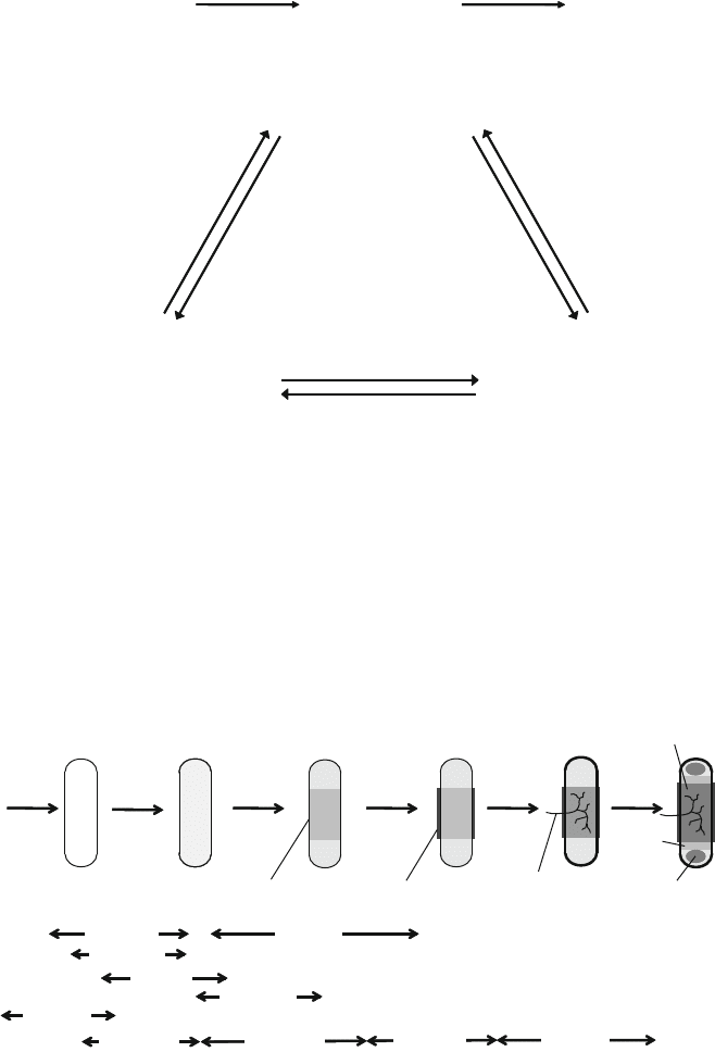
Fig. 1 Interaction of cellular differentiation, growth and differentiation factors, and specific
matrix molecules during histogenesis. Morphogenesis and histogenesis result from the interaction
of specific growth and differentiation factors acting in a stage dependent manner. Growth factor
actions are modulated by binding to ECM molecules and the expression of their receptors
depending on the developmental stage of the cells
proliferation and
differentiation
terminal
differentiation
calcification /
vascularization
ossificationproliferation
and migration
FGF-10
FGF-2,4,8
FGFR1
condensation
TGF-ß
FGF-2
shh
FGF-2/FGFR2
IGF-II
BMP-2,4,7
FGF-18/FGFR3
PTHrP
BMP-2,7
FGF-2/FGFR1
BMP-2,7
collagen I
collagen IIA collagen IIB
collagen X
collagen I
diaphysis
epiphysis
perichondral
bone collar
fibronectin
blood vessel
cartilaginous
model
N-
cadherin
aggrecan
tenascin
hyaluronan
growth
plate
Fig. 2 Development of the long bones under the control of growth factors. The different develop-
mental stages of the skeletal primordia are shown together with the specific growth and differenti-
ation factors (above the arrows) which promote the stage specific synthesis of matrix molecules
(below)[6, 144]
Cartilage Engineering from Mesenchymal Stem Cells 177
2.1 Migration and Proliferation
In the initial phase of limb development, mesenchymal cells of the lateral plate
mesenchyme and of the somitic mesenchyme migrate towar ds the limb field [2].
The ectodermal cells are induced by the mesoderm to form a specialized epithelial
structure termed the apical ectodermal ridge (AER). The AER supports migration
and proliferation of the mesench ymal cells providing the cell mass for the forma-
tion of precartilage condensations [46, 47].
2.2 Cell Condensation
Cell condensation is characterized as a transient stage during the early morphogen-
esis which can be detected by PNA (peanut agglutinin) staining [48, 49]. During
cell condensation, cell density in the prospective limb regions is increased leading
to cell–cell contacts mediated by cell adhesion molecules such as N-cadherin and
N-CAM [50–52] and the formation of gap junctions [53, 54]. Cell adhesion
molecules are expressed specifically during the condensation phase and down
regulated subsequently upon chondrogenic differentiation of the prechondro genic
cells. Prior to cell condensation, the extracellular matrix in the prospective limb
regions contains high amounts of collagen type I and hyaluronan [55, 56]. Hyaluronan
is supposed to prevent cell–cell interactions prior to the condensation phase [56].
During the condensation phase, hyaluronidase activity is detected, suggesting that
matrix remodeling takes place allowing for cell–cell interaction. During the con-
densation phase, a specific splice variant of fibronectin, FnEIIIA, is detected
throughout the condensations [57, 58]. Fibronectin was shown to be essential
for condensation and subsequent chondrogenesis. Furthermor e, fibrone ctin distri-
bution during the condensation phase indicates the localization of skeletal elements
formed later on [55].
2.3 Chondrogenic Differentiation
Cellular differentiation is characterized by the increased synthesis of transcription
factors sox-5 and sox-6, and the appearance of the cartilage specific transcription
factor sox-9 [59–61]. Collagen type I and fibronectin are synthesized in the ECM
prior to condensation and reach a maximum density at the time of cellular differen-
tiation [55]. Chondrogenic differentiation of the condensing cells is characterized
by the appearance of collagen types II, IX, and XI, the characteristic components of
collagenous network of cartilage tissue. As a result of chondrogenic differentiation,
178 C. Goepfert et al.