Kasper C., van Griensven M., P?rtner R. (Eds.) Bioreactor Systems for Tissue Engineering II: Strategies for the Expansion and Directed Differentiation of Stem Cells
Подождите немного. Документ загружается.

into cells from distinct germ layers like mesoderm and ectoderm. Another indica-
tion is that a large subset of the cultured umbilical cord matrix cells remain in the
quiescent state which is related to self renewal ability [10].
Stem cells from extraembryonic tissues are furthermore expected to express low
immunogenicity and may, therefore, potentially serve as allogeneic donor cells in
thefuture[10]. However, trials proving the allogeneic applicability of extra-embryonic
tissue derived stem cells in regenerative medicine are still missing. Hoynowski
et al. [10] evaluated the expression of markers related to immunogenicity such as
HLA-ABC, HLA-1AG and MHC-11. Unfortunately, they were unable to confirm
whether the lack of significant expression was truly negative or if equine cells
simply do not cross react with the reagents developed for human cells.
All these findings indicate that the umbilical cord matrix seems to be a good
alternative to bone marrow. MSCs from the umbilical cord can be collected in a
non-invasive manner at birth and stored for future use [10, 36]. The only downside
is that the maternal perineum and the delivery environment are certainly not sterile
[36], particularly referring to animals. Therefore, a sterile collection is challenging
and only samples cultured in a medium containing a relatively high amount of
antibiotics can be considered suitable for experiments and clinical applications.
2.7 Synovial Membrane
The synovial membrane (synovium) lines the inside of joint cavities, bursae and
tendon sheaths and regulates the content of the synovial fluid which is contained in
these cavities. The first successful extraction of MSCs from the synovium was
performed in humans by De Bari et al. [39]. According to Fan et al. [12] and
Yoshimura et al. [14], stem cells from the synovial membrane of humans and rats
excel other sources of MSCs in higher ability of proliferation and superiority in
chondrogenesis and adipogenesis. The achievable colony numb er per nucleated
cells was reported to be 100-fold higher than that of bone marrow derived rat MSCs.
Compared to other MSC sources, synovium-derived stem cells were also highest in
colony forming efficiency, fold increase and growth kinetics [14]. On the other
hand, they seem to be inferior in osteogenic capability compared to periosteal-
derived MSCs and bone marrow derived, but still superior in comparison to fat and
muscle derived stem cells [12, 27].
In addition to the in vitro results, multipl e reasons why it is assumed that
synovium derived MSCs are especially superior in chondrogenesis compared to
MSCs from other sources were reported.
Synovium derived MSC have a higher hyaluronan receptor expression and
express enzymes involved in hyaluronan synthesis, the synovial membrane is further
believed to contribute to repair of partial thickness cartilage defects [ 12] and
cartilage can be formed in pathological synovial tissue (synovial chondromatosis)
and synovial pannus of rheumatoid arthritic knee joints [12, 14].
230 I. Ribitsch et al.
Moreover, synovium and cartilage originate from a common source of progeni-
tor cells and synovial tissue expresses a variety of cartilage specific markers [12].
A very interest ing finding in particular for the treatment of tendon injuries is that
synovium derived MSCs can serve as hyaluronic acid blasters, avoiding adhesion, a
common complication in tendon injuries that can lead to scar tissue formation [12].
Although they can also be extracted from pathological synovium [12] and only a
minimal amount of synovial tissue is necessary for the extraction of a reportedly
high amount of MSCs, and in spite of the high regeneration rate of the synovium
which leads one to expect few complications at the donor site [12, 27], the cell
recovery process by arthroscopy requires general anaesthesia and is therefore
associated with a relatively high risk for the patient, especially in large animals.
Furthermore, it would also be expensive. In addition, preparation of the synovial
tissue for stem cell extraction is not easy. It was reported that the separation of the
subsynovial tissue from the synovial tissue – which is required in order to obtain
homogenous cell culture – is difficult. Another question regarding the quality of
MSCs from the synovium that needs to be addressed is the fact that some of them
seem to retain their fibroblastic characteristics even after differentiation induction
[12]. Therefore, synovium might be a practicable MSC source in human but not
necessarily in veterinary medicine.
2.8 Periodontal Ligament
In equine orthopaedics, MSCs have attracted much notice because of promising
results of MSC treatments of superficial digi tal flexor tendonitis. However, recov-
ered tendons have inferior biomechani cal properties compared to healthy tendons.
Consequently, a source of MSCs is needed which guarantees a high tenogenic
differentiation capacity. In this regard, the periodontal ligament (PDL) earns much
attention. Under physiological conditions, the equine PDL combines two remark-
able characteristics. It withstands high biomechanical strains presenting character-
istics similar to a tendon, and at the same time it possesses a high regenerative
capacity [40].
The periodontial ligament is situat ed between the tooth and the jaw bone and is
part of the complex that keeps a tooth in place in its alveolar cavity. In veterinary
medicine the PDL as source for MSCs was only described in rodents and horses.
In order to compensate for rapid surface attrition the hypsodont equine cheek
tooth erupts continuously which is inevitably associated with permanent remodel-
ling of the periodontium. Therefore, the periodontium shows a rapid cell turnover
compared to other soft connective tissues. The equine PDL for example shows a
proliferation index of 1–3% [41]. In addition, the functional requirements of the
PDL depend on ample capacity for dynamic changes regarding tissue synthesis
remodelling and repair. This is only possible because of the inherent capacity of the
periodontial cells to differentiate into osteoblasts, collagen-forming fibroblasts or
cementoblast [42, 43].
Basic Science and Clinical Application of Stem Cells in Veterinary Medicine 231
It has been propos ed that the periodontium comprises a population of undiffer-
entiated progenitor cells which migrate either towards the cementum to differenti-
ate into cementoblasts or towards the alveolar bone to become osteoblasts [41].
Gould [44] and McCulloch [42] showed typical characteristics of these cells in mice
which strongly indicate that they indeed are stem cells. A population of progenitor
cells which may be stem cells was also found in the paravascular sites of the mouse
molar PDL [42].
Only recently these suggestions were confirmed. MSCs in the periodontial
ligament of sheep and pigs – which were able to differentiate into a large vari ety
of cell lineages in vitro – were detected [45, 46]. At the same time it was shown that
there seems to be a considerable difference between PDL derived and bone marrow
derived MSCs based on their higher expression of tenocyte specific transcription
factors [47]. Periodontal cells also showed significantly higher capacities for self-
regeneration, i.e. number of CFUs, than cells from the subcutis, whereas the
population doubling time of subcutane ous cells seems to be significantly faster
than those of PDL cells. All cells showed osteogenic and adipogenic differentiation.
Marker mRNA for chondrogenic differentiation (Aggrecan, Collagen 2, COMP)
was highly expressed by cells from the middle and apical areas of the PDL. In
contrast, in subcutaneous cells and PDL cells from the subgingival area the expres-
sion of chondrogenic marker mRNA was limited to Aggrecan and COMP. The
equine PDL contains cell populations that exhibit typical properties defined for
MSCs. Cells from the apical and the middle areas showed higher differentiation
capacities than subgingival cells and subcutaneous cells [40].
Equine PDL cells might be a promising source for MSC-therapies in equine
musculoskeletal disorders [40]. However, the PDL contains only a small number of
progenitor cells [44], suggesting that the rol e as practicable MSC source in regen-
erative medicine needs to be questioned.
2.9 Skin
MSC derived from skin would be very easily accessible (regardless of the species)
with low costs and a low risk for both the patient and the veterinarian.
In juvenile and adult rodents it was shown that stem cells can be isolated from
the dermis. Interestingly, these cells seem to be able to differentiate into neuroec-
todermal and mesenchymal lineages, including neurons, glia, smooth muscle cells
and adipocyt es. Based on these findings, rodent dermi s derived stem cells are
distinct from MSCs. They can be passaged for at least one year witho ut losing
their differentiation capacity and therefore probably represent a novel multipotent
adult stem cell type. They also clearly differ from adherent bone marrow derived
stem cells in the way that they require different growth factors to proliferate and
their selective ability to express proteins typical for neuronal precursors, as well as
their morphology and habit to grow in spheres. It is suggested that these precursor
cells represent a novel multipotent adult stem cell capable of generating cells from
232 I. Ribitsch et al.
more than one embryonic lineage [48]. However, further investigations will be
necessary to confirm these findings.
2.10 Other Potential MSC Sources
Other potential sources of MSCs are muscle, brain [15, 49], synovial fluid [50 ],
tendon [51] or periosteum [14]. Although rodent models for these tissues exist, the
practicable isolation of these cells as a common stem cell type used in regenerative
medicine is doubtful.
Regarding the differentiation ability, it is remarkable that MSCs derived from
muscle produce only tiny pellets after chondrogenic differentiation [34]. However,
they show a good calcification potential after osteo genic differentiation [14] as well
as an easy differentiation to adipocytes [52]. According to Koga et al. [34] muscle-
derived MSCs have a higher proliferation potential than other stem cells [34].
Although the isolation of brain-derived MSCs after enzymatic digestion of the
whole brain is not a practicable way to harvest MSCs for tissue engineering
purposes, it is interesting to see that this stem cell type has a less efficient adipogenic
proliferation potential [52].
MSCs isolated from the cambium layer of the periosteum have a high chondro-
genic proliferation potential which results in a greater production of cartilage
matrix. Regarding the calcification ability, it is not surprising that the periosteum-
derived MSCs have a high osteogenic proliferation potential [14].
3 Immunogenicity
The immunogenicity of adult MSCs is not completely understood. MSCs are said
to be hypo-immunogenic and to suppress T-cell activity and dendritic cell function
[8, 11] in humans and animal models [8].
Normally, allogeneic cells would be rejected by immune response. Surprisingly,
immunologists found that MSCs do not seem to obey the normal rules of allogeneic
rejection. Evidence indicates that the use of mismatched MSCs does not provoke a
proliferative T-cell response, thus suggesting an immunosuppr essive potential [8].
Krampera et al. [53] found that murine MSCs lack MHC class II and inhibit
T-cell activity. Furthermore, Tse et al. [54] showed in humans that MSCs do not
elicit allogeneic T-cell response even when MHC class II was upregulated. It was
also reported that allogeneic baboon MSCs suppress lymphocyte activity in vitro
and prolong graft survival, indicating the anti inflammatory and pro healing effect
of MSCs which was later confirmed by Di Nicola et al. [55], Tse et al. [54] and
Krampera et al. [53].
MSCs appear to evade allogeneic rejection by being hypoimmunogenic, inter-
fering with maturation and function of dendritic cells, modulating CD4 and CD8
Basic Science and Clinical Application of Stem Cells in Veterinary Medicine 233
T-cell phenotype and response as well as natural killer cell activity and creating an
immunosuppressive milieu based on secretion of a variety of soluble factors [8].
Recently, allogeneic MSC transplantations were carried out in rabbits and
horses, showing no significant difference compared to autologous transplants.
Autologous and allogeneic bone marrow derived MSCs in a fibrin carrier were
implanted into rabbit Achilles tendons. In accordance with Guest et al. [56] it was
found that the distribution of inflammatory cells was similar in the allogeneic and
the autologous group. No apparent immune reaction such as lymphocyte infiltration
associated with the allogeneic transplantation was observed. Viable allogeneic
MSCs were detectable at 8 weeks post implantation [57].
In another study, autologous and allogeneic green fluorescent protein (GFP)
labelled mesenchymal progenitor cells (MPCs) were injected into artificially cre-
ated superficial digital flexor tend on (SDFT) lesions in horses. A very interesting
finding was that no differences in either the number or distribution of autologous
and allogeneic cells as well as in the density of leukocytes at the respective injection
sites were observed. Injection of allogeneic MSCs did not lead to any immune
response from the host. Neither external nor histological signs of increased inflam-
mation were found compared to the autologous injection site [56].
Therefore, MSCs really seem to be immunoprivileged and one could possibly
provide a readily available source of allogeneic MSCs for regenerative medicine
purposes at least in veterinary medicine. What needs to be kept in mind, though, is
the inherent risk of disease transmission from donor to recipient [5]. However,
some day regenerative veterinary medicine might be able to rely on allogeneic cells
to repair or replace tissue [8].
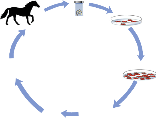
4 Clinical Applications of Stem Cells in Veterinary Medicine
Stem cell therapy in veterinary medicine is gradually turning into clinical reality.
Especially in equine orthopaedics and small animals stem cell treatments are being
commercially offered (Fig. 8).
It was demonstrated that special chemokine receptors enable MSCs to respond to
signals produced by damaged tissues [56]. As a response to these signals MSCs
migrate into the damaged tissue and seem to induce regeneration of the respective
tissue. Therefore, a lot of hope and research emphasis is put into the newly evolving
field of human as well as veterinary regenerative medicine.
However, not every treatment that is being advertised as stem cell therapy is
actually what it promises to be.
In many cases direct injection of crude bone marrow or nucleated cells isolated
from fat without further culture expansion is performed. This treatment is often
wrongly referred to as stem cell therapy, which might lead to misunderstandings [58].
As a matter of fact the transpl ant mainly consists of nucleated cells rather than
actual stem cells.
234 I. Ribitsch et al.
Herthel [59] for example reported that direct bone marrow injection for the
treatment of suspensory ligament (SL) injuries led to significantly better results and
a decreased reinjury rate 92% of the bone marrow treated horses went back to
work compared to 84.8% that did not become sound or did not go back to work that
had received conventional treatment.
For multiple reasons the success of this treatment is questionable.
First of all, bone marrow contains only a small number of actual stem cells.
Therefore, the treatment cannot be referred to as actual stem cell treatment [5, 24,
58]. Convincing studies show that only about 0.001–0.01% of mononuclear cells
isolated from bone marrow aspirate using Ficoll density gradient are MSCs. Hence
the number of MSCs in crude bone marrow would actually be even less than 0.001–
0.01%. In horses under 5 years of age it was shown that only 1–2 10
5
adherent
cells can be obtained from 10 mL of bone marrow aspirate after 3 days in culture [5].
Second, crude bone marrow might contain bone spicules and fat cells which can
be deleterious to tissue regeneration [24].
In contrast, injection of in vitro expanded MSCs provides a larger number of MSCs
than endogenously available or delivered by direct bone marrow injection and addi-
tionally avoids the risk of adverse effects of other bone marrow constituents [24].
Therefore, it is important to interpret cautiously results from studies using stem
cell treatment because the term “stem cell therapy” is not always used in the correct
way and might be misleading.
Passaging and
further
expansion
Stem cell isolation
Proliferation
Cell
har-
vest
Biopsy
MSCs
Genetic Engineering
Tissue Engineering
Implantation
Adherent cells
Fig. 8 MSC therapy principle
Basic Science and Clinical Application of Stem Cells in Veterinary Medicine 235
4.1 Tendon Injuries
In addition to in vitro studies and small animal experiments, it is certainly the horse
that veterinary research is focussed on concerning stem cell treatment of tendon
injuries.
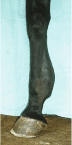
Tendon injuries are a frequently occurring problem in the equine athlete (Fig. 9).
Due to the equine quadruped-specific anatomy characterised by the proximally
located muscles and the distally located long SDFT, deep digital flexor tendon
(DDFT) and SL in combination with the hyper-extended metacarpophalangeal
joint, equine tendons and ligaments are exposed to enormous forces during athletic
workout. Maximal strains in the SDFT are reported to be at 16%, which comes up to
the functional limit, during galloping in thoroughbreds [60].
After suffering a clinical injury, a short inflammatory phase is observed in the
tendon, followed by the creation of fibrous scar tissue. This scar tissue lacks
elasticity compared to healthy tendon and therefore the risk of re-injury is high
[61]. Outcomes following conventional treatment regimes unfortunately are rather
poor [62]. As stem cell therapy encourages the regeneration of functional tendon
tissue rather than scar tissue, it is expected to reduce re-injury rates [56]. Two
possible theories regarding the effect of stem cells are discussed. One possibility is
that they differentiate into tenocytes within the tendon environment and support
healing via collagen production and remodelling activi ties. The second possibility
is that the injected cells supply growth factors rather than differentiate terminally
into the required tissue [57, 60].
It is proposed that the introduction of MSCs into the tissue which contains the
required cell type, in addition to the adequate mechanical environment, provides the
best stimulus for appropriate differentiation [24]. In case of stem cell treatments for
tendon or ligament lesions it is suggested that tensional mechanical load is neces-
sary for an optimal formation of organised tendon and ligament matrix [63].
Therefore, equine tendinopathy, with its typical centrally-positioned damage
surrounded by relatively intact tendon tissue or at least the thick paratenon offers
Fig. 9 Equine tendinitis of
the SDFT (courtesy of Dr.
Johannes Edinger)
236 I. Ribitsch et al.
perfect conditions for stem cell applications (Fig. 10, 11 and 12). Abundant growth
factors are also involved in early tendon healing and provide a perfect graft bed for
the injected MSCs. However, treatments of other forms of injuries are more
problematic mainly because accurate placement of the cells and cell retention is
more difficult [24].
In many cases direct intratendinous injection of crude bone marrow to support
tendon healing is performed, which was first reported by Herthel [59]. Although the
results of this study were favourable com pared to conventional treatment, the
success of this technique is questionable for multiple reasons which have already
been discussed above. In addition, injection of large volumes of bone marrow might
even exacerbate the tendon injury, due to disruption of remaining intact tendon
tissue [24].
There are two different approaches of stem cell therapy that are clinically used
for the treatment of equine tendon disease: one is to apply isolated and expanded
bone marrow-derived MSCs, the other is to implant adipose-derived nucleated cell
(ADNC) fractions [60] or adipose derived expanded MSC.
The latter technique was tested in a small controlled experimental study with
eight horses suffering from collagenase-induced tendinitis. Five days after creation
of the SDFT lesions, adipose tissue was harvested from the paraxial caudodorsal
gluteal region under standing sedation and local anaesthesia. Collagenase digestion
and serial centri fugation was used to isolate and purify the ADNC fractions which
were then resuspended in phosphate buffered saline (PBS) solution, in order to be
injected only 2 days after adipose tissue harvest and 7 days after the lesions were
created. Four horses obtained ADNC-treatment, the others served as control.
Ultrasonographic and, 6 weeks later, gross and histologic examination revealed
an improvement in structural organisation and a reduction of inflammation in the
ADNC-treated tendons compared to the controls. Gene expression for COMP was
also significantly increased (concentrations of COMP, a noncollagenous glycopro-
tein, are positively correlated with ultimate tensile strength and stiffness in equine
tendons). However, analysis of collagen revealed no significant differences
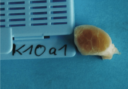
between the two groups [64]. Unfortunately, although this technique is widely
Fig. 10 Core lesion in an
equine SDFT
Basic Science and Clinical Application of Stem Cells in Veterinary Medicine 237
used in the USA, there seems to be little information concerning the clinical
outcomes available so far. Dahlgren [6] reported that a total of 78% of sport horses
have returned to their previous level of performance and 69% of race horses have
returned to race more than once. Also Leppa
¨
nen et al. [65] showed some promising
results after the application of enriched adipose derived stem cells in treatment of
equine tendon and ligament injuries. Significant improvement in ultrasonographic
fibre alignment scores and echogenicity scores were found during the follow-ups at
1, 3 and 6 months after the treatment. After a year from the injury 85% of the horses
in the recovery population (n ¼ 31) were back to competing and 75.9% of all
patient owners included in a survey (n ¼ 44) reported excellent or good satisfac-
tion, no significant adverse effects being reported [65].
These results are promising indications of good clinical success using the
procedure. The full potential of adipose-derived adult stem cell technology will
become evident in the coming years [6].
The major advantage of using ADNCs would be the immense reduction of the
interval from tissue harvest until cell application which minimises the cost and
simplifies laboratory procedures. Studies have revealed that approximately 80% of
the cells isolated from human lipid aspirates are multipotent MSCs [ 64 ]. However,
this has not yet been confirmed for the horse, and therefore this kind of treatment
should not be referred to as stem cell therapy in the narrow sense.
Another approach, using BM-derived MSCs, is performed according to a tech-
nique reported in [24]. By now, some aspects of this technique have been modified.
To name the most consider able ones, first the number of injected cells rose from
500,000 cells mentioned by [24] to approximately 10 10
6
cells [61, 66, 67].
Pacini et al. [66] observed that a cell number of less than 1 10
6
was insufficient
for tendon healing. Second, while Smith et al. [24] used fresh autologous plasma to
resuspend the cells before injection, nowadays citrated bone marrow supernatant is
applied, which has stimulatory effects on the injected cells and, due to the diffusion
of the citrate, clots after injection [68]. Nevertheless, there are also other
approaches, such as using PBS [67], autologous serum [66] or fibrinogen [69].
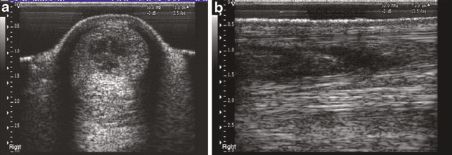
Fig. 11 Sonography of a core lesion in an equine SDFT. (a) Transversal view. (b) Longitudinal
view (courtesy of Prof. Roger Smith)
238 I. Ribitsch et al.
In a more recent study, eight horses with naturally occurring SDFT injury were
used. Autologous bone marrow derived MSCs were expanded in vitro, suspended in
citrated bone marrow supernatant and 1 10
7
implanted into the damaged SDFT
of four horses under ultrasound guidance. Saline was injected into four controls.
Horses received controlled exercise and were euthanised after 6 months. However
markers of regeneration in tendon were not identified but a normalisation of
biomechanical (reduced stiffness), histological (lower scores) and compositional
parameters (lower GAG content) towards those levels in normal (or less injured)
tendon could be considerable surrogate markers of regeneration. MSC implantation
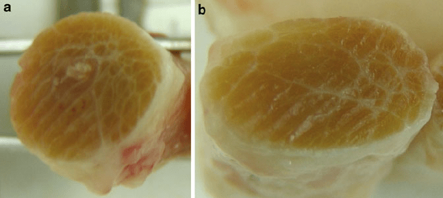
results in a tissue more like normal matrix rather than fibrous scar tissue formed
after natural repair (Fig. 12a, b). MSC-treated SDFT had greater elasticity than
saline-treated SDFT ( p < 0.05). Cross-sectional area of MSC-treated tendons was
lower than saline-treated tendons ( p < 0.05). Histologically, MSC-treated tendons
had improved cellularity and organisation scores at the injured site and were
comparable to uninjured sites of the treated tendon. In the MSC-treated SDFT,
collagen-linked fluorescence was higher and DNA content lower than the saline-
treated SDFT ( p < 0.05). Collagen and GAG content was lower in MSC-treated
SDFT but not significantly. The evidence of optimised healing seen experimentally
is supported clinically where a reduction in re-injury rate was found [70].
To date, initial reports describing long-term results of stem cell treatment of
tendinous lesions in horses as clinical patients have been published [61, 66]. Results
are favourable. Pacini et al. [66] reported a success rate of 90% following MSC
treatment of SDFT lesions in 10 race horses, showing that horses successfully
returned to their previous level of competition without re-injuring for more than
2 years, while in the non-MSC-treated control group, re-injury occurred in all
horses after a median time of 7 months [66]. The biggest clinical trial – with 500
cases of MSC-treated SDFT lesions involved, with long-term follow-up in 82 race
horses and in 24 other sports horses – was presented by Smith [61 ]. Investigating
re-injury rates after a 48-week rehabilitation, only 13–36% of the horses re-injured,
Fig. 12 (a) Equine tendon after conservative treatment – obvious scar tissue formation. (b) Equine
tendon after MSC therapy – no scar tissue formation (courtesy of Prof. Roger Smith)
Basic Science and Clinical Application of Stem Cells in Veterinary Medicine 239