Kasper C., van Griensven M., P?rtner R. (Eds.) Bioreactor Systems for Tissue Engineering II: Strategies for the Expansion and Directed Differentiation of Stem Cells
Подождите немного. Документ загружается.

including injuries to the contralateral untreated limbs. These results were compared
to success rat es of 23–66% in all horses after more than 2 years of full work
following conventional treatment, published by Dyson [71] and Smith [61]. Own
experiences with MSC-treatment of equine tendinous lesions are based on 120
cases, whereof 35% had SDFT lesions and 56% were affected in the SL; success
rates for the first group were nearly 80% and ov er 70% for the second group, these
being horses that had returned to their previous level of performance and horses that
were in full training [72].
Considering these promising results, it is important to point out that the time of
cell injection plays an important role in the success of the treatment. Based on
clinical experience, it is suggested that the optimal time for implantation of MSCs is
1–2 months after injury, whe n a suitable granulation bed has formed and before
fibrosis is dominating [68].
Besides the encouraging clinical outcome, the ultrasonographic and post mortem
examinations of either clinical [61, 73] or experimental studies [56, 64, 67, 69] also
provide promising results. In most cases, ultrasonography revealed that MSC-
treated lesions filled in more quickly [73] and showed a linear striated pattern in
the longitudinal view [61]. However, Schnabel et al. [67] could not find any
significant differences between the treated and their control groups.
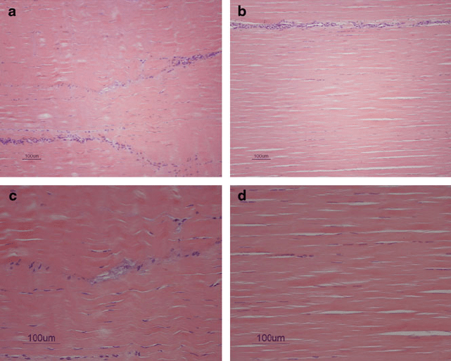
Histological findings showed that treated lesions appear to heal excellently and
organised collagen fibres in a crimp pattern were found [61, 67] (Fig. 13a, b, c, d).
In the study conducted by Schnabel et al. [67], the effect of Insulin-Like Growth
factor I gene transfer to the MSCs was tested additionally, but no sign ificant
differences between tendons treated with IGF-I gene enhanced MSCs (AdIGF-
MSCs) and unmodified MSCs could be detected. Schnabe l et al. [67] also examined
mechanical properties, anabolic and catabolic gene expressions, as well as DNA,
glycosaminoglycan and total collagen content. Although the treated tendons were
stiffer than the controls, and AdIGF-MSC-treated tendons showed an increased
gene expression of the catabolic MMP-13, there were no significant differences in
all of these parameters. These results suggest that the predominant effect of MSCs
on tendon healing is administered through structural organisation.
Another interesting study investigated the possibility of allogeneic MSC appli-
cation. Autologous and allogeneic GFP labelled MPCs, isolated from bone marrow,
were injected separately into SDFT lesions which had been artificially created using
a synovial resector blade. At 10 and 34 days after the treatment, no gross and
histological qualitative differences between the control lesions and those treated
with MPCs could be found in post mortem examinations, which might be due to the
short period of time after cell injection. In both cases large aggregations of
disorganised cells as well as completely acellular areas within the lesions were
detected with haematoxilin and eosin staining. Most labelled cells were located
within the MPC-treated lesions, and some were well integrated into the crimp
pattern of adjacent healthy tendon areas. A very interesting finding was that
no differences in either the number or distribution of autologous and allogeneic
cells as well as in the density of leukocytes observed at the respective injection sites
were obser ved, and neither external nor other histological signs of increased
240 I. Ribitsch et al.
inflammation were found compared to the autologous injection site. This indicates
that injection of allogeneic MSCs did not lead to any immune response from the
host [56].
In a similar small animal study, autologous and allogeneic bone marrow derived
MSCs in a fibrin carrier were implanted into rabbi t Achilles tendons. In accordance
with Guest et al. [56] it was found that the distribution of inflammatory cells was
similar in the allogeneic and the autologous group. No apparent immune reaction
such as lymphocyte infiltration associated with the allogeneic transplantation was
observed. This seems to confirm that MSCs do not cause alloresponses due to
attributed mechanisms such as hypoimmunogenicity and the prevention of normal
T-cell responses. Viable allogeneic MSCs were detectable at 8 weeks post implan-
tation. At 3 and 6 weeks following implantation, the cells were shown to migrate
around the repair site but in contrast to Guest et al. [56] no migration into the
proximal or distal normal tendon was found. Collagen fibres seemed more orga-
nised with denser collagen I structures and better biomechanical properties in early
tendon healing. At 6 and 12 weeks, however, no differences were detected com-
pared to the group treated with the fibri n carrier alon e [57].
Crovace et al. [74] also evaluated the efficacy of local injection of allogenic
MSC but in an ovine Achilles tendinitis model. The tendons injected with
Fig. 13 (a, c) Histology of an MSC treated tendon. (b, d) Histology of a normal tendon
Basic Science and Clinical Application of Stem Cells in Veterinary Medicine 241
allogeneic red fluorescent protein labelled stem cells in fibrin glue showed better
architecture of collagen fibres and higher expression of Collagen I compared to
control tendons. Moreover, no red fluorescent protein labelled cells were detected
in control tendons [74].
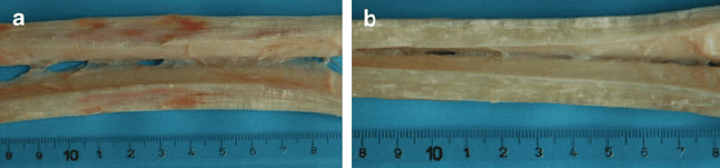
As in the studies mentioned above, current investigations of tissue engineered
tendons are based on histological and mechanical properties (Fig. 14a, b), due to a
lack of specific markers that characterise tendon fibroblasts. A recent study is now
analysing a panel of marker genes, which are, in combination, characteristic for
adult tendon tissue. Suggested markers are COL1A2, scleraxis and tenascin-C,
whereof low expression of tenascin-C and high expression of the former ones
distinguish tendon tissue from bone or cartilage [75].
Based on this knowledge, it might be possible to evaluate objectively tendon
neogenesis after stem cell application. Further controlled studies will reveal the
treatment success with different progenitor cell types, with or without modification,
so that optimal tendon tissue promoting MSCs can be identified.
4.2 Osteoarthritis
Osteoarthritis (OA) is a degenerative joint disease with intermittent inflammatory
episodes. It is induced by mec hanical and biological factors interfering with the
normal balance between cartilage synthesis and degradation. These factors,
together with inflammatory episodes, lead to softening, fibrillation and degradation
of the cartilage surface, as well as to a loss of articular cartilage and sclerosis of the
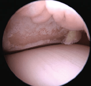
subchondral bone in conjunction with osteophyte formation [146] (Fig. 15a, b, c).
The disease can be inherited or induced by one major trauma, several microtraumas
or strenuous exercise [76], and leads to pain and decreased range of motio n.
OA is the most common human and animal joint disease encountered world-
wide. Therefore, MSC therapy for OA is of interest for both human and veterinary
medicine and results obtained from research in animals will serve as baseline for
clinical trials in humans. Unfortunately, no experimental models that reall y resem-
ble the pathology of spontaneous OA are available.
However, for single site cartilage defects, several animal models are available.
Fig. 14 (a) Core lesion in an equine SDFT – typical haemorrhage. (b) Normal equine SDFT
242 I. Ribitsch et al.
When choosing a certain species for an OA research study one needs to consider
anatomical, physiological and biomechanical aspects as well as availability,
handling, ethical concerns and, last but not least, economic aspects.
Rodents are rarely used for cartilage defect models, due to their knee joint size
and physiology (growth plates do not close). Rabbits are a useful species for early
cartilage defect research; however , important differences in size and physiology
minimise their applicability (spontaneous cartilage regeneration in young indivi-
duals). Furthermore, dogs can be used for OA research, since they can have defects
exclusively involving cartilage tissue (without damaging the subchondral plate),
second look arthroscopy can be performed and anatomy and weight bearing is
similar to human conditions. But relatively small defect volumes and ethical issues
make the dog a less often used species. Small ruminants (sheep and goat) are more
commonly employed for preclinical studies as joint anatomy and biomechanical
aspects resemble the human situation. A debatable issue is the sheep ’s variable
cartilage thickness (0.4–1.68 mm in different studies) that can produce variable
results within the same study. Cartilage thickness seems to be less variable in goats,
allowing partial and complete thickne ss defects. Pigs are a seldom used species for
research, due to difficulties with handling and behaviour. The horse represents the
largest available animal model and probably the species with most anatomical
similarities to humans [77]. Comparable to humans, horses tend to develop sponta-
neous joint disorders. This is an essential aspect for clinically relevant OA as there
might be differences between spontaneous long-lasting and experimentally induced
development of OA [78]. Furthermore, cartilage thickness in the equine stifle joint
approximates 1.75–2 mm and is therefore comparable to cartilage thickness in the
knee of humans, which approximates 2.2 mm. Nev ertheless, the differences in
human and equine body weights might result in different weight bearings and
biomechanical properties within the knee joints [77]. Frisbie et al. [76] compared
different animal models with regard to cartilage thickness in the knee joint and
observed that the horse is most similar to the human, followed by goat, sheep, dog
and finally rabbit.
Fig. 15 Extensive cartilage
defect (courtesy of
Dr. Johannes Edinger)
Basic Science and Clinical Application of Stem Cells in Veterinary Medicine 243
In veterinary orthopaedics in general, but especially in horses and dogs, joint
disease plays a major role. Joint diseases are the most prevalent causes of lameness
in horses [79, 80]. Degenerative forms of arthritis constitute approximately one
third of all equine lameness, and OA is certainly the most important one [81]. The
reasons for the development of OA are not yet fully understood. It is assumed that
injury, age and genetics are some of the risk factors [82].
Several epidemiologic studies have shown that lameness due to joint disease is
the most significant factor responsible for inability to race and loss of performance
in horses [83]. Thus, OA not only has a major impact on equine performance [ 84,
85], causing morbidity and pain, but is also a major cause of economic loss [86].
Unfortunately, articular cartilage shows only minimal regenera tion potential as
there is a limited response of cartilage to tissue damage and an inability of natural
repair response from adjacent tissue s to produce cartilage tissue with morphologic,
biochemical and biomechanical properties of healthy articular cartilage. Current
treatments include a wide range of non-pharmacological, pharmacological and
surgical modalities. Evidence to support the effectiveness of individual treatments,
however, is variable [87]. Therefore, the prognosis for patients suffering from OA is
still poor. The goals of contemporary management of the OA patient remain control
of pain and improvement of joint function as well as of quality of life. However,
there are no effective pharmacological therapies available that alter the path obio-
logic course of the disease [88]. Therefore, major attemp ts have been made during
recent years to assess the efficacy of regenerative treatments for OA.
For cartilage repair, chondrocytes seem to be the preferred cell type. It is
possible to harvest cartilage, isolate the chondrocytes and expand them in vitro.
These cells can later be transplanted as fresh or cryopreserved cells. Seddighi et al.
[89] found that cartilage engineered with fresh chondrocytes contains more cells
and extracellular matrix than constructs engineered with cryopreserved cells. The
chondrocytes can be implanted into an existing cartilage defect under a periosteal
covering graft [90] or seeded on a collagen membrane which is then transplanted
into the cartilage defect [91]. Litzke et al. [92] performed autologous chondrocyte
transplantation (ACT) in an equine large animal model. They could show that in
comparison to untreated defects, ACT-treated defects had a significantly improved
defect filling with well integrated neocartilage.
However, because of the limited cell amount in donors, terminated life span and
possible de-differentiation of chondrocytes during the culture period, alternative
cell types with chondrogenic potential need to be found.
Currently, adult MSCs are being evaluated for various therapeutic approaches in
OA treatment [1, 93].
Well known and practicable sources for MSCs with promising chondrogenic
potential are bone marrow [3] and UCB [13, 18]. PB was also used but, in compari-
son to the other sources, yields were much lower and chondrogenic differentiation
was difficult to achieve [15].
It has been shown that MSCs in general – when exposed to TGF ß (Transforming
growth factor) – are capable of chondrogenic differentiation and production of
collagen type 2 and proteoglycan – two major factors needed for cartilage repair
244 I. Ribitsch et al.
[1, 3, 4]. Furthermore, Hegewald et al. [94] found that hyaluroni c acid and autolo-
gous synovial fluid induce chondrogenic differentiation and collagen type 2-produc-
tion of equine MSCs. Chondrogenic differentiation is also supported by bone
morphogenetic protein-4 (BMP-4). In a study conducted by Kuroda et al. [95],
muscle derived stem cells, transduced to express BMP-4, were mixed with fibrin
glue and implanted into cartilage defects. The results of this treatment showed
improvement of cartilage repair up to 24 weeks after transplantation. All these
findings suggest that MSCs may be used as a therapeutic agent in OA.
Currently, different techniques exist for the transplantation of MSCs into carti-
lage defects.
There is the possibility of transplanting differentiated [96] or undifferentiated
MSCs, with or without a scaffold. For the implantation without a scaffold, cells can
be suspended in various fluids and injected blindly or arthroscopically. Further-
more, they can be fixed with fibrin glue to a certain location. Scaffolds consist of
natural or synthetic materials and are usually fixed onto the defect site by suturing,
press-fit and/or fibrin glue. MSCs are loaded onto the scaffold either before or
immediately after the implantation into the defect and are supposed to expand,
differentiate, and produce cartilage matrix.
Nowadays, the intra-articular injection of suspended cells is the most practicable
way and therefore most common cell application mode for veterinarians. Agung
et al. [1] injected fluorescent-labelled MSCs into rat knee joints with multiple
injured tissues (anterior cruciate ligament, medial meniscus and articular cartilage
of the femural condyles). Four weeks after injection, they found that MSCs mobi-
lised into some or even all inj ured tissues depending on the initial number of
injected cells. When 1 10
6
MSCs were injected cells migrated only into the
injured anterior cruciate ligament (ACL). When 1 10
7
MSCs were injected, cells
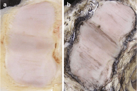
Fig. 16 (a) Naturally occurring osteoarthritis – extensive cartilage degeneration with typical
wearlines. (b) Naturally occurring osteoarthritis – extensive cartilage degeneration with typical
wearlines after Indian ink staining
Basic Science and Clinical Application of Stem Cells in Veterinary Medicine 245
were also found in the injured meniscus and articular cartilage, with extracellular
matrix present adjacent to the injected MSCs. However, it was shown that injection
of larger numbers of MSCs led to the formation of free scar tissue within the joint,
which might have adverse effects on cartilage regeneration. Therefore, determining
the optimal number of cells to be injected is essential to minimise problems
resulting from unrequested tissues. Regardless of the questions that still need to
be solved, Ferris et al. [97] reported about their results of a clinical evaluation of
bone marrow derived MSCs in naturally occurring joint disease in horses. Of the 40
horses integrated in the study, 72% returned to work. About half of them returned to
or even exceeded their previous level of work. They also found that age, sex, breed
and discipline were not significantly associated with outcome. Only the severity of
the injury, as classified by the attending veterinarian, was significantly associated
with a return to work as four horses who had severe cartilage damage were unable
to return to performance. This study confirms anecdotal reports of good clinical
outcome post MSC treatme nt for joint related lesions. Results of this study support
future controlled trials to be undertaken for the use of MSCs in horses [97]. Also
dogs suffering from OA in their elbow and hip joints were treated with adipose
tissue derived cells resulting in an overall clinical improvement of the patients [147,
148]. However, in an equine OA model no difference in lameness improvement
between horses treated with MSCs from bone marrow and nucleated cells from
adipose tissue [98] was found.
Oshima et al. [99] transplanted undifferentiated green fluorescence protein-
marked mesenchymal cells (MCs) rigidly into an osteochondral defect in rats
using fibrin glue. It was shown that there were still some marked MSCs in the
defect for as long as 24 weeks after transplantation. Also the defects showed better
repaired with hyaline-like cartilage than untreated defects.
Wilke et al. [100] implanted undifferentiated MSCs arthroscopically in a self-
polymerising autologous fibrin vehicle. The advantage of this technique is the one-
step surgical procedure, requiring only one arthroscopy under general anaesthesia.
They observed that MSC grafts in horses did improve early healing (1 month) of
full thickness cartilage lesions, but the long-term healing (8 months) did not
improve compared to untreated defects.
Recently, another new technique of transplanting cells into rabbit cartilage
defects, called local adherent technique, was described [34]. Undifferentiated
MSCs in suspension were directly placed on the cartilage defect. The defect
is pointing upwards and is held stationary for approximately 10 min to allow
cell adherence. This easy technique can also be performed via minimal invasive
surgery.
Although some studies have provided promising results [1, 93, 101], the efficacy
of MSCs in the treatment of OA is still controversial.
To date it is unknown whether tissue regeneration after MSC transplantation
originates from the transplanted cells themselves, or whether the transplanted
MSCs initiate and support local cells in regenerating the damaged tissue [102].
Another important factor which might contribute to therapeutic success is the anti-
inflammatory function assigned to MSCs [103].
246 I. Ribitsch et al.
4.3 MSCs in Bone Regeneration
The natural repair process of fractured bone occurs via prima ry and secondary bone
union. It is a complex process in which local MSCs generate various essential
progenies: chondroblasts, chondrocytes, fibroblasts and osteoblasts forming a frac-
ture callus. Cellular events during regeneration include MSC chemoattraction,
migration, proliferation and differentiation into osteoblastic, chondroblasic or
fibroblastic lineages depending on the local fracture environment [104]. New
extracellular matrix (ECM) is formed and comprises osteoid s and cartilage that
undergo enchondral ossification and bone formation until the fracture gap is
bridged [105].
This natural repair process is efficient for most fractures since the mechani cal
environment is maintained or created by internal fixation or adjustment. However,
specific situations such as tumour resection, trauma, arthrodesis, spinal fusions,
metabolic disease or insufficient healing capacities lead to substantial loss of bone.
They require augmentation of the natural healing process to regenerate larger
quantities of bone. The tissue engineer ing process of osseous tissue delivers some
or all elements required for the natural repair process directly to the site of the large
defect. Based on that, three general approaches have been applied to the art of tissue
engineering of bone: matrix based therapies that use scaffolding implants to replace
the missing bone, factor based therapies that directly provide osteoinductive stimuli
such as the family of BMPs and cell based therapies that transfer cells with
osteogenic potential directly to the repair site [106]. The latter is based on the
implantation of unfractionated fresh bone marrow, culture expanded MSCs, MSCs
differentiated towards osteoblastic and chondrogenic lineages or cells that have
been modified genetically to express a rhBMP [106]. In general, less differentiated
cells are easier to expand in vitro due to their high proliferation rate, while
differentiated cells are more effective in vivo due to their higher and rapid produc-
tion of mineralised ECM.
For both humans and animals, expanded MSCs derived from various tissues
(e.g. bone marrow, adipose tissue, periosteum, skeletal muscle) are confirmed to
possess osteogenic potential after culture in the presence of dexamethas one, ascor-
bic acid and glycerophosphate in vitro [15, 18 , 107, 108]. Among all adult stem
cells, bone marrow-derived stem cells remain the most commonly used cell source
for bone regeneration and repair in studies using different animal models [109].
After in vitro findings, the first animal studies were conducted and indicated that
MSCs maintain their osteogenic capacity in vivo. Therefore, isolated and expanded
MSCs were loaded into porous scaffold matrices and implanted into the subcutane-
ous tissue of athymic murine hosts where the cells induced the formation of
vascularised bone [19]. Next steps comprised the implantation of expanded MSCs
and scaffolds into segmental defects in the femur of small animal models (e.g. rats)
as shown by Kadiyala and coworkers [19]. By 8 weeks, substantial new bone
formation occurred at the interface between the host tissue and the implant, leading
to a continuous span of bone across the defect. Furthermore, Richards and
Basic Science and Clinical Application of Stem Cells in Veterinary Medicine 247
coworkers injected murine MSCs into distracted femoral bones of rats. After
5 weeks they observed significant increase of new bone volume, formation of
new trabecular bone with marked osteoblastic activity and osteoid production
[110]. These studies established the proof of principle for MSC based tissue
regeneration therapy in bone.
So far, the bone regeneration capacity of MSCs to repair various damaged bone
tissues such as long bones, cranial bone, mandibular bone and alveolar bone as well
as for the enhancement of spinal fusion was examined.
For large segment defects of long bones, Bruder et al. [111] studied the healing
of critical-sized osteoperiosteal defects using porous ceramic implants loaded with
expanded MSCs. At 16 weeks, radiographic union was established at the interface
between the host bone and the implants in samples that had been loaded with MSCs.
Significantly more bone was found in the pores of the implants loaded with MSCs
than in the cell free implants. In addition, a large bone collar formed around the
MSC loaded implants which became integrated and contiguous with callus that
formed in the region of the periosteum of the host bone [111]. Other investigators
used sheep as alternative species for the segmental bone defect model and con-
firmed that, after a 2 months period, MSC loaded implants resulted in increased
bone formation and accelerated repair compared to unloaded scaffolds [112].
The group of Cui applied adipose derived stem cells and coral scaffolds to
repair a cranial bone defect in a canine model. Three-dimensional CT scans after
12 weeks showed that MSC loading of the scaffold resulted in new bone formation
while unloaded scaffolds were found partially degraded. Furthermor e, radiographic
analysis after 24 weeks showed that MSC loaded scaffolds led to more than
threefold higher percentages of repair volume than unloaded scaffolds. This study
substantiates the potency to apply MSCs and coral scaffold for cranial bone
regeneration [113].
In terms of mandibular regeneration, Yuan and coworkers seeded osteogenically
induced bone marrow derived MSCs onto a porous beta-TCP scaffold. The cell-
scaffold-construct was implanted into critical-sized mandibular bone defects in
dogs. New bone formation was observed from 4 weeks after implantation and
bony union was achieved after 32 weeks. More importantly, the engineered bone
achieved a satisfacto ry biomechanical property in terms of bending load strength,
bending displacement and bending stress [114].
Alveolar bone resorption that is caused by periodontal disease is another field of
interest for the application of MSCs in bone regeneration. Weng and coworkers
mixed osteogenically induced bone marrow derived MSCs with calcium alginate to
create a cell-scaffold-construct in gel form. Those were implanted into alveolar
defects in dogs. After 4 weeks bone nodule structures were observed via histology
in the tissue. The engineered bone became more mature over 12 weeks, which was
similar to normal bone. At 24 weeks the repair level of the alveolus reached nearly
half of the height of the normal alveolus showing the applicability of MSCs for
alveolar bone regeneration [115].
Next to the described treatments for acute fractures, fracture nonunions and bone
defects, MSCs can further be used to achieve therapeutic arthrodesis as necessary
248 I. Ribitsch et al.
for spinal fusions. Muschler and coworkers developed a rapid, simple and effective
method to prepare cellular grafts containing enriched populations of bone marrow-
derived MSCs in an implantable matrix of demineralised cortical bone powder.
Afterwards, the MSCs enriched cellular graft was implanted into an established
canine spinal fusion model. The study showed that a simple aspirate of bone
marrow plus demineralised cortical bone powder resulted in an improvement in
bone union score, fusion area, and fusion volume compared to matrix alone and
matrix with pure bone marrow [116].
Crovace [117] also reported about enhanced bone healing using a resorbable
bioceramic based on silicon stabilised tricalcium phosphate and bone marrow
mononuclear cells, in a sheep model with a large-sized (4.8 cm), experimentally
induced defect in a weight-bearing long bone.
Gardel et al. [118] and McDuffee [119] on the other hand used MSCs which had
been differentiated into osteoblasts prior to dir ect injection into the fracture site in
canine patients and a horse model. The former successfully implanted osteoblasts
resuspended in PBS into a tibial fracture of a cat. The osteogenic behaviour of the
implanted cells was shown by the increased activity of serum ALP after the first and
second week of cell application and was in good agreement with the excellent
regeneration and bone healing characteristics of the fracture site. Based on the
results, MSC application may be considered a possible adjuvant therapy for a quick
and successful treatment of long-bone fracture in orthopaedic surgery of small
animals but requires further investigation [118].
According to McDuffee [119], periosteal tissue turned out to be the tissue of
choice to be used in the in vivo study in a large animal fracture model. Twenty
million labelled cells, stimulated to differentiate into osteoprogenitors, combined
with a fibrin glue were transplanted into the treatment limb. Fibrin glue alone
served as control. Results form five horses demonst rated enhanced bone formation
in simulated fractures which received the osteoprogenitor cell-based therapy.
Radiographic data showed an increase (p < 0.05) in the bone density and histolog-
ical data a greater percentage of bone area in the limbs which received osteopro-
genitor cells compared to control limbs [119].
Another possible indication for the application of MSCs in bone regeneration is
Legg–Calve
´
–Perthe
´
s disease in dogs. Legg–Calve
´
–Perthe
´
s syndrome, also known
as aseptic necrosis of the femoral head, is a degenerative disease of the hip joint,
characterised by loss of bone mass which may lead to a deformity of the femur head
and the surface of the hip socket. The disease is characterised by idiopathic
avascular osteonecrosis of the capital femoral epiphysis of the femoral head leading
to an interruption of the blood supply of the head of the femur close to the hip joint.
Small breeds are typically affected. Clinical symptoms are usually seen at a young
age (6–8 months). Radiographically the patients show increased opacity and focal
lysis in the head of the femur and, later in the disease, collapse and fracture of the
neck of the femur. The recommended treatment is surgical removal of the femur
head.
Lately Crovace and coworkers reported about the implantation of autologous bone
marrow mononuclear cells as a minimal invasive therapy of Legg–Calve
´
–Perthe
´
s
Basic Science and Clinical Application of Stem Cells in Veterinary Medicine 249