Kenneth E. Gonsalves, Craig R. Halberstadt, Cato T. Laurencin, Lakshmi S. Nair. Biomedical Nanostructures
Подождите немного. Документ загружается.

426. Merisko E. Liversidge nanocrystals: resolving pharmaceutical formulation issues
associated with poorly water-soluble compounds. In: Marty JJ, editor. Particles.
Orlando: Marcel Dekker; 2002.
427. Lee J. Drug nano- and microparticles processed into solid dosage forms: physical
properties. J Pharm Sci 2003;92:20572068.
428. Lee J. Intrinsic adhesion force of lubricants to steel surface. J Pharm Sci
2004;93:23102318.
429. Liu R. Water-Insoluble Drug Formation. Buffalo, IL: Inter-Pharm Press; 2000.
p 455.
430. Fromming KS and Fromming J. Cyclodextrines in Pharmacy. Dordrecht: Kluwer
Academic Publishers; 1993.
431. Mu
¨
ller RH. Dispersions for the formulation of slightly or poorly soluble drugs.
PCT/EP01/08726, PharmasSol GmbH Berlin; 2001.
432. Choi JY, Yoo JY, Kwak HS, Nam BU, Lee J. Role of polymeric stabilizers for
drug nanocrystal dispersions. Curr Appl Phys 2005;5:472474.
433. Sucker H and Gassmann P. Improvements in pharmaceutical compositions.GB
patent 2,269,536. Sandozy Ltd;1994.
434. Buchmann S, Fischli W, Thiel FP, Alex R. Aqueous microsuspension, an
alternative intravenous formulation for animal studies. In: 42nd Annual Congress
of the International Association for Pharmaceutical Technology; Mainz;1996.
p 124.
435. Mu
¨
ller RH, Becker R, Kruss B, Peters K. Pharmaceutical nanosuspensions for
medicament administration as systems with increased saturation solubility and
rate of solution. US patent 5,858,410. 1999.
436. Mu
¨
ller RH, Mader K, Krause K. Verfahren zur schonenden Herstellung von
hochfeinen Micro-/Nanopartikeln. PCT application PCT/EP00/06535. Germany;
2000.
437. Rabinow BE. Nanosuspensions in drug delivery. Nat Rev Drug Discov
2004;3:785796.
438. Mu
¨
ller RH and Peters K. Nanosuspensions for the formulation of poorly soluble
drugs I. Preparation by a size-reduction technique. Int J Pharm 1998;60:229237.
206
BIOMEDICAL NANOSTRUCTURES

CHAPTER 8
Bioconjugated Nanoparticles for
Ultrasensitive Detection of Molecular
Biomarkers and Infectious Agents
AMIT AGRAWAL, MAY DONGMEI WANG, and SHUMING NIE
8.1 INTRODUCTION
Infectious diseases are one of the major health problems in the world and have
in recent years led to several epidemics including SARS, the West Niles virus,
and the avian flu. An urgent need is to develop new technologies for detection
and identification of infectious agents with high sensitivity and specificity. At
the community level, early detection of communicable infections helps
implement better quarantine measures. For individuals, early detection leads
to effective treatment while at the cellular level sensitive detection and imaging
of microbehost cell interactions inside live cells helps to reveal infection
mechanisms and to develop better drugs and vaccines. Several technologies
based on DNA amplification and serol ogical assays are currently available for
detection, but target amplification (such as the polymerase chain reactions)
often suffers from contamination, while serological assays are slow and are not
as sensitive.
The ultimate limit in biomedical diagnostics is the ability to detect and
identify single biomarker molecules and single intact viruses in complex
samples. Recent advances have allowed detection of single dye molecules and
fluorescent proteins, but these fluorescent tags are not bright or stable enough
for routine single-molecule studies. Another problem is that target molecules
often need to be chemically derivatized with a fluorophore, a difficult task for
low abundance genes and proteins. A further challenge is the need to
discriminate bound targets from excess unbound probes in complex mixtures
or inside living cells.
BiomedicalNanostructures, EditedbyKennethE.Gonsalves,CraigR.Halberstadt,CatoT.Laurencin,
and Lakshmi S. Nair
Copyright # 2008 John Wiley & Sons, Inc.
207
In this chapter, we discuss the use of bioconjugated nanoparticles and two-
color fluorescence coincidence for rapid detection of single native biomolecules
and intact viruses. Recent research by us and others has shown that
nanometer-sized particles such as quantum dots (QDs) can be covalently
linked with biorecognition molecules such as peptides, antibodies, or nucleic
acids for use as fluorescent probes [1–6]. In comparison with organic dyes and
fluorescent proteins, quantum dots and related nanoparticles exhibit unique
optical and electronic properties such as size and composition-tunable
fluorescence emission, large absorption coefficients, and improved brightness
and photostability [7–9]. By taking advantage of these properties, we have
developed a nanoparticle ‘‘sandwich’’ assay in which two nanoparticle probes
of different colors simultaneously recognize two binding sites on a single
target molecule. This two-site sandwich method relies on a ‘‘double-selection’’
process to improve both detection sensitivity and specificity. Indeed, a number
of powerful diagnostic technologies are based on this two-site sandwich format
such as latex agglutination tests (LATs) [10], enzyme-linked immunoabsorbent
assays (ELISAs) [11], luminescent oxygen channeling immunoassay (LOCI)
[12, 13] (in which light emission arises from proximal diffusion of single t
oxygen specie s between two adjacent particles after target binding), and
fluorescence cross-correlation spectroscopy (FCCS) [14, 15]. In this format,
target molecules do not need to be chemically deriva tized, but the bound
targets must be differentiated from excess probe.
8.2 NOVEL PRO PERTIES OF NANOPA RTICLES
Fluorescent nanoparticles such as QDs have several unique properties that
make them excellent labels for ultrasensitive optical detection (Fig. 8.1).
Quantum dots have very large molar extinction coefficients on the order of
0.55 10
6
M
1
cm
1
[16], which makes them 1050 times brighter probes
than organic dye molecules. Quantum dots are also several thousand times
more photostable than organic dye molecules [17, 18] so that a single nano-
particle can be imaged and tracked over a long period of time with continuous
excitation (Fig. 8.1b). This allows one to use single nanoparticles as probes for
ultrasensitive detection. The ability to collect higher number of photons from
bright nanoparticles has allowed us to locate their position with errors less than
1 nm (discussed in the next section). Since QDs also have high absorption rates,
they require lower excitation powers when used as a probe for biological
systems. This minimizes photodamage of the biological sample in experiments
that involve long-term, continuous observations.
Quantum dots have size and composition-tunable fluorescence emission
from visible to infrared wavelengths (Fig. 8.1d) [21], with very broad
absorbance profiles [22] (Fig. 8.1a). This leads to very large Stokes spectral
shifts (up to 30 0400 nm, measured by the distance between the excitation and
emission peaks) so that single excitation source can be used to obtain multiple
208 BIOMEDICAL NANOSTRUCTURES
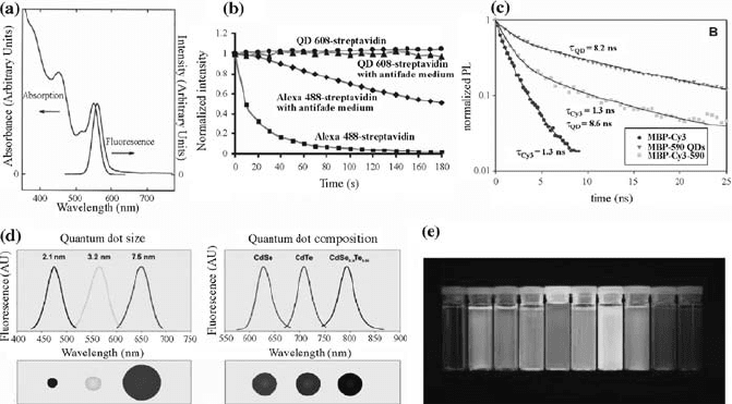
colors of fluorescence emission [7]. As a result, it is possible to prepare QD
probes with maximum emission offset with the background signal resulting in
improved signal-to-noise ratios (SNRs). In single-molecule detection (SMD),
this eliminates the requirement for the expensive two-laser optical setup that
is required to focus two different wavelengths in overlapping probe volumes
[23, 24]. The large Stokes shift also becomes important for in vivo molecular
imaging due to the high autofluorescence background often seen in complex
biomedical specimens. Orga nic dye signals with a small Stokes shift are often
buried by strong tissue autofluorescence, whereas QD signals with a large
Stokes shift are clearly recognizable above the background. This ‘‘color
contrast’’ is only available to QD probes because the signals and background
can be separated by wavelengt h-resolved or spectral imaging [25].
To improve the SNR, one can either increase the signal or decrease the
background noise. In SMD, a major challenge is to minimize the background
noise. In this regard, the longer fluorescence lifetimes of QDs may be utilized
to improve the SNR. As shown in Fig. 8.1c, the fluorescence lifetimes of QDs
are roughly 10 ns while that of the dye molecules is 12 ns. Thus, if we excite
the QD-labeled sample with a laser pulse and wait more than a nanosecond
before collecting emission from the sample, the QD signal would still be strong
FIGURE 8.1 Novel optical properties of quantum dots. (a) Broad absorption and
narrow and symmetric emission of quantum dots (reprinted from Murray et al. (1993),
with permission of the American Chemical Society). (b) Photostability of QDs
compared with the dye molecule Alexa-488 (reprinted from Reference [18], with
permission from the Nature Publishing Group). (c) Fluorescence lifetime of protein-
coated QDs compared with Cy3 (reprinted from [19], with permission of the American
Chemical Society). (d) Size-or composition-based tuning of emission wavelength in QDs
(reprinted from Reference [20]). (e) Bright QDs with emissions spanning the entire
visible spectrum (reprinted from Reference [7]).
BIOCONJUGATED NANOPARTICLES FOR ULTRASENSITIVE DETECTION 209
while the autofluorescent species would have decayed, resulting in improved
SNRs.
Dye-doped nanobeads are another class of fluorescent nanoparticles that
provide similar benefits as QDs. Since several thousands of dye molecules are
doped into each bead, these particles are very bright and photostable. Multiple
color emissions with the same excitation are enabled by fluorescence resonance
energy transfer (FRET) between the doperacceptor dye pairs inside the bead.
These beads are available in the size range of 20 nm to 1 mm, and with various
surface functions suitable for bioconjugation (Invitrogen, Inc.).
It is worth noting that quantum dots and color-coded nanobeads are in the
same size regime as the biological molecules and are amenable to biological
conjugation [1, 2]. The small size ensures minimal interference with the native
biological system and allows tailoring of the QDs as a target-specific probe for
protein, nucleic acid, or small molecule detection. By controlling the number of
ligands on their surface, it is also possible to control the stability of the target-QD-
probe binding strength. Further, by tailoring their surface chemistry, one can tune
their biocompatibility and toxicity to suit the specific application [25, 26].
8.3 SINGLE-MOLECULE DETECTION
8.3.1 Instrumental Setup and Principles
Optical methods for SMD have been reported for nearly 40 years. In 1961,
Rotman detected conversion of a nonfluorescent substrate into a fluorescent
molecule by b-galactosidase enzyme encapsulated in microdroplets and
measured the reaction rate for a single enzyme molecule [27]. In 1976,
Hirshfield reported detection of 80100 molecules of fluorescein isothiocyanate
dye bound to an antibody molecule [28]. These pioneering experiments shared
several features such as an optical setup to probe a very small volume, low
concentration of fluorophores, and time-gated optical detection using a
photomultiplier. SMD systems employ these basic principles even today. In
1990, Shera et al. reported the first efficient detection of individual rhodamine-
6G dye molecules at 100 FM concentration [29]. In the following years, two
broad approaches were devised for detection of single biomolecules: fluorescence
correlation spectroscopy [30] and coincidence-based SMD [31]. Both of these
approaches use a confocal optical setup and rely on photon burst analysis.
A typical setup for SMD is shown in Fig. 8.2a. A high numerical aperture
objective focuses a laser beam into a small volume (<10 fl) in the solution.
When the dye-labeled biomolecule enters this probe volume (Fig. 8.2b), it is
repeatedly excited by the tightly focused laser beam and emits an intense
fluorescence burst. The fluorescence signal is allow ed to pass through a
pinhole and optical filters to reduce background noise and is detected using a
sensitive photon detector (such as a CCD, a photomultiplier tube, or an
avalanche photodiodeAPD).
210 BIOMEDICAL NANOSTRUCTURES
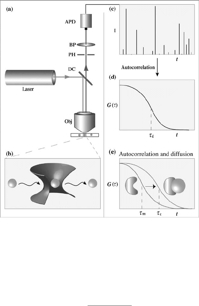
The train of photon bursts can be analyzed for autocorrelation (GðtÞ) using
the following equation and a correlation curve is plotted.
GðtÞ¼
hdFðtÞ:dFðt þ tÞi
hFðtÞi
2
ð8:1Þ
Here, FðtÞ is the fluorescence burst signal obtained over time (t), and dFðtÞ is
the difference between the average of FðtÞ and FðtÞ. In summary, the
FIGURE 8.2 Instrumental setup and basic principles of single-molecule detection.
(a) A laser light source excites a confocal volume (b) in solution containing dye-labeled
molecules. The signal is filtered optically and detected on an avalanche photodiode
(APD), and the photon burst data (c) are analyzed by using an autocorrelation device
(d). (e) Target binding decreases the diffusion kinetics of the fluorescent probe and is
measured as an increase in the autocorrelation time.
BIOCONJUGATED NANOPARTICLES FOR ULTRASENSITIVE DETECTION 211
autocorrelation function GðtÞ represents the extent to which the photon burst
signal is related to itself when shifted by time t. The correlation curve is used to
obtain the time (t) the fluorophore spends in the probe volume. This time (t)
depends on diffusion and blinking characteristics of the fluorophore. Since
biomolecular binding results in formation of a complex with higher mass and
lower diffusion constant, the above methods can be used to detect bio-
molecular complex formation.
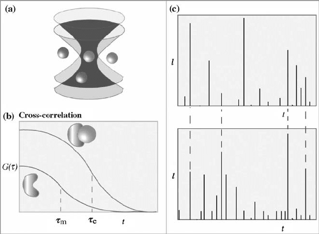
By using two-color labels and cross-correlation analysis, one can also measure
biomolecular interactions and binding behavior, as shown schematically in
Fig. 8.3. For a detailed account on fluorescence correlation spectroscopy, the
reader is referred to the work of Schwille and coworkers [32]. Recently,
coincidence of two-color photon bursts has been used to study biomolecular
interactions and to detect disease biomarkers in solution (Fig. 8.3c) [23, 24]. This
is discussed in greater detail in a later section.
In the dual-color detection sch emes, two complementary, target-specific
probes are labeled with fluorescent dyes of different emission wavelengths.
Because organic fluorophores usually have narrow excitation profiles, each dye
needs to match a specific excitation wavelength. Weiss and coworkers [33] have
FIGURE 8.3 Single-molecule detection by two-color fluorescence correlation. (a)
Difficulties in achieving par-focality and probe volume overlapping with two color
lasers are used to excite two dyes, due to spherical and chromatic aberrations of the
microscope objective. (b) Signals from the two color fluorophores are analyzed by cross-
correlation or (c) by coincidence of arrival on two APD detectors.
212
BIOMEDICAL NANOSTRUCTURES
shown that the cofocusing of two laser beams (par-focality) is an exceedingly
difficult task due to both chromatic and spherical aberrations of the
microscope objective. Klenerman and coworkers [24, 34] reported that even
under carefully matched conditions, the volume overlap for tw o excitation
laser beams was less than 30%.
8.3.2 Color-Coded Nanoparticles
The basic principles of single-molecule counting using nanoparticle probes
are shown in Fig. 8.4. In this scheme, two bioconjugated nanoparticles are
designed to recognize the same target molecule at two different sites (anti-
genic sites or nucleic acid sequences). This sandwich-type binding brings two
color-coded nanoparticles together to form a nanoparticle pair (Fig. 8.4a).
This pair moves in solution as a single complex, and when excited by a laser
beam, emits green and r ed fluorescence light simultaneously (i.e., spatial
colocalization of two particles leads to time coincidence of their fluorescence
signals). In contrast, unbound green and red particles move in a random
fashion and are unlikely to pass through the laser beam at the same time
(Fig. 8.4b). Thus, coincident green and red light emission allows one to
discriminate bound targets from excess unbound probes in a homogeneous
solution mixture.
Both QDs and energy-transfer nanoparticles are well suit ed for SMD. A
major advantage is that a single light source can be used to excite two or more
fluorescence colors (Fig. 8.4c). A single excitation beam produces only one
probe volume, and this overcomes the difficulties in focusing two color laser
beams into a small confocal volume (femtoliter or 10
15
l). QDs also have more
symmetric and narrower emission spectra than single-color organic fluor-
ophores, a feature that is important for minimizing spectral overlaps between
two or more colors.
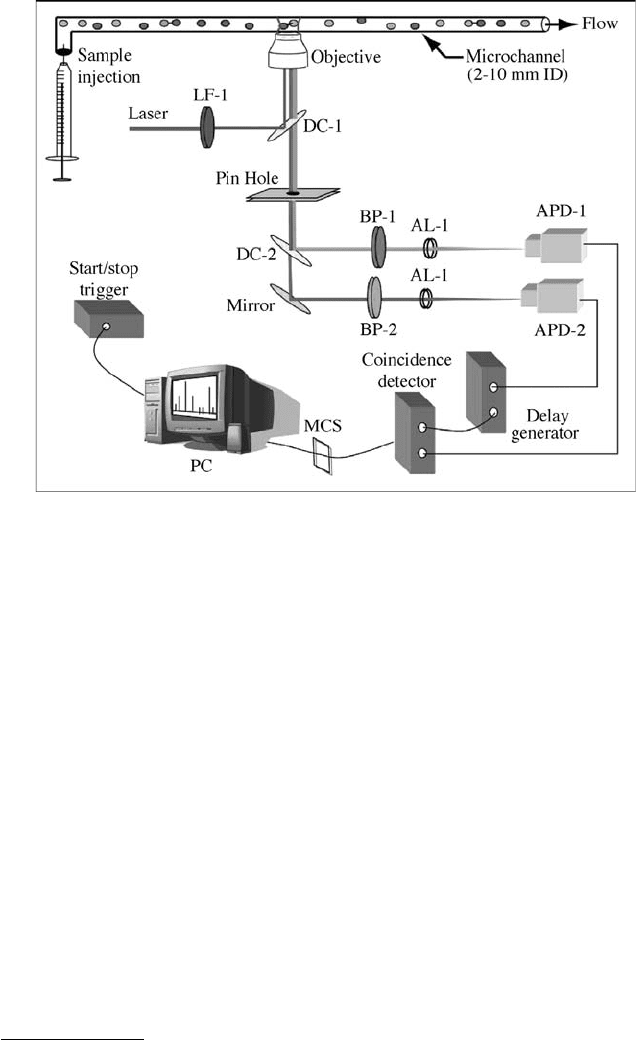
Single-molecule instrumentation is based on an inverted single-point
confocal microscope, equipped with two photon-counting avalanche photo-
diodes (APD-1 and APD-2), a single-photon counting and coincidence
analysis module (attached to the microscope side port), and a capillary flow
channel placed on the microscope stage (Fig. 8.5). In the photon analysis
module, fluorescence light emitted from green and red nanoparticles is
separated by a dichroic filter (DC-2) and is detected in real time by APD-1 and
APD-2. The single-photon output signal from APD-1 is used to trigger a delay
generator, which produces a voltage pulse with a controllable width. This
modified pulse is fed into a coincidence event detector, and its preset width is
used as a time window to determine whether one or more photons are detected
by APD-2 during this time period. Using a standard photon-counting device
such as a m ultichannel scalar (MCS), the coincidence output signals are
recorded over a period of time. At an integration time of 1 ms, this system
permits high speed detection of single molecules in a flow channel at 1000 data
points per second.
BIOCONJUGATED NANOPARTICLES FOR ULTRASENSITIVE DETECTION 213
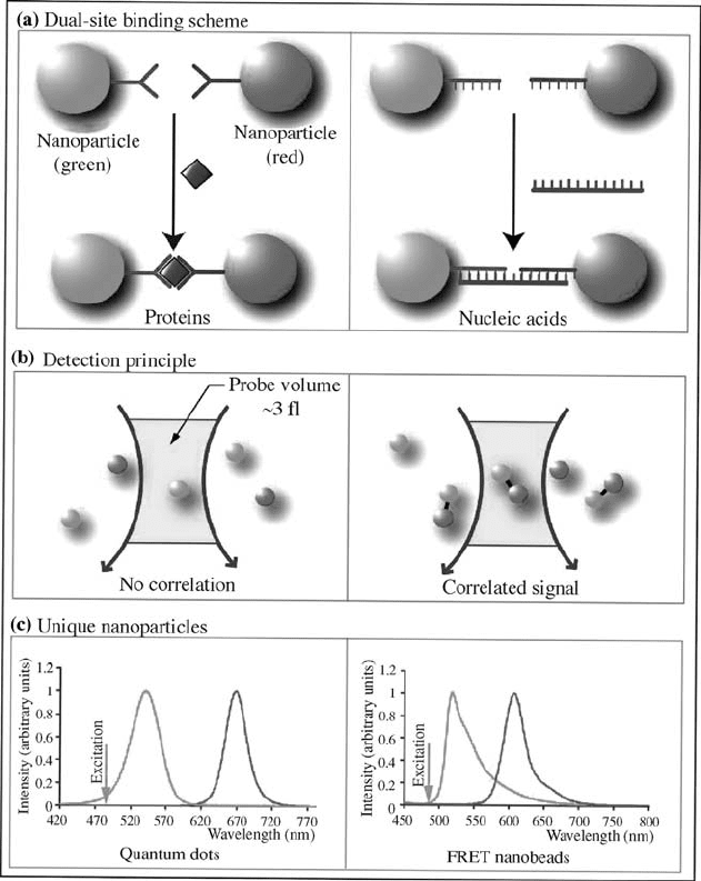
FIGURE 8.4 Single-molecule sandwich assays using color-coded nanoparticles.
(a) Simultaneous double-site binding for protein and nucleic acid detection; (b) free
nanoparticle probes and bound sandwich pairs moving across a tightly focused laser
beam; and (c) fluorescence emission spectra of color-coded quantum dots and energy-
transfer nanoparticles. The left panel shows green and red QDs simultaneously excited
with a single light source at 420 nm, and the right panel shows green and red energy-
transfer nanoparticles excited at the same wavelength. The arrows indicate the relative
position of the excitation laser wavelength (488 nm) used in single-molecule detection.
214
BIOMEDICAL NANOSTRUCTURES
8.3.3 Single-Molecule Imaging
The main concept is to determine the location of color-coded nanoparticles at
nanometer precision. This is done by using a 2D Gaussian kernel convolution,
local maxima-based centroid determination in the convolved image, followed
by a Gaussian point spread function (PSF) fitting step to locate the position of
the nanoparticles. The proced ure is adapted from an astrophotometry package
named DAOPHOT
1
developed by P.B. Stetson in 1987 [35]. Recent research by
several groups has shown that the location of a single molecule can be
determined at nanometer accuracy [33, 36–40], far beyond the ability to resolve
objects located within the diffraction limit. Weiss and coworkers demonstrated
highly accurate nanoparticle localization (error < 10 nm) and measured
FIGURE 8.5 Instrumental diagram showing real-time detection of single nanoparti-
cles and correlated sandwich pairs flowing in a small capillary. See the text for detailed
discussion. LF-1 = laser filter; DC-1 = dichroic filter in the microscope filter cube;
DC-2 = dichroic filter outside the microscope side port; BP-1 = bandpass filter (HQ514
M10); BP-2 = bandpass filter (D670 M40); AL-1 and AL-2 = aspheric focusing lenses;
ADP-1 and APD-2 = single-photon counting avalanche photodiodes; MCS = multi-
channel scalar; PC = personal computer.
1
DAOPHOT was developed at the Dominion Astrophysical Observatory for the photometric
analysis.
BIOCONJUGATED NANOPARTICLES FOR ULTRASENSITIVE DETECTION 215