Kenneth E. Gonsalves, Craig R. Halberstadt, Cato T. Laurencin, Lakshmi S. Nair. Biomedical Nanostructures
Подождите немного. Документ загружается.


CHAPTER 11
Cellular Behavior on Basement
Membrane Inspired Topographically
Patterned Synthetic Matrices
JOSHUA Z. GASIOROWSKI, JOHN D. FOLEY, PAUL RUSSELL,
SARA J. LILIENSIEK, PAUL F. NEALEY, and CHRISTOPHER J. MURPHY
11.1 INTRODUCTION
The basement membrane (BM) is a specialized layer composed of a complex
web of extracellular matrix (ECM) proteins. It is through the basement
membrane that a wide array of cells (e.g., epithelial, vascular endothelial cells)
interact with their underlying stromal elements. Basement membranes have a
number of intrinsic characteristics that can serve to regulate the behaviors of
cells associated with them. These features include the presentation of ligand
sequences for specific cell receptors, serving as a reservoir for soluble cytoactive
factors such as polypeptide growth fact ors, and possessing physical character-
istics that can modulate cell behaviors. Two key physical characteristics of the
basement membrane shown to dramatically influence cell behaviors are
compliance and the inherently complex three-dimensional topography that
consists of feature types in the nano- through submicron-size scale [1].
This chapter will briefly review the biology of basement membranes and
their biochemical and physical attributes. We then address the primary focus of
this chapter, namely, the development of basement membrane inspired
topographically patterned synthetic matrices for in vitro cell and tissue culture
systems. A summary of cellular behaviors influenced by the topographic
features of synthetic matrices is provided with an emphasis on the use of
anisotropically grooved surfaces. Such surfaces allow for the rapid evaluation
of cellular responses to topographic cues.
BiomedicalNanostructures, Edited by KennethE.Gonsalves, CraigR. Halberstadt,CatoT. Laurencin,
and Lakshmi S. Nair
Copyright # 2008 John Wiley & Sons, Inc.
297
11.2 THE BASEMENT MEMBRANE
The evolution of the BM is likely driven by the need for the simple cell to
interact with its environment. The earliest life forms on the planet were most
likely acutely sensitiv e to changes in their environment. If the primordial milieu
of simple molecules from which the first living cells arose did not supply
enough nutrients at the appropriate pH, the earliest cells could not have
survived. However, in order for life to evolve into complex organisms, cells
needed to develop the ability to not only survive in their provided environment,
but also alter and shape it to be more advantageous for cell growth and
development. For this reason, secretion would appear to be a major step in
evolution. With secretion, cells could utilize chemical signals to co mmunicate
with each other as well as deposit proteins onto surfaces to create attachment
sites. By laying a protein coat down on a surface to creat e an anchor site, cells
began to shape their environment to provide them with a selective advantage.
Adherence to surfaces and to other cells could, in many instances, improve
survival, change cellular behaviors, and lead to the evolution of multicellular
organisms. This is still evident today as an individual bacterium that exists in
our current world has different resistances and expression profiles than the
same bacterium living within a thick, densely populated biofilm [2–4]. Without
a doubt, the extracellular matrix and basement membrane have evolved to
become integral to development and disease.
11.2.1 Significance of Basement Membranes in Disease
Besides providing cellular structural support, the functional impor tance of
basement membranes is readily evident by their role in development and
maintaining tissue homeostasis. One of the most well-known BM diseases is
thin basement membrane disease, which is exactly what it implies, a decrease in
basement membrane thickness. Thin basement membrane disease is character-
ized by a 100200 nm glomeruli BM in the kidney, which ranges from half to a
quarter of the normal size [5]. The decreased thickness can lead to blood in the
urine as well as blood pressure complications [6].
The ECM and BMs also play roles in the orchestration of normal wound
healing processes. Humans and rats with diabetes tend to have abnormally
thick basement membranes in the cornea and glomerulus [7, 8]. Though not
thoroughly investigated, thicker BMs may have altered topographical features
and compliance. These features could contribute to improper wound healing, a
common pathologic feature of diabetic patients. There are other cases,
especially within the skin and bone, where extracellular matrix proteins are
mutated or autoimmune reactions arise against matrix epitopes [9–12]. The
result of these mutations and immune reactions are alterations of basement
membrane function that can lead to disease. As such, it is important to
characterize and understand the biochemical and physical properties of
basement membranes.
298 BIOMEDICAL NANOSTRUCTURES
11.2.2 Biochemical Attributes
Cells secrete many different fibrous proteins into the extracellular space and are
constantly shaping and remodeling their surrounding environment. Collectively,
this mix of secreted proteins is known as the extracellular matrix. A
specialization of ECM proteins on the basal surface of many cells within a
given tissue becomes the basement membrane. This layer provides tensile
support, serves as a reservoir for signaling molecules, and presents biochemical
and physical cues for cellular structures and tissues (Fig. 11.1). Transmission
electron micrographs reveal that the basement membrane is comprised of an
electron dense (lamina densa) and an electron lucid layer (lamina lucida) [13–15].
The biochemical composition of these layers will vary from tissue to tissue, but
most BMs share the same fibrous protein components such as collagen, laminin,
and proteoglycans [13, 16–18]. Type IV collagen is the main building block of all
basement membranes and is highly conserved across species from C. elegans to
humans. Collagen will self-assemble with itself, nidogen, the laminin family, and
other proteins to form BMs of various functions and thicknesses that usually
range from 50 to 150 nm in most tissues and up to 300400 nm in glomerular
BM or 8 mm in the human lens capsule [19–22, 55]. Many ECM proteins have
whole domains or short conserved amino acid sequences that act as signaling
antagonists or adherence ligands for cell membrane receptors. The slight
biochemical differences that make up each BM account for many of the changes
seen in cellular structures from one tissue to the next.
The majority of studies to date that have analyzed the impact of BMs on cell
behavior have focused on the cellular response to the biochemical composition
of the BM. The biochemical properties of ECMs and BMs that regulate cell
behavior are better understood than the physical propert ies because they have
historically been easier to manipulate and test. Typical experimental designs
fall into three broad categories. First, most tests on ECM proteins have
characterized changes in cells after they were transiently transfected with the
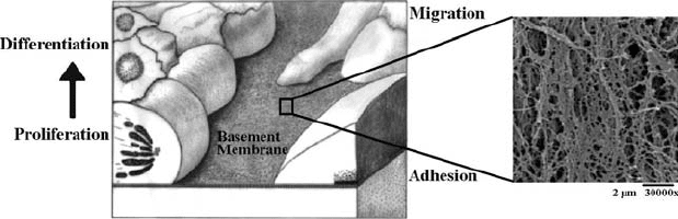
FIGURE 11.1 Schematic of cells on a basement membrane. The cartoon depicts cells
adhering, proliferating, and migrating on a basement membrane. As cells expand in a
basal to apical direction, they often begin to differentiate, as indicated by the arrow. The
enlargement on the right is a 30,000 scanning electron micrograph of a rhesus aorta
basement membrane.
CELLULAR BEHAVIOR ON BASEMENT 299
protein in question, or subjected to various doses of recombinant protein
added to the cell media. These experiments are often performed with cells
plated onto flat surfaces and can reveal visual phenotypic changes. They also
allow researchers to work out cell signaling pathways, such as how the
proteolytic release of endostatin from collagen inhibits angiogenesis pathways
[23–25]. Second, studies have been performed to characterize the effect of a
growth factor or peptide on cellular migration using a Boyden chamber or
experiments involving wound healing. These experiments typically measure the
chemotactic potential of a signal protein to control immunocyte invasion or
direct cells into a wound bed [26, 27]. Third, studies to measure the effect of a
peptide signal on adherence are usually performed by seeding cells onto a
surface coated with the protein being tested. Perhaps the best characterized
ECM peptide signal from these types of experiments that has been shown to
elicit a cellular adherence response is the arginine glycineasp artic acid
(RGD) signal peptide. The RGD signal motif, found within many different
ECM proteins, is a common ligand for several integrins, and can alter the
ability of different cell types to adhere to a given surface [28, 29]. Experiments
are commonly designed to measure RGD-dependent changes in cellular
adhesion by coating a plastic surface with RGD signal peptides or full-length
ECM proteins that contain the RGD domain, like fibronectin. However, while
each of the aforementioned experimental strategies is designed to characterize
the biochemical effects of individual ECMs, they rarely take into account the
impact of the physical micron, submicron, and nanotopographic environments
that cells are exposed to both in vivo and in vitro.
In addition to biochemical differences, BMs have unique physical
characteristics: thickness, compliance, pore size, fiber width, and three-
dimensional organization. These are all physical features that define a BM.
Provided below is a concise summary of compliance followed by a presentation
of the focus of this chapter, the topographic features of basement membranes,
and their cellular consequences.
11.2.3 Physical Characteristics: Compliance
To date, the majority of studies on cells have been conducted on rigid surfaces
such as silicon, polystyrene, and polyurethane. These surfaces have been coated
with various ligands such as the RGD peptide to increase cell attachment or to
alter cell behavior. It has beco me clear that mechanical compliance of cellular
matrix may be as significant as ligand functionalization in impacting
cell behavior [30, 31]. The compliance of a material relates the extent of
deformation (strain) of the material to an imposed stress (force/unit area). A
more rigid substratum has been shown to promote cell spreading and induce
phosphorylation pathways [32]. The effects of compliance on different iation
have also been demonstrated with breast epithelial cells [33]. When these cells
are cultured on floating gels, the cells differentiate into tubules. If the gels are
attached to a substrate, this differentiation does not occur. Similarly, work on
300 BIOMEDICAL NANOSTRUCTURES
human umbilical vein endothelial cells grown on polyacrylamide/gelatin
supports has shown that by decreasing the rigidity of the support, cellular
differentiation into tube-like structures will occur [34]. Increases in the traction
forces of fibro blasts and the contractility of smooth muscle cells have been
reported by altering the compliance of the substrata [31, 35–39]. Cortical
neurons and glial cells are greatly influ enced by compliance [40]. On hard gels,
astrocytes will outgrow neurites and possess actin stress fibers. On soft matrix,
astrocytes were rounded and showed few stress fibers similar to results
observed with fibroblasts [31]. These characteristics are very different from
their behavior on hard surfaces. However, neurites have robust actin filaments
and enhanced F-actin protrusions on soft as well as hard surfaces.
Compliance of the matrix is probably vital in the organization of cells and
tissues in living organisms. Neurons have been shown to grow into hyrdogels in
injured areas while astrocytes will not [41]. Certainly, the results with the brain
cells suggest that the distribution and population sorting in the central nervous
system may be closely related to compliance in particular areas. Work with
stem cells has indicated that soft matrices similar to that of the brain induced
neurogenic phenotypes [42]. Stiffer matrices similar to muscle were myogeni c
and rigid supports were osteogenic. Thus, the physical properties of cellular
matrices as well as the biochemical ones are extremely important determinants
of cell behavior. Despite the recent recognition that compliance is an essent ial
property on the ECM, the compliance of native basement membranes has yet
to be reported.
11.2.3.1 Physical Characteristics: Topography Basement membranes
possess a complex ‘‘felt-like’’ three-dimensional topography. Individual
features can range in scale from approximately 10 to 400 nm with the majority
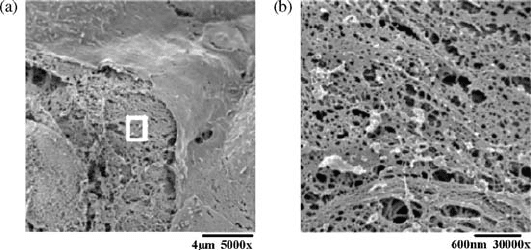
of features smaller than 100 nm [21, 22, 43, 44] (Fig. 11.2). The diameter of a
primary epithelial cell in culture can average between 20 and 50 mm, depending
FIGURE 11.2 Electron micrographs of native basement membrane topography.
(a) An SEM micrograph of cells covering the fibrous basement membrane. (b) An SEM
magnified image of the window in (a) showing complex topography of the basement
membrane.
CELLULAR BEHAVIOR ON BASEMENT 301
on the cell type. This represents several orders of magnitude difference in scale
between a cell and the basement membrane structural components and ensures
that a single cell interfaces with thousands of nanoscale and submicron
topographic features. Thus, in addition to a wide range of biochemical cueing,
epithelial and endothelial cells are exposed to a complex infor mation-rich
topographic environment. Altogether, the integration of biochemical and
physical cues provided by a BM plays a large role in development and disease
by regulating cell behavior.
The physical features of BMs are believed to exert great control over cell
behavior and development; however, it has been technically challenging to
design controlled experiments to test these hypotheses. The ability to separate
inherent chemical signals from purely physical ones for in vitro topography
experiments has been a difficul t task for cell biologists to overcome.
Nevertheless, there has been nearly a century’s worth of studies investigating
the influence of topography on cellular behavior. As early as 1911, R.G.
Harrison was using spider webs to study cellular responses to complex surface
topography [45, 46]. What Harrison lacked was the ability to create controlled
surfaces with precisely defined physical features for his experi ments.
The interdisciplinary merging of engineering and cell biology has lead to
revolutionary bioengineering techniques, such as the use of photolithography
and reactive ion etching, to create substrates to test the effects of topography
on cell behavior. Due to recent innovative fabrication strategies, researchers
can now produce the large number of nanostructured surfaces with controlled
feature sizes required to conduct statistically robust cell behavior studies [1, 47].
Different surface features (e.g., surface roughness, grooves, pores, etc.) with
dimensions ranging from tens to hundreds of nanomete rs have been reported
to affect proliferation, alignment, adhesion, and cell viability [48–53]. However,
the effects of physical features on cellular behaviors remain poorly understood
for a number of reasons. First, the topographic layouts of many BMs have not
been quantitatively analyzed. To date, the best characterized native BMs are
skin, cornea, urothelium, vascular endothelium, and the glomerulus [19, 21, 22,
54, 55]. While some morphologic aspects of the BMs from other tissues have
been characterized, a detailed quantitative description of their surface
topographic features has not be en reported. Second, research groups have
been experimenting with different manufacturing protocols to produce the
same type of topographical surfaces, with techniques such as UV or electron
beam lithography, to fabricate nanogrooves from titanium or silicon chips.
There are drawbacks and limitations for each and the result can lead to
inconsistent surface types and feature sizes. This ultimately makes it difficult to
draw direct conclusions between earlier studies. Third, bioengineers still face
challenges in developing surfa ces that reach the lower limit of physical features
needed to recreate the full nanometer to submicron range of topographic
features that define native basement membranes. Despite these limitations and
shortcomings, biologists have made significant advances in understanding the
interactions between the basement membrane and the cell. However, there is
302 BIOMEDICAL NANOSTRUCTURES

still a strong need for further development of artificial surfaces that incorporate
defined topographic features in order to fully characterize the role of the
physical characteristic s of BMs in development, disease, and maintenance of
cell and tissue hom eostasis.
11.3 HISTORY OF BIOMIMETIC SYNTHETIC MATRIC ES
Biological systems are comprised of entities whose scales range at least 10 orders
of magnitude from the size of organisms down to the individual molecules that
regulate cellular behaviors. At the level where cells interact with a basement
membrane, most information is found at the submicron, nanoscale, and
subnanoscale levels. Early attempts to create biomimetic synthetic matrices were
limited by fabrication methods so that the manufactured surfaces had only
micron-scale features. With the aid of new nanofabrication techniques, we have
now been able to develop a generation of artificial surfaces that incorporate
nano- through micron-scale topographies. The nano- and submicron features
closely mimic the biologically relevant scale of physical features found in living
tissues and the micron-scale features provide a connect to the bulk of the
literature of topographic cueing.
To understand whether the scale of topographic features can influence
cellular responses, one must first determine the relevant feature sizes of a cell’s
native microenvironment. Quantitative measurements of BM features such as
pore diameter and fiber width are similar in scale for many tissues studied and
also conserved across different species (Table 11.1). As more tissues have been
analyzed, some differences have emerged. One of the best known exceptions to
TABLE 11.1 SEM Physical Feature Measurements of Several Basement Membranes
Species Tissue BM
Mean feature
height
(nm)
Mean fiber
diameter
(nm)
Mean pore
diameter
(nm) Reference
Rhesus Cornea 191 72 77 44 72 40 [22]
Human Cornea 182 49 46 16 92 34 [21]
Human Descemet 131 41 31 93815 [21]
Human Foreskin 24 840 17
a
Porcine Aortic valve 27 12 38 24
a
Rhesus Aorta 30 11 62 37
a
Rhesus Carotid 31 11 60 42
a
Rhesus Saphenous 27 838 16
a
Rhesus Bladder 178 57 52 28 82 49 [43]
Bovine Glomerulus 9 3 [20]
Synthetic Matrigel 162 52 69 35 105 70 [44]
Most features are consistent in scale across different basement membranes and species. One
noticeable difference is the average pore size of the glomerulus BM, which has a specialized role
regulating solute transport.
a
Liliensiek SJ, Murphy CJ, personal communication.
CELLULAR BEHAVIOR ON BASEMENT 303
date is the glomerulus BM, which has smaller pore diameters [19, 20]. This
difference may be attributed to its specialized role as a selective diffusion
barrier. In order to test the roles these physical features play in guiding cellular
behavior, the features must be altered and arranged in a controlled fashion to
produce a useful analytic end point. Unfortunately, the ability to manipulate
the native BM of a tissue in vivo is limited with current biological techniques,
hence the value of artificial surfaces that can be designed with varying physical
features to exact specifications.
11.3.1 Matrigel and Randomly Ordered Arrays
Cells behave differently on flat tissue culture plastic than they will on a
complex basement membrane in vivo. There is currently a demand for a more
realistic in vivo-like environment that can be used for in vitro studies; however,
this has proved to be a technically challenging task. Isolation of extracellular
matrices from living tissue is difficult, often does not produce useful amounts
of experimental material, and often contains contaminating cells. The first
major breakthrough to address this prob lem was the development of Matrigel.
Researchers discovered a tumor that was rich in basement membrane
components and grew rapidly, but remained benign in mice [56, 57]. Extracts
of that tumor were isolated, purified, and sterilized for in vitro use. It was
found that the resultant gel would alter the morphology, behaviors, and
expression patterns of cells that were plate d onto it [58, 59]. For example,
endothelial cells form a monolayer on tissue culture plastic, but arrange
themselves into tube-like structures on Matrigel [59]. Although Matrigel is a
commonly used reagent, the actual components cannot be easily manipulated
and defined, demonstrating a need for alternative artificial BMs. Matrigel still
remains one of the most useful artificial basement membrane-like complexes to
study behaviors, but new nanotechnology manufacturing techniques are
beginning to provide different options for cell biologists searching for in vitro
ECM alternatives.
There have been other attempts to recreate the topographical environment
of a basement membrane, with silicon grooves, ridges, and tubes replacing
protein fibers and pores (Fig. 11.3). These projects have utilized micro- and
nanomanufacturing techniques to produce surfaces that have randomly
ordered arrays, such as carbon fibers, nanotubes, or nanocolumns. These
arrays have been shown to influence the morpholog y and expression of certain
cell types [60–62]. Artificial matrices that have isotropic randomly ordered
arrays of physical features are valuable research tools to create tissue culture
environments that more closely resemble in vivo environments. They are ideal
for experiments where cells can respond to a drug or protein in question within
a background more similar to in vivo conditions. Commercial stochastic
surfaces are available using electrospun fibrillar structures as well as
membranes containing specified feature dimensions that have been shown to
impact cell behaviors [63, 64]. A stochastic surface has the advantage of better
304 BIOMEDICAL NANOSTRUCTURES
mimicking the surface order of the native basement membrane. These systems,
however, are not ideal for studying the direct impact that physical ECM
features have on cells. The drawback to these artificial systems is that there are
too many variables to control in an attempt to isolate the effect of one
individual feature. Pore sizes, fiber sizes, and depth are all randomly ordered
and cannot be easily changed in these systems. With stochastic surfa ces it is not
possible to observe anisotropic cellular behaviors (such as alignment) that
provide a rapid assessment of the cellular consequences of a surface structure.
In order to answer specific questions about the individual physical features of
BMs and their effects on cell behavior, new artificial systems had to be created.
For this reason, several laboratories have focused on the modulation of cellular
behaviors by nanogrooves.
Nanogrooves are repeating anisotropic ridges and channels that can be
created with specified lengths and depths. While they do not simulate the
surface order of the native BM, nanogrooves can be used to test a cellular
response to a single anisotropic feature. For example, they can be used to test
whether or not cells will align parallel to a topographic feature of a specific
ridge size. The advantage of the nanogroove system is that it allows biologists
to characterize cell behavior in response to a single topographic variable, thus
defining basement membrane features through a reductionist approach.
11.3.2 Nanogroove Synthesis
Several laboratories have developed slightly different modifications to similar
protocols in order to create grooved surfaces [65–72]. Most surface designs
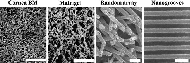
FIGURE 11.3 Scanning electron micrographs of native basement membrane,
Matrigel, and bioinspired synthetic matrices. The topographical features of the corneal
basement membrane compared to artificial surfaces. Matrigel biochemically and
topographically mimics native BMs, but it is difficult to alter individual physical
features. Synthetic isotropic surfaces, such as gold-coated ‘‘grass-like’’ fibers, can have
some physical features altered, but still retain the random array appearance of native
BMs. The physical features of anisotropic nanogrooves can be controlled and sized to
study the effects of individual topographic features on cell behavior. Scale
bars = 600 nm.
CELLULAR BEHAVIOR ON BASEMENT 305