Kenneth E. Gonsalves, Craig R. Halberstadt, Cato T. Laurencin, Lakshmi S. Nair. Biomedical Nanostructures
Подождите немного. Документ загружается.


Technique
Topographic
features Merits Shortcomings Cell type used
Observed
change in
cellular
behaviors References
3 Electrospinning
(aligned
nanofibers)
Aligned fibers
in the nanometer
dimension and
can be extended
to several micron
thick fibers
Simple, facile, wide
range of materials
available for
spinning ability to
incorporate
bioactive
molecules
Can create only
nonwoven
fiber structures
Endothelial,
neural stem
cells, ligament
fibroblast, smooth
muscle cells,
chondrocyte
(HTB-94)
osteoblast
(MG-63)
Spreading,
proliferation,
phenotype
expression,
orientation,
alignment
[70–77]
4 Nanoimprinting Wide range of
structures such as
pillars, grooves,
ridges possible in
nanodimensions
Multilayer three-
dimensional
structures at
comparatively
lower cost than
electron beam
lithography
Expensive
equipment
and time
consuming
Osteoblasts,
smooth
muscle cells
Strong
alignment,
spreading,
migration,
phenotype
expression
[78–80]
Unordered nanotopographic surfaces
5 Self-assembly Can create fibers of
any dimensions
based on the
start-up material
Process is simple,
facile, low tech,
can create complex
functional structures.
Structures are
defect-free and
self-healing
No direct control
over fabrication,
compared to
lithography
complex structure
fabrication is
difficult.
MC3T3-E1,
neural,
bladder, smooth
muscle, aortic
endothelia and
neural progenitor
cells. In vivo
studies in Syrian
hamsters
Adhesion,
increased
proliferation,
differentiation,
and phenotype
expression.
[81–95]
TABLE 10.1 (Continued )
266
(continued )

Technique
Topographic
features Merits Shortcomings Cell type used
Observed
change in
cellular
behaviors References
6 Phase separation Wide range of
geometry
and dimensions
include pits, islands,
fibers, and irregular
pore structures
Simple, facile, no
equipment needed,
highly porous
scaffolds with
control on porosity,
and scalable
No organized
patterns
OCT-1 osteoblast-
like cells, nerve
stem cells,
MC3T3-E1
preosteoblasts
Adhesion,
proliferation,
differentiation
[81, 96–99]
7 Colloidal
lithography
Columns and islands
can be created in
nanometer ranges
Relatively cheap and
less time consuming,
can pattern large
surface area with
less effort
Specific feature
geometries
hard to design
Epitenon cells,
pancreatic
epithelial cells
(AR4-2J),
mammary
epithelial
cells (HC11),
human bladder
carcinoma,
HTB-4, primary
human
osteoblasts,
macrophages/
monocytes
human
fibroblast
Interaction,
spreading,
alignment,
migration,
phenotype
expression,
and
proliferation
(37, 43, 44,
100–105)
8 Electrospinning
(random
nanofibers)
Fibers in the
nanometer
dimension
and can be
extended
to several micron
thick fibers
Simple, facile, wide
range of materials
available for spinning
ability to incorporate
bioactive molecules
Can create
only fibrous
structures
Fibroblasts,
MC3T3-E1
osteoblast-like,
SMCs, NIH3T3,
bone marrow
stromal cells
Spreading,
proliferation,
phenotype
expression,
orientation,
[106–123]
267
(continued )

Technique
Topographic
features Merits Shortcomings Cell type used
Observed
change in
cellular
behaviors References
9 Chemical etching Depends on the
nature and time of
etching agent used
Fast, simple, and
inexpensive
Difficult to get
required geometry
and features
Human
osteoblasts
CRL-11372,
aortic smooth
muscle cells,
primary rat
smooth muscle
cells from
thoracic aorta,
bladder and
cortical cells,
LRM55
CNS tumor
Adhesion,
migration,
proliferation
and phenotype,
expression
[124–127]
10 Carbon
nanofibers/
nanotubes
Fibers in
nanodimensions
5 nm to several
100 nm in diameter
and several microns
in length. Various
shapes straight,
spiral, fishbone,
and so on
Excellent mechanical,
electrical, and surface
properties. Relatively
easy to fabricate and
no costly equipment
needed
Can create only
fibers and tubes
and specific
geometries not
possible
Human
osteoblasts,
epidermal
keratinocytes,
macrophages
and skin
fibroblast rat
astrocytes,
aortic smooth
muscle cells,
cardiac muscle
cells and cortical
cells, mouse skin
fibroblasts, bovine
bladder smooth
muscle cells
Compatibility,
adhesion,
proliferation,
differentiation,
morphology
[128–138]
TABLE 10.1 (Continued )
268

nano
fib
ers
/
nanotubes
nano
di
mens
i
ons
5 nm to several
100 nm in diameter
and several microns
in length. Various
shapes straight,
spiral, fishbone,
e
l
ectr
i
ca
l
,an
d
sur
f
ace
properties. Relatively
easy to fabricate and
no costly equipment
needed
fib
ers an
d
tu
b
es
and specific
geometries not
possible
osteo
bl
asts,
epidermal
keratinocytes,
macrophages
and skin
fibroblast rat
astrocytes,
aortic smooth
muscle cells,
cardiac muscle
cells and cortical
cells, mouse skin
fibroblasts, bovine
bladder smooth
muscle cells
a
dh
es
i
on,
proliferation,
differentiation,
morphology
11 Polymer demixing Nanotopographic
features such as
pits, islands, or
ribbons of wide
range of height or
depth possible
Can pattern large
surface area, low
cost, high
efficiency,
less effort
Can only create pits,
islands, or ribbons.
No other specific
geometries possible
Human fibroblasts,
osteoblasts,
endothelial cells
Adhesion,
morphology,
proliferation,
differentiation,
gene expression
[139–149]
269
nanofabrication techniques in brief while discussing the importance of
nanotopographic features on cell behavior.
10.2.1 Cell Behavior Toward Nanotopographic Surfaces Created
by Electron Beam Lithography
Electron beam lithography is an attractive computer-controlled techn ique to
create precise geometries and patterns on nanoscale without the use of any
mask. In brief, either positive or negative resists are used to create programmed
nanotopographic features. Positive resists break down into lower molecular
weight fragments when irradiated by a high energy electron beam, whereas
negative resists form insoluble cross-linked networks. These irradiated resists
are developed in a suitable developer. The preprogrammed nanotopographic
features were created by leaching out of the resists in a developer. However, the
use of negative controls limits the resolution of these features due to swelling
while developing. Further, this technique requires expensive equipment and is
time consuming. Many nanotopographic features including square grooves,
ridges, and nanopillars, created using the current technique, were used to study
in vitro cellular behavior to elicit the importance of nanoscale features for a
variety of biomedical ap plications [52, 55 –62].
Micro- and nanotopographic features containing grooves and ridges with
dimensions varying from 400 to 4000 nm directed the alignment and migration
of SV40-transformed human corneal epithelial cells along the grooves and
ridges of all the dimensions [55]. It was observed that cell colonies migrated out
along grooves and ridges in circular zones and migrated perpendicular to ridges
in some cases. On flat controls equal migration was observed in all directions.
Stress fibers and focal adhesions were also aligned along the ridges and groves
[55]. Keratocytes showed a stronger alignment than epithelial cells to these
nanotopographic surfaces [20]. Keratocytes presented fewer stress fibers and
focal adhesions on nanodimensions when compared to micro and flat control
surfaces [56]. Nanotopographic features containing varying widths of grooves
and ridges ranging from nano- to micron dimensions provided stimulus for
human corneal epithelial cells [57]. Cells were found to align along the grooves
and the ridges of nanodimensions with elongated structures while rounded cells
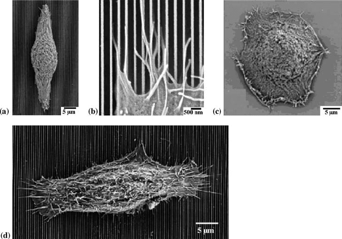
were observed on smooth surfaces as seen in Fig. 10.2. On the nanopatterned
surfaces, cells periodically extend filopodia along the direction of the
nanofeature as shown in Fig. 10.2c [57]. When the culture medium was
changed from DMEM/F12 to Epilife medium, human corneal epithe lial cells
aligned perpendicular to the nanotopographic features as evident from Fig.
10.2d whereas parallel alignment was observed on microscale topographies
[58]. Such an observed change in alignment is presumably due to a change in
focal adhesion structure. Human gingival fibroblasts and rat neurons on
patterned surfaces with grooves showed focal adhesion contacts and cells
aligned to the grooves [59, 60]. Xenopus neuritis aligned parallel to all groove
sizes whereas hippocampal neuritis aligned perpendicular to narrow, shallow
270 BIOMEDICAL NANOSTRUCTURES
grooves [60, 61]. Nanotopographic surfaces containing ordered features of
pillars, pits, cliffs, and random gold colloid particles showed topography
dependence in their adhesion. Ordered cliffs showed enhanced adhesion at the
cliff, edges. Reduction in cell adhesion was observed on pits and pillars when
compared to flat and colloid particle surfaces [52]. Anisotropic features in the
size range of 2002000 nm on polyurethane surfaces showed decreasing
proliferation of corneal epithelial cells with decreasing dimensions when
compared to planar surfaces [62].
10.2.2 Cell Behavior Toward Nanotopographic Surfaces Created
by Photolithography
Photolithography is one of the popular techniques to create nanotopographic
surfaces containing many geometric features including grooves, ridges, and
round nodes from nanometers to several microns in size. Photolithography
involves a series of processes in brief; cleaned surfaces of silicon wafers are
coated with photoresist and soft baked to remove the solvents from photoresist
FIGURE 10.2 SEM micrographs of human corneal epithelial cells cultured in
DMEM/F12 on (a) patterned nanotopography elongated and aligned (b) patterned
surfaces filopodia extend in the direction of features. (c) Flat substrates with rounded
morphology. (d) Patterned surfaces cultured in Epilife medium showing perpendicular
alignment to nanotopographic features. (Reprinted with permission from (a, b and c)
Reference [57] and (d) Reference [58].)
CELL BEHAVIOR TOWARD NANO STRUCTURED SURFACES 271
that converts the resist photosensitive. The photomask containing previously
defined geometric features aligned with the silicon surface and exposed to light
source followed by etching and developing leaves the predetermined patterned
surfaces. The hard baking process improves the adhesion of photoresist to
silicon wafer and hardens the photoresist. Photolithography can create precise
geometries and patterns but require expensive complicated instrumentation,
and mostly microsized topographies can be created easil y. Thus created
nanotopographic features elicit changes in cellular functions including
orientation, interaction, morphology, and differentiation based on the
topography and dimensions they are exp osed to [63–69].
Implant surfaces containing nodes of 2 and 5 mm diameter and having
different heights in nanometer length showed fewer mononuclear cells and
thinner fibrous capsules when implanted in a rabbit model than the control
planar and 8-mm-diameter grooved surfaces [68]. Also, cells appeared to be
elongated and with more number of filopodia on nanotextured surfaces, whereas
cells assumed a rounded shape with less number of filopodia indicating less
interaction with the implant surfaces [68]. Nanotopographic features containing
rough surfaces were created by reactive ion etching, and smooth wet etched
surfaces were created on silicon wafers showing surface-dependent adhesion
behavior to rat astrocytes [69]. Transformed astrocytes showed preferential
attachment and a spread morphology on wet etched surfaces. Columnar
nanotopographic features created by reaction ion beam etching resulted in round
morphology, loose attachment, and exhibition of complex surface projections of
transformed cells [69]. Rat fibroblasts on square grooves with submicron
dimension oriented and elongated along grooves [63]. P388D1 macrophages, rat
peritoneal macrophages, and chick embryo cerebral neurons showed increasing
orientation and spreading with increasing groove depth [64, 65]. Well-
characterized cytoskeleton and F-actin and vinculin accumulation was observed
along the edges of the grooves [64, 66]. Uromyces appendiculatus fungus cells
showed a high degree of orientation to the polystyrene nanoridge spacing of
0.56.7 mm, whereas ridge height of 500 nm showed maximum cell differentia-
tion compared to ridges of height greater than 1 mm or less than 0.25 mm [67].
10.2.3 Cell Behavior Toward Nanotopographic Surfaces Composed
of Aligned Nanofibers by Electrospinning
Polymeric nanofibers are created using a variety of techniques such as template
synthesis, phase separation, drawing, self-assembly, and electrospinning.
Among these techniques, electrospinning is extensively studied since the
process is simple, elegant, and facile, and can create polymeric fibers in the
range of a few nanometers to several micron thicknesses using the same
experimental setsup [70–77]. During the process of electrospinning, fibers
randomly deposited on the grounded collector create unordered surface
topographies. Aligned nanofibers that could present ordered nanotopographies
can be created by using a high speed rotating collector, applying an auxiliary
272 BIOMEDICAL NANOSTRUCTURES
electric field, and using a sharp edged thin wheel collector or a frame collector
in place of the stationary grounded collector [71–76]. Thus created
nanostructured surfaces both aligned and random were used as tissue
engineering constructs, and cells respond differently to the surfaces they were
exposed to [71–76].
Human coronary artery endothelial cells on aligned gelatin-modified
poly(caprolactone) (PCL) nanofibers in the diameter range of 2001000 nm
provided improved adhesion, spreading, and proliferation than the control
PCL and random nanofibers [71]. These aligned nanofibers strongly cause the
orientation of endothelial cells parallel to the nanofibers with spindle-like
structures; also well-defined cytoskeleton and enhanced phenotypic expression
were observed. Shear stress caused by the blood flow orients the endothelial
cells in the flow direction in vivo, and surface-modified aligned nanofibers
simulate the actual in vivo condition in a static culture that provides an
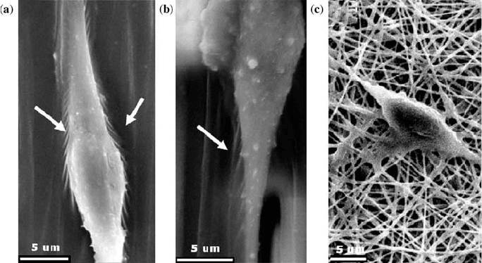
alternative to complex dynamic culture [71]. Neonatal mouse cerebellum C17.2
stem cells (NSCs) on PLLA-aligned and random nanofibers attached well and
changed their original round shape to elongated spindle-like sh ape that
provided morphological evidence for NSC differentiation. The direction of
NSC elongation and its neurite outgrowth was exactly parallel to the direction
of aligned nanofibers with a higher rate of NSC differentiation than the
control. Increased interaction between the filament-like structures produced by
NSCs was observed on aligned fibers that were absent on random nanofibers as
seen from Fig. 10.3 [72]. Such aligned nanofi bers could be used as a potential
FIGURE 10.3 NSCs on PLL-aligned fibers had an apparent bipolar elongated
morphology with neuritis, and filament-like structures marked with arrows (a) attach to
nanofibers and (b) microfibers, and (c) absence of filament-like structures on random
nanofibers. (Reprinted with permission from Reference [72].)
CELL BEHAVIOR TOWARD NANO STRUCTURED SURFACES 273
cell carrier for neural tissue engineering. Human ligament fibroblast (HLF) on
aligned polyurethane nanofibers also oriented in the direction of aligned
nanofibers and had a spindle shape [73]. Fibroblasts synthesized significantly
more collagen on aligned nanofibers, and furt her HLFs were more sensitive to
the stress applied in longitudinal direction resulting in an increased production
of collagen in response to applied stra in [73]. Human coronary artery smooth
muscle cells (SMCs) on poly(
L-lactid-co-e-caprolactone)-aligned nanofibers
attached and migrated along the direction of the nanofiber orientation and
maintained a spindle-like structure [74, 76]. SMCs resulted in increased
adhesion and proliferation on aligned nanofibers, and cytoskeleton proteins
inside SMCs were parallel to the direction of the nanofibers. Chitosan
nanofibers and aligned na nofibers promoted the attachment of human
osteoblasts and chondrocytes and maintained characteristic morphology
throughout the study [75]. The observed cell orientation on aligned nanofibers
presumably follows the contact guidance theory, which states that cells have a
higher probability of migrating in directions of chemical, structural, and/or
mechanical properties of the substratum [77].
10.2.4 Cell Behavior Toward Nanotopographic Surfaces Created
by Nanoimprinting
Nanoimprint lithography can create ordered nanotopographic features such as
pillars, grooves, and ridges at the required dimensions. In brief, the technique
utilizes a hard mold containing previously defined nanoscale patterns. Wafer
substrates covered by polymer cast under the controlled temperature and
pressure will be imprinted by the hard mold. Hard mold embossing creates a
thickness contrast on the polymer surfa ce with the required nanotopographic
features. It is possible to create multilayer three-dimensional structures using
nanoimprint lithography at comparatively lower cost than electron beam
lithography. However, the present technique also utilizes expensive equipment,
and the process is time consuming. Thus, created ordered nanotopographic
features elicit changes in c ellular functions in vitro [78–80].
Nanopatterned gratings with 350 nm line width, 700 nm pitch, and 350 nm
depth created on poly(methyl methacrylate) and poly(dimethylsiloxane)
showed decreased proliferation of bovine pulmonary artery SMCs than the
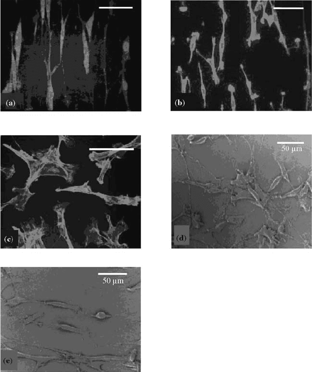
flat control [78]. More than 90% of the cells were aligned and assumed an
elongated structure in both cytoskeleton and nuclei on nanopatterned surfaces
as seen in Fig. 10.4 [78]. Also, the polarization of microtubule organizing
centers of the SMCs associated with cell migration showed a preference toward
the axis of cell alignment in an in vitro wound healing assay on nanopatterned
surfaces. Polystyrene surfaces containing nanopillars in the diameter range of
1601000 nm and 1000 nm height were tested as an alternative tissue culture
plate by studying the in vitro behavior of epithelial-like cell line (HeLa) on the
said topography [79]. HeLa cells adhered only to the heads of the nanopillar
sheet with a significantly lower number of cells than the control and acquired
274 BIOMEDICAL NANOSTRUCTURES
rounded morphology on nanopillars in comparison to a spread structure on
the flat control. Also, actin molecules appeared to localize to the circumference
of the nanopillar heads, whereas vinculin molecules were distributed
homogeneously in regions away from the nanopillar heads [79]. Thus, the
low adhesion properties exhibited by the cells might lead to alternative tissue
culture plates that do not require conventional trypsin treatment to lift the
cells. Polystyrene nanopatterned surfaces with grooves having two different
depths of 50 and 150 nm with a periodicity of 500 100 nm showed a strong
FIGURE 10.4 Confocal micrographs of F-actin stained SMCs on (a) nanoimprinted on
PMMA, (b) PDMS, and (c) nonpatterned PMMA surfaces (scale bar 50 mm). SEM
micrographs on (d) nonpatterned PMMA and (e) nanoimprinted surfaces show elongated
structures on patterned surfaces. (Reprinted with permission from Reference [78].)
CELL BEHAVIOR TOWARD NANO STRUCTURED SURFACES 275