Kenneth E. Gonsalves, Craig R. Halberstadt, Cato T. Laurencin, Lakshmi S. Nair. Biomedical Nanostructures
Подождите немного. Документ загружается.

alignment and orientation of primary osteoblasts in the direction of grooves
[80]. The exposed patterned surface topography promoted the cell elongation,
cytoskeleton actin, and actinin molecules’ orientation in the direction of the
grooves. However, vinculin, a protein associated with focal adhesions,
appeared to concentrate at the opposite end of aligned cells presumably due
to the balancing tension generated from the surface anisotropy [80]. The
observed anisotropic behavior of osteoblasts on the nanogrooves might
enhance cell settlement on the nano topographic surface.
10.2.5 Cell Behavior Toward Nanotopographic Surfaces Created
by Self-Assembly
Self-assembly is a powerful technique to create nanostructures from various
polymers and biomolecules via molecular self-assembly. Molecules self-
assemble by virtue of weak noncovalent bonds. Such interaction forces may
be hydrogen bonds, electrostatic interactions, hydrophobic interactions, and
van der waals forces. All the biomacromolecules interact and self-assemble to
create complex functional defect-free and self-healing nanostructures. The
molecular self-assembly is simple, facile, and scalable. However, no direct
control over the fabrication process is possible, and also it is not possible, to
design specific geometric features. Using the bottom-up approach, many
nanostructures created were studied as scaffold materials for various
biomedical applications [81–95].
Self-assembled peptide nanofiber scaffolds (SAPNS) of branched and linear
peptide-amphiphile molecules as coating on poly(glycolic acid) scaffolds
resulted in preferential attachment of primary human smooth muscle cells on
branched than on linear peptide amphiphile and control [88]. The SAPNS as
sciatic nerve grafts could partially restore the optic tract and functional vision
in brachium transected experimental adult animals in vivo [94]. Rat
mesenchymal stem cells on SAPNS with RGD sequence resulted in increased
attachment, alkaline phosphatase (ALP) activity, and osteocalcin content
than the SAPNS without RGD sequence and tissue cultur e pol ystyrene [95].
Hybrid SAPNS scaffolds also supported MSC and resulted in homogeneous
bone formation in vivo in a rat subcutaneous model. In perfusion, in vitro
culture registered higher alkaline phosphatase activity and osteocalcin
content indicated enhanced osteogenic differentiation of MSC than control
and static culture [84]. The pentapeptide epitope isolucinelysinevaline
alaninevaline (IKVAV) containing SAPNS supported the neural progenitor
cells and caused selective rapid differentiation into neurons with lesser number
of astrocytes [87]. The IKVAV-incorporated SAPNS combine bioactive
epitope and nanotopography elicits the observed advantages [87]. MC3T3-E1
cells not only survive and proliferate on SAPNS but also possibly utilize
peptide molecules in their metabolic pathways [89]. Functionalized and
biomimetic SAPNS can elicit cellular events such as wound healing and tissue
regeneration by creating ECM recognition domains with nanofeatures [86].
276 BIOMEDICAL NANOSTRUCTURES
10.2.6 Cell Behavior Toward Nanotopographic Surfaces Created
by Phase Separation
The phase separation technique used for the fabrication of highly porous
scaffolds with controllable porosity can create a wide range of geometries and
dimensions including pits, fibers, and irregular pore structures [81, 96– 99].
Phase separat ion either solidliquid or liquidliquid can be induced by
lowering the solution temperature. In brief, to a polymer solution in a low
melting point solvent, addition of a small quantity of water generates polymer-
rich and polymer-poor phases. Such a system cooled below solvent melting
point followed by vacuum drying to sublime the solvent produces the porous
scaffolds of both micro- and nanostructures. The technique can create highly
porous scaffolds with controllable porosity. The process is simple, facile, and
scalable and does not require any costly equipment. The inability to fabricate
well-organized patterns and specific geometric features is the limiting factor of
this technique.
Nanostructures formed by phase separation technique favored the adhesion,
proliferation, and differentiation of different cell lines studied. MC3T3-E1
showed better response on PLL A nanofiber scaffolds than the solid walled
controls created using reverse solid freeform fabrication and thermal phase
separation technique [96]. Signific antly higher cell number, and osteocalcin and
bone sialoprotein expressions were observed on nanofiber scaffolds. However,
observed lower expression of type I collagen on nanofibers was presumably due
to the quicker differentiation of preosteoblasts [96]. PLLA nanofiber scaffolds
with an average diameter of 200 nm fabricated by phase separation technique
supported the nerve stem cell differentiation and neurite outgrowth [99].
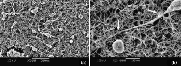
Figure 10.5 present s the neonatal mouse cerebellum C17-2 stem cells on PLLA
nanofiber scaffolds presenting a differentiated cell with neurite penetration into
the scaffolds [99]. Thus, the nanofiber structures could mimic the ECM
FIGURE 10.5 SEM micrographs of PLLA nanofiber scaffolds fabricated by the phase
separation technique presenting neonatal mouse cerebellum stem cells seeded on these
materials after 1 day in culture (a) at a magnification of 500 and (b) differentiated cells
with neurite penetration into the nanofiber scaffolds identified by arrows at 2000.
(Reprinted with permission from Reference [99].)
CELL BEHAVIOR TOWARD NANO STRUCTURED SURFACES 277
structure and support the adhesion, proliferation, and phenotype expression of
the cells.
10.2.7 Cell Behavior Toward Nanotopographic Surfaces Created
by Colloidal Lithography
Colloidal lithography provides a means to pattern surfaces containing columns
and pits with controllable dimens ions. This technique utilizes nanocolloids as
an etch mask that is dispersed and self-assembled electrostatically into a
monolayer on the substrate surfaces. The area surrounding the nanocolloids is
etched away along with the surface itself leaving a patterned substrate using a
suitable etching technique. For example, surface nanotopographies containing
columns can be created using chemically assisted ion beam etching or directed
ion beam bombardment and pits with film evaporation technique. Patterned
surface properties can be varied changing colloid size, colloid ionic strength,
and the monolayer coverage on the substrate surface. Colloidal lithography is
relatively cheap and less time consuming, and can pattern large surface area
with less effort; however, it is difficult to create specific feature geometries.
Cells on these columns and grooves in nanoscale showed interesting structural,
morphological, an d phenotypic behaviors on these nanotopographies [37, 43,
44, 100–105].
Columns with an average height of 160 nm, diameter of 100 nm and with a
230 nm spacing between the features produced by colloidal lithography
resulted in less spreading of human fibroblasts (h-tert BJ1) compared to
control plane surfaces [100]. Microarray analysis showed a shift in gene level
expression when compared to microtopographic surfaces. Upregulation of
several genes responsible for cellcell signaling, wound healing, proliferation,
and cell differentiation, and downre gulation of several genes such as collagen,
and cytoskeleton were observed. Such observed changes in gene level
expression were presumably due to the decreased cell spreadi ng on colloidal
nanotopography [100]. The fibroblasts assumed more stellate and less well-
spread morphology on these nanocolumns. Fibroblasts produced increased
filopodia and were observed to interact with nanocolumns regularly with
filopodia [103]. An attempt to endocytose the nanocolumns by fibroblasts
failed; however, some cells appeared to try and go further producing high levels
of Rac at sites of pseudopodia formation [37]. Human fibroblasts cultured for a
short duration of time, 180 min, showed decreased cell adhesion and spreading
on these nanocolumns than the planar control [101]. Fibroblasts showed less
well-organized actin and vimentin cytoskeletons on these nanocolumns;
however, tubulin cytoskeleton was well organized with least number of
microtubules [101]. Silicon wafer surfaces modified with 50-nm colloidal gold
particles resulted in the direct interaction with the primary rat epitenon cells at
the peripheral cell membrane [102]. Nanocolumns with diameters of 58, 91,
111, and 166 nm resulted in increased spreading of rat pancreatic epithelial cells
when compared to flat surfaces and spreading increased with increase in
278 BIOMEDICAL NANOSTRUCTURES
column diameter [44]. Epithelial cells (human bladder carcinoma, HTB4) on
nanotopographies containing continuous edged grooves with 184 nm depth
were seen to become align ed [43]. Cells on nanotopographies containing
hemispheres with 100 nm height and 167 nm diameter assumed less spread, less
round, and more stellate morphology than the control. Cells registered no
change in the production of cytokine on both the nanofeatures and a decreased
production of IL-6 and IL-8 on the nanofeatures than the control [43]. Mouse
mammary epithelial cells (HC11) on nanotopographies containing either
continuous or discontinuous edges of various depths ranging from 40 to
400 nm were seen to align on the grooves than the control [104]. Grooved
surfaces with continuous edges favored the orientation of cells with more
elongated cells than grooves with discontinuous edges [104]. Nanotopographies
containing heminanosp heres features with a height of 110 nm and various
packing densities of these features were coated with Ti oxide thin films to
obtain a single chemistry [105]. These nanotopographies induced the release of
chemotactic macrophage activation agents and caused stress fiber and
fibronectin formation from the primary human macrophages. Primary human
osteoblasts were seen to migrate away from these nanofeatures [105]. The
nanotopographies present physical cues for the cells that can elicit responses to
align the cells, increase or reduce cell adhesion and proliferation, alter
differentiation, and increase motility.
10.2.8 Cell Behavior Toward Nanotopographic Surfaces Com posed
of Random Nanofibers Created by Electrospinning
Cells identify the exposed surface topo graphy and random nanofiber porous
matrices influence the adh esion, spreading, proliferation, and gene expression
of various cell types seeded on them. Mouse fibroblasts adhered, migrated
through the pores, and integrated well with PLAGA nanofibers, and the
development of the cell growth was guided by nanofiber architecture [106].
Fibroblasts changed morphology from a spread and flat to a long and spindle
shape on nanofibers when adhered to the nanofibers detaching the flat surfaces
[107]. Cross-linked gelatin nanofibers with improved mechanical properties and
thermal stability supported human dermal fibroblast proliferation, and a linear
increase in cell number was observed with time [108]. Scaffolds of polystyrene
nanofiber resulted in a significant increase in smooth muscl e cell attachment
than control, and the ECN produced by oriented nanofibers was similar to
native bladder tissue [109]. MC3T3-E1 cells showed significantly increased cell
number and osteocalcin, alkaline phosphate production with increa sing culture
time on silk fibroin nanofibers [110, 111]. The silk fibroin nanofiber scaff olds in
vivo showed good biocompatibility and enhanced bone regeneration without
any inflammatory reaction [110]. Human coronary artery SMCs showed
normal phenotypic shape on polycaprolactone and collagen nanofibers, and
nanofibers coated with collagen resulted in the migration of SM Cs inside the
nanofiber scaffolds and formation of smooth muscle tissue [112–114]. NIH 3T3
CELL BEHAVIOR TOWARD NANO STRUCTURED SURFACES 279
fibroblasts and normal rat kidney cells showed the morphology and
characteristics of their counterparts in vivo on polyamide nanofibers [115].
Bone marrow stromal cells’ (BMSCs) attachment and proliferation was
favored on gelatin/PCL nanofibers, and cells were able to migrate inside the
scaffold [116]. Increased proliferation of H9c2 cardiac rat myoblasts was
observed on polyaniline gelatin blend nanofibers with different cell morphol-
ogies at the initial time point; however, similar cell density and morphology
were attained after 1 week on all the nanofiber scaffolds as the cultures reached
confluence [117, 118]. Cellnanofiber interaction and orientation is more
pronounced in the initial culture time, and once the confluence is reached it is
difficult to identify morphological changes. Hepatocytes cultured on the
galactosylated poly(e-caprolactone-co-ethyl ethylene phosphate) (PCLEEP)
nanofibers exhibited similar functional profiles as on flat control surfaces;
however, morphologicalchangeswere observed. Hepatocytesformed 50300-mm
spheroids on flat surfaces whereas smaller aggregates of 20100 mm were
formed on nanofiber surfaces [119]. Random and fused surfa ce topographies
having fiber diameters in the range of 140 nm to 2.1 mm showed increased
MC3T3-E1 osteoprogenitor cell density in the presence of osteogenic factors
on the fibers than the smooth surfaces. The cell density increased with
increasing fiber diameter while ALP expression was independent of surface
topography [120]. Osteoblast-like cells on starch/polycapolactone micronano-
fibers showed interesting morphological and phenotype expression behavior
[121]. With the introduction of nanofiber structures on microfiber scaffolds,
osteoblasts organized to bridge between microfibers and possessed much more
spread and stretched morphology in contrast to continuous monolayer on
microfibers. Additionally, the presence of nanostructure resulted in increased
ALP activity than the microfibers. Nanomicroscaffolds were fabricated by
electrospinning nanofibers on PLAGA knitted scaffolds, and porcine bone
marrow stromal cells showed improved cellular behavior on nanofiber
combined scaff old than the control [122]. Increased cell attachment, faster
cell proliferation, and higher expression of collagen I, decorin, and biglycan
genes were observed on nanofiber-included scaffolds. Increased adhesion and
proliferation of human um bilical vein endothelial cells were observed on
poly(
L-lactide-co-e-caprolactone) nanofiber matrices (0.31.2 mm) whereas a
decline in cell adhesion and restricted spreading was observed on larger fiber
diameter (7.0 mm) [123].
10.2.9 Cell Behavior Toward Nanotopographic Surfaces Created
by Chemical Etching
Chemical etching i s a surface modification process to create surf ace roughness
in nanometer scale length. In a typical procedure, material surfac es are soaked
in a variety of etchants such as sodium hydroxide (NaOH), nitric acid
(HNO
3
), and hydrofluoric acid (HF) to name a few and the selection of the
etchants depends on the material properties [124–127]. The material surface
280 BIOMEDICAL NANOSTRUCTURES
disintegrated as they were exposed to etchants, and the surface became rough
with pits and projections in nanometer scale length. It is possible to vary the
surface roughness b y varying exposure time, na ture, and concentration of
etchants to get various surface roughness dimensions in nanoscale . This
process is fa st, sim ple, and inexpensive; however, it is not possible to get
required geometry features since it is a surface treatment phenomenon. Thus,
created nanometer-scale roughness can potentially affect cellular b ehavior
[124–127].
Compacts of selenium (Se) metal particles in micron- and nanometer-size
range were tested as an anticarcinogenic orthop edic material [124]. These
compacts were chemically etched with different con centrations of sodium
hydroxide to create surfa ce roughness in nanometer scale. Osteoblast densities
increased on both nano- and microparticles with nanoscale roughness when
compared to reference wrought titanium and Se microparticles after 24 h of cell
culture under standard in vitro conditions [124]. The pillar patterned silicon
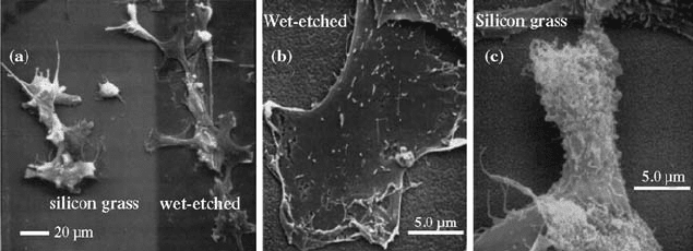
wafers were subjected to wet ch emical etch to produce nanometer-scale surface
roughness, and astrocytes showed preferential attachment to these surfaces
[125]. Transformed astrocytes preferred the wet etched surface to the
unmodified silicon substrate as seen in Fig. 10.6 [125]. The degree of selective
adhesion and spreading increased with duration of wet etch presumably due to
the increased surface area. Primary cortical astrocytes from neonatal rats
showed a preference for silicon glass over the wet etched surface. Such
observed cellular behaviors are presum ably due to the different cell types in the
study [125]. Nanostructures created on PLAGA and PU surfaces by chemical
etching showed improved adhesion and proliferation of bladder smooth muscle
cells [126]. Surface chemistry and increased nanometer surface roughness
created after prolonged chemical etching brought changes in the observed
cellular behavior. Nanostructures produced after longer duration of etching on
FIGURE 10.6 Transformed astrocytes cell line (LRM55) on (a) unmodified silicon
glass and wet etched glass surface, and (b) the same at higher magnification. Cells
appear flat and well attached showing very few projections on wet etched surfaces,
whereas cells appear not closely adhered to the surface that contains the complex
projections and ruffles. (Reprinted with permission from Reference [125].)
CELL BEHAVIOR TOWARD NANO STRUCTURED SURFACES 281
PLAGA surfaces registered significantly higher cell number compared to
submicron surfaces, flat untreated and control glass surfaces. While
nanostructured surfaces of PU did not show increasing proliferation trend
with increasing nanometer surface roughness, chemically treated surfaces and
submicron dimension surface roughness showed higher cell number than the
control and conventional flat surfaces [126]. Further nanometer surface
roughness on PLAGA resulted in absorption of significantly more vitronectin
and fibronectin from serum compared to untreated flat PLAGA surfaces [127].
Significantly higher amoun ts of proteins, na mely fibronectin and vitronectin
adsorptions, enhanced the vascular smooth muscle cell and endothelial cell
density presumably due to the cellular recognition sites present on the proteins.
It is also observed that blocking of cell-binding epitopes of fibronectin and
vitronectin on nanometer surface roughness resulted in significantly decreased
vascular cell adhesion on nanostructured surfaces [127].
10.2.10 Cell Behavior Toward Nanotopographic Surfaces Created
by Incorporating Carbon Nanotubes/Nanofibers
Carbon nanostructures such as carbon nanotubes and nanofibers possess
outstanding physical and chemical properties and have a diversified application
range. Carbon nanotubes can be eithe r multiwalled nanotubes (MWNTs) or
single-walled nanotubes (SWNTs). Carbon nanofibers are essentially of
filamentous structure and can assume various shapes such as straight, spiral,
or fishbone depending on the metal catalyst used. These carbon nanostructures
are fabricated in industrial scale by three main methods, namely electric arc
discharge, laser ablation, and catalytic chemical vapor deposition (CVD).
Usually these processes simultaneously produce SWNTs, MWNTs, fullerenes,
and a considerable amount of soot and carbon nanoparticles and need further
purification to isolate each component. However, the yield of individual
nanostructures varies depending on the method and fabrication condition
used. Thus, produced carbon nanostructures are 5 nm to several 100 nm in
diameter and several microns in length and have excellent mechanical,
electrical, and surface properties and a potential utility in various biomedical
applications including tissue engineering scaffolds [128–138].
Osteoblasts registered size-dependent behavior on multiwalled carbon
nanofibers with diameters ranging from 60 to 200 nm in an in vitro culture of
21 days. Increased osteoblast proliferation, ALP synthesis, and calcium
depositions were observed on carbon nanofibers with lesser diameter than the
control (larger borosilicate glass) [129]. Ost eoblasts, fibroblasts, chondrocytes,
and smooth muscle cells showed dimension-dependent behavior on carbon
nanofibers with the diameter range of 60200 nm [130]. Osteoblasts adhesion
increased with decreasing fiber diameter as previously observed [129], whereas
other cells were not influenced by fiber dimension. Adhesi on of fibroblasts,
chondrocytes, and smooth muscle cells decreased with a decrease in nanofiber
diameter and were dependent on carbon nanofiber chemistry. Further,
282 BIOMEDICAL NANOSTRUCTURES
PLAGA carbon nanofiber composites enhanced the osteoblast adhesion that
increased with lower diameter nanofiber dispersions [130]. SWNTs showed
biocompatibility with cardiomyocytes in culture; however, slight modification
in cell shape was observed microscopically due to the binding of carbon
nanotubes to cell membranes that affected the cell count and viability only
after 3 days of culture [128]. Polycarbonate urethane composites with different
weight percentages of carbon nanofibers were used to study the adhesion
behavior of astrocytes and osteoblasts on these composites and register ed a
size-dependent adhesion behavior [131, 132]. Astrocytes preferentially adhered
and proliferated on carbon nanofibers with higher fiber diameter. Osteoblasts
preferentially adhered and proliferated well on greater weight percentages of
carbon nanofibers with least fiber diameter composites, whereas astrocytes
showed reduced adhesion and proliferation on these composites [131, 132].
Neurons also showed improved adhesion and proliferation on these
composites [131]. Such observed phenomenon is presumably due to the high
degree of surface roughness in the nanometer scale. Osteosarcoma ROS 17/2.8
cells cultured on chemically modified SWNTs and MWNTs supported
osteoblast proliferation. Nanotubes carrying neutral electric charge resulted
in increased cell growth and produced plate-shaped crystals. Osteoblasts
presented a dramatic change in cell morphology on MWNTs, which was
correlated with changes in plasma membrane functions [133]. Osteoblasts and
fibroblasts exhibited better viability for high purity MWNTs, and nanotubes
induced an increase in collagen expression by these cells [135]. High purity
SWNTs and fullerenes offered a very low toxicity to human macrophage cells
and did not simulate nitric oxide release from murine macrophage cells in vitro
[134]. MWNTs induced the release of the proinflammatory cytokine interleukin
8 from human epidermal keratinocytes [136]. Composites of collagen-SWNT
gels showed biocompatibility and maintained smooth muscle cell viability more
than 85% in vitro for 7 days [137]. MWNTs’ and nano-onions’ exposure
resulted in increased apoptosis/necrosis of skin fibroblasts that indicated a
strong immune and inflammatory response, which was further evident from the
changes in the expression of genes involved in cellular transport, metabolism,
cell cycle regulation, and stress response [138].
10.2.11 Cell Behavior Toward Nanotopographic Surfaces Created
by Polymer Demixing
Polymer demixing is a widely studied nanofabricating techni que to pattern
large surface area with relatively low cost and high efficiency [139, 140]. In
brief, in a typical experiment, polystyrene (PS) and poly(4-bromostyrene)
blends undergo spontaneous phase separation during spin casting on silicon
wafers. Various topographic features including pits, islands, or ribbons can be
created with a wide range of height and depth by varying the blend
concentration and composition. Different geometric shapes were fabricated
by changing the blend ratio while the blend concentration changed the sizes of
CELL BEHAVIOR TOWARD NANO STRUCTURED SURFACES 283
these shapes. Thus, created nanotopographic features are unordered and
covered randomly on the surfaces. This technique has precise control on
patterning in the vertical direction as opposed to the horizontal, and thus
allowed creating nanostructures with defined heights or depths. These
unordered randomly placed nanotopographic features such as islands, pits,
or ribbons of different heights or depths were utilized to evaluate cellular
behavior [38, 41, 53, 139–149].
Dalby et al. have done extensive studies on fibroblast interaction and their
behavior by changing heights of the islands [141–145, 149]. A detailed study
was performed to know the effect of cell nanotopography interactions on
different gene expressions. Human fibroblasts on 13-nm-high polymer demixed
islands were studied for various gene expressions using a 1718-gene microarray
analysis. Many changes in genes involved in signaling, cytoskeleton, ECM gene
transcription, and protein translation were observed. A total of 584 genes
showed an upregulation in their expression when compared to flat surfaces as
control. The upregulation of these genes suggests that the fibroblasts were well
differentiated on the studied nanotopography than the control. Fibroblasts on
similar surface topography resulted in more spreading with the aid of many
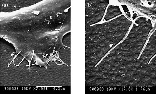
filopodia projecting from the cell membranes as seen in Fig. 10.7 [53]. Increased
cell attachment, spreading, and well-developed cytoskeleton were also
observed. Fibroblast interaction with nanoislands of different height s 13, 35,
and 95 nm resulted in interesting cellular behavior [38]. Fibroblasts interaction
seemed to increase with increasing island size, which is evident by the observed
large pseudopodial projections on 95 nm. The 13-nm islands identified in the
FIGURE 10.7 Scanning electron micrographs of human fibroblasts on 13-nm
nanoislands created by polymer demixing of polystyrenepoly(4-bromostyrene) blends.
Strong interaction between filopodia and nanoislands was seen on nanotopographies:
(a) low magnification; (b) high magnification. (Reprinted with permission from
Reference [53].)
284
BIOMEDICAL NANOSTRUCTURES
most proliferative cell population had well-developed actin, tubuli n, and
vimentin cytoskeletons, whereas the 95-nm islands were characterized by a low
level proliferative population and poorly developed cytoskeleton [38]. The
nylon tubes of internal diameters 0.5 and 1.5 mm were used to create
nanotopographic features with heights of 90 and 40 nm by polymer demixing
using the blends of polystyrene poly(n-butylmethacrylate) (PS/PnBMA) [142].
Fibroblasts on control tubes showed better spreading, well-arranged b-tubulin,
whereas nanotubular topog raphies assumed high stellate morphology with
rounded cell struc ture and were poorly organized. The cell membranes on
nanotubular topographies appeared ruffled with exceptionally long filopodia
interacting with nanoislands. However, no significant differences in cellular
responses were observed for both the diameters with varying island heights.
Primary human fibroblasts’ interaction with the nanoislands of 10 nm height
and controls resulted in increased cell adhesion, spreading, well-defined
cytoskeleton formation, the fibroblas tic morphologie s were maintained, and
produced lamellipodia with visi ble stress fibers at any given time point studied
[143], whereas nanoislands with a height of 50 nm resulted in poor cell
adhesion, low percentage of spreading, poorly defined cytoskeleton with very
few lamellae, and developed no stress fibers at all the time points studied [143].
These topo graphies studied might help in designing surfaces with reduced cell
adhesion and could be utilized in biomaterial design for stents and heart valves.
Fibroblasts cultured for 4 days on the 27-nm nanoislands resulted in
significantly greater areas than flat controls used in the study [144]. Well-
organized cytoskeleton characterized by organized acti n and tubulin on both
the surfaces and stress fibers were more often observed in cells on the 27-nm
islands. By day 30, vimentin was well organiz ed on the control whereas poorly
organized on the 27-nm islands. Fibroblasts on nanoislands of 95 nm height
registered temporal changes in both morphology and cytoskeleton [145].
Fibroblasts on these islands started producing lamellae and filopodia after
5 min of seeding and with progressing time filopodiaisland interactions
increased. Fibroblasts assumed different morphologies with increasing time
points and attained more stellate morphology with the large pseudopodial
processes. Cultures even after 3 weeks had few patches of grouped cells and a
large number of isolated cells. Isolated cells appeared to have a strong
interaction with these nanoislands. On the contrary, flat control surfaces
maintained normal fibroblast morphology and by 1 week cells were confluent.
Many stress fibers around the cell periphery were observed, and these fibers
were seen stretched across the cytoplasm on the cells on the na noislands at the
initial time points. At the later time points, controls had a matured
cytoskeleton with distinct actin stress fibers and organized tubulin whereas
cells on the nanoislands had less organized cytoskeleton [145]. Fibroblast
interaction with the nanoislands can be summarized as presented in Fig. 10.8
that indicates that controlling the island height has a direct effect on different
cellular activity.
CELL BEHAVIOR TOWARD NANO STRUCTURED SURFACES 285