Kenneth E. Gonsalves, Craig R. Halberstadt, Cato T. Laurencin, Lakshmi S. Nair. Biomedical Nanostructures
Подождите немного. Документ загружается.

observed to form bridges similar to focal contacts with the nearest fibers as
seen in Fig. 15.7b. The expression and accumulation of cytoskeletal proteins
such as a-actin (Fig. 15.7c) and myosin (Fig. 15.7d) inside the SMC in relation
to the nanofiber orientation revealed the oriented fiber matrix effect on SMC
expression. This is further assumed that cytoskeletal protei ns expressed may
have guided SMCs to orient along the fiber direction. In a native blood vessel,
tunica intima consists of ECs aligned parallel to blood flow and in tunica media
SMCs aligned perpendicular to the blood flow. Therefore, this particular study
is very important as it could successfully align SMCs in a preferred direction so
that the tissue engineered vascular grafts could better mimic the structure and
also the cellular functionality of a native blood vessel.
Adding a cellular recognition layer to the internal surface of a vascular
construct could significantly reduce intimal hyperplasia following scaffold
implantation. Chemical and topographical simulation of the ECM may control
ECs spreading, proliferation, and phenotype retention that are of great interest
when designing biodegradable nanostructures for vascular graft applications.
Therefore, an effective approach would be modifying the nanofiber mat surface
with biocompatible layers such as gelatin and collagen to impart biofunction-
ality that ultimately prevent thrombosis and improve graft patency rate.
Several studies thus far addressed mimicking the ECM topographically;
however, Ma et al. grafted gelatin on to PCL nanofibers and created artificial
basal lamina with both ECM-mimicking structure and cell-compatible surfaces
[89]. Gelatin immobilization on random and oriented PCL nanofiber scaffolds
was achieved via air plasma treatment to introduce COOH groups on PCL
nanofiber surface, followed by covalent grafting of gelatin molecules. Gelatin
grafting increased the wettability of PCL nanofiber mats and significantly
improved EC spreading and growth, and also maintained their phenotypic
character. Endothelialization of the vascular graft is one of the best ways to
avoid coagulation and make these scaffolds ideal for blood vessel tissue
engineering. For this reason, human coronary artery endothelial cells
(HCAECs) were seeded onto the collagen-blended [P(LLA-CL), 70:30]
electrospun nanofibers [90]. Cells on collagen-blended fibers showed better
phenotypic spreading as compared to P(LLA-CL) nanofibers. Further,
HCAECs observed to interconnect well with the nanofibers and also the
pseudopods of the cells were oriented along the collagen-blen ded nanofibers.
This study has confirmed that biocompatible layers such as gelatin and
collagen in conjunction with nanofiber topography induce better phenotypic
retention for longer time periods, which is a desirable feature for a vascular
graft scaffold.
Angiogenesis, the process of forming new blood vessels from preexisting
vessels, is essential in the repair and regeneration of tissues. For this reason,
various scaffolds have been designed with the ability to deliver high
concentrations of angiogenic growth factors locally [91]. Another important
strategy is using heparin as a delivery agent for growth factors where vascular
endothelial growth factor (VEGF) and fibroblast growth factor-2 (FGF-2) are
396 BIOMEDICAL NANOSTRUCTURES
bonded through their he parin-binding domains [92, 93]. To better understand
and control molecular-scale matrixheparin microstructure, Rajangam et al.
used heparin nucleated PA self-assembled nanofibrous scaff olds for effective
binding of angiogenic growth factors [94]. In this process, aqueous solution
containing heparin and growth factors is mixed with PA (specially designed to
bind heparin) aqueous so lution to form nanofibrous scaffold. Positively
charged PA molecule aggregate is nucleated by negatively charged heparin and
later bond to the resultant cylindrical nanostructure. The growth factor that is
bonded to the rigid heparin on the nanofiber causes less Brownian motion that
leads to a high ligandreceptor binding. This hypothesis was proven in vitro by
a slow release of FGF-2 (57.1% in 10 days in presence of heparin) from a
network of PAheparin nanofibers compared to a faster release from
PANa
2
HPO
4
gel (98% in 10 days in the absence of heparin). A rat cornea
angiogenesis assay was performed to test the angiogenesis capacity of
heparinPA system in vivo. It was observed that heparin-binding PA
nanostructures combined with growth factors VEGF and FGF-2 promoted
vascularization that is significantly higher than all the controls involved in this
study. Thus, the self-assembled nanostr uctures cause extensive new blood
vessel formation and might have potential as scaffolds for vascular tissue
engineering.
15.5.3 Neural Tissue Engineering
Neural tissue regeneration is necessary in cases where scar tissue is formed after
injury, gaps in the nervous system formed during phagocytosis, and failure of
growth and extension of axons in the mature mammali an central nervous
system [95]. As described in Section 15.2.3, a peptide hydrogel consists of
standard amino acids (1% w/v) and 99% water. Under physiological
conditions, the peptide component self-assembles into a 3D hydrogel that
exhibits a nanometer-scale fibrous structure. Self-assembled peptide nanofiber
scaffolds (SAPNS) are superior over the currently available scaffolding
materials because they form a network of nanofibers that mimic the ECM,
break into natural amino acids that can be effectively used by surrounding
tissue, and are immunologically inert since they are free from chemical and
biological contaminants.
Self-assembled scaffolds support the growth of PC12 cells and the formation
of functional synapses in vitro with rat hippocampal neurons on arganine,
alanine, aspartate, and alanine nanofiber scaffolds [96]. Extensive neurite
outgrowth and active synapse formation encouraged similar studies in an
in vivo model to demonstrate the suitability of these scaffolds for nano neuro
knitting [97]. In this model, a wound was created in the mid brain superior
colliculus (SC) region and SAPNS is inflicted to regenerate an optic tract in
young and adult hamsters. The cavity that is seen with the control group
(where 10 ml of saline solution is injected) at 30-day time point showed the
failure of tissue healing (Fig. 15.8a), whereas the site of the lesion has healed as
NANOSTRUCTURES FOR TISSUE ENGINEERING/REGENERATIVE MEDICINE 397
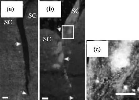
evident from Fig. 15.8b with the injection of 10 ml of 1% SAPNS. In the case of
SAPNS, axons have grown through the treated area and the tissue appeared to
be knitted with little sign of the original lesion. Figure 15.9c clearly shows the
innervation density, where the white regions correspond to axons or axon
terminals labeled from the retina. This study has successfully demonstrated the
feasibility of using SAPNS for axon repair and regeneration.
After CNS trauma, inhibition of astrocyte proliferation is important in
preventing glial scar tissue formation. This is an important step since the newly
formed scar tissue acts as a barrier to axon elongation [98]. Recently, many
studies have aimed at achieving neural progenitor cell (NPC) differentiation
into neurons while suppressing astrocyte differentiation. Silva et al. studied
selective differentiation of NPCs using nanofibrous neurite promoting laminin
epitope isolucinelysinevalinealaninevaline (IKVAV-PA) self-assembled
scaffolds [99]. Murine NPCs were mixed with 1 wt% of IKVAV-PA in 1:1
volume ratio in media or physiological fluids. The resulting transparent gel-like
solid containing NPC clusters were encapsulated within a 3D network of
nanofibers formed via self-assembly of PA molecules. NPCs survived the self-
assembly process and showed differentiated neurons that were significantly
larger than those of controls at both 1 and 7 days. In addition to the
nanofibrous structure that simulates the ECM-like morphology, high content
of water (99.5%) present in these scaffolds serves as a medium for nutrient
diffusion and for cell migration. Differentiation and migration of NPC
FIGURE 15.8 Dark field images of parasagittal sections from animals at day 30 after
lesion and treatment. (a) Cavity in the retinal projections shows the failure of the tissue
healing. (b) A similar lesion section injected with 10 ml of 1 % SAPNS. Axons that
are indicated by light color have grown through the lesion and helped in tissue healing.
Enlarged view of boxed area in (b) is shown in (c), which is an area of dense termination
of axons (traced with cholera-toxin labeling) that have crossed the lesion. Scale bars,
100 mm (SC, superior colliculus). (Reproduced from Reference [99] with permission,
from The National Academy of Sciences of the USA.)
398
BIOMEDICAL NANOSTRUCTURES
neurospheres encapsulated in IKVAV-PA gel wer e observed to be significantly
higher than the nonphysiological sequence glutamic acidglutamineserine
(EQS) gel and poly(
D-lysine) controls. In addition to the rapid selective
differentiation, these scaffolds were further proven to be in vivo compatible and
might find applications in neural tissue engineering where controlled neural
differentiation and migration are required for treating CNS trauma conditions.
15.5.4 Cardiac Tissue Engineering
Myocardial infarction, fibrotic scar tissue formation (massive cell loss due to
ischemia), and impaired cardiac function are common problems that lead to heart
failure. This occurs because of the fact that cardiomyocytes (CMs) are terminally
differentiated cells and unable to regenerate into myocardial tissue after
infarction. Although heart transplantation is the ultimate cure to heart failure,
lack of organ donors and complications associated with immune suppressive
treatments are forcing scientists and surgeons to look for new strategies to
regenerate the wounded heart [100]. Gene delivery techniques followed by cell
transplantation techniques were considered in the mid-1990s to spark the failing
heart [101, 102]. Recently, cardiac tissue engineering has emerged as a novel
approach to repair and reinforce wounded cardiac tissue using embryonic or
neonatal cardiomyocytes in combination with natural and synthetic biodegrad-
able scaffolds [103]. This approach has been further modified by developing
nanofeatured scaffolds that mimic certain features of the natural ECM and
improve the regenerative capability of the tissue engineered cardiac constructs.
Collagen and gelatin-based tissue engineered patches have been recently
reported [104, 105]. They exhibited good contraction and function, but are not
popular for use due to the lack of stability required for handling. An alternative
approach is using synthetic polymers that require physical properties such as
stability and resistance to contractions [103]. Zong et al. seeded cardiomyocytes
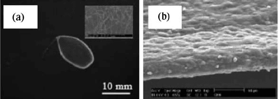
FIGURE 15.9 (a) A 10-mm-thick fibrous mesh suspended across a wire ring that acts
as a passive load to condition cardiomyocytes during in vitro culture. Inset shows the
SEM of an electrospun mesh with an average fiber diameter of 250 nm. (b) Cross-
sectional SEM recorded on a cardiac nanofibrous mesh after 14 days of in vitro culture
showing cardiomyocyte presence throughout the entire mesh. (Reproduced from
Reference [107] with permission, from Elsevier)
NANOSTRUCTURES FOR TISSUE ENGINEERING/REGENERATIVE MEDICINE 399
on electrospun nanofiber scaffolds made of PLLA, PLGA (PLA10/
PGA90) + PLLA, and PLGA(PLA75/PGA25) + PEGPLA [106]. This study
compared growth of cardiomyocytes on randomly oriented fiber scaffolds, and
also on locally oriented fiber scaffolds where the microscale fiber orientation was
achieved by uniaxial stretching. It was observed that the fiber orientation
provided guidance for CM growth and showed sensitivity to the composition
and degradation rate of the electrospun PLGA-based scaffolds. CM cell density
was observed to be higher on relatively hydrophobic surfaces that in general
exhibited slower degradation. Myocardial functional studies of CMs on different
synthetic scaffolds showed PLLA as a better scaffold as compared to other
candidates involved in this study.
Shin et al. electrospun PCL nanofiber meshes and seeded with cardiomyo-
cytes isolated from neonatal Lewis rats [107]. ECM-like mesh with an average
fiber diameter of 250 nm and a mesh thickness around 10 mm (inset of
Fig. 15.9a) was suspended across a wire ring as shown in Fig. 15.9a.
Cardiomyocytes attached well to these scaffolds and started contracting after 3
days of culture. After 14 days of culture, cardiomyocytes retained their
phenotype, penetrated the entire scaffold, and stained positive for cardio
typical proteins such as actin, tropomyosin, cardiac troponin-I, and connexin
43. Also, a cross-sectional view clear ly confirms the presence of cells through
the entire nanofibrous scaffold as presented in Fig. 15.9b. This study has
established a versatile in vitro system to develop functional myocardial patches
that may be used for healing and regeneration of wounded hearts.
As described in the previous sections, nanotechnology for tissue engineering/
regenerative medicine is actively perused recently for multiple applic ations
including the regeneration of bone, cartilage, neural, v ascular and cardiac
tissues. In addition, research is currently underway to regenerate skin, bladder
and liver tissues using nanostructured scaffolds [108, 109, 110].
There have already bee n successful tissue engineering products that are
clinically relevant, have achieved FDA approval, and are currently being
marketed for tissue replacement. The most successful product is tissue
engineered skin that is currently in clinical use (Dermagraft
1
and Apligraf
1
)
for treating burn victims and patients with diabetic ulcers. Some of the
products at advanced clinical stage are contractile pa tches for damaged heart,
autologous chondrocytes in a polyme r gel for treating urinary reflux in
children, and urinary stress incontinence in adult women [111]. In addition,
various other tissue engineered products are at preliminary stages of
investigation (in vitro or animal studies) for regenerating tissues/organs such
as bone, ligament, heart valves, heart muscles, blood vessels, and bladders
[112]. Thus far, efforts have been on designing materials in combination with
cells and factors to meet the targeted clinical application. The success of this
approach can be further enhanced by creating cellular recognition nano/
microtopographical features that aid in the control of cell functions and may
lead to better tissue engineer ed products that meet future clinical needs.
400 BIOMEDICAL NANOSTRUCTURES
15.6 CONCLUSIONS
The need for mimicking the ECM has led to the development of various
scaffolding methodologies. These methods have helped in the creation of 3D
scaffolds consisting of fibers, grooves, pits, and pillars in nanoscale for tissue
engineering and biomedical applications. Due to the in vivo-like environment,
cells seeded on nanofeatured scaffolds showed improved adhesion, prolifera-
tion, migration, differentiation, and phenotypic expression. These scaffolds
with improved cellular compatibility can be directly translated into successful
tissue constructs for restoration, repair, and regener ation of bone, cartilage,
neural, vascular, and cardiac tissues. Even though scaffolds with nanofeatures
have generated lot of interest and excitement, the future of tissue engineering
will incorporate scaffolds with topographical and biochemical cues that would
exactly duplicate the in vivo environment. Nanostructured scaffolds are still at
their infancy stage and further work is required to answer questions such as
what size is right for a desired cell response and how different cells react to the
same topography.
REFERENCES
1. Data derived from American Association of Tissue Banks, Eye Bank Association
of America, National Marrow Donor Program, National Kidney Foundation,
Inc., and UNOS Scientific Registry; 2005.
2. Laurencin CT, Amborsio A, Borden MD, Cooper JA. Tissue engineering:
orthopedic applications. Anu Rev Biomed Eng 1999;1:1946.
3. Atala A and Lanza RP. Methods of Tissue Engineering. New York: Academic
Press; 2002.
4. Kofron MD and Laurencin CT. Bone tissue engineering by gene delivery. Adv
Drug Deliv Rev 2006;58:555576.
5. Yu X, Botchwey EA, Levine EM, Pollack SR, Laurencin CT. Bioreactor-based
bone tissue engineering: the influence of dynamic flow on osteoblast phenotypic
expression and matrix mineralization. Proc Natl Acad Sci USA 2004;101:
1120311208.
6. Langer R and Vacanti JP. Tissue engineering. Science 1993;260:920926.
7. Williams DF, The Williams dictionary of Biomaterials. Liverpool UK: Liverpool
University Press; 1999.
8. Williams DF. Revisiting the definition of biocompatibility. Med Device Technol
2003;14:1013.
9. Dankers PY, Harmsen MC, Brouwer LA, van Luyn MJ, Meijer EW. A modular
and supramolecular approach to bioactive scaffolds for tissue engineering. Nat
Mater 2005;4:568574.
10. Livingston T, Ducheyne P, Garino J. In vivo evaluation of a bioactive scaffold for
bone tissue engineering. J Biomed Mater Res 2002;62:113.
11. Griffith LG. Polymeric biomaterials. Acta Mater 2000;48:263277.
NANOSTRUCTURES FOR TISSUE ENGINEERING/REGENERATIVE MEDICINE 401
12. Hayashi T. Biodegradable polymers for biomedical uses. Prog Polym Sci
1994;19:663702.
13. Mikos AG, Thorsen AJ, Czerwonka LA, Bao Y, Langer R. Preparation and
characterization of poly(
L-lactic acid) foams. Polymer 1994;35:10681077.
14. Mooney DJ, Baldwin DF, Suh NP, Vacanti JP, Langer R. Novel approach to
fabricate porous sponges of poly(
D,L-lactic-co-glycolic acid) without the use of
organic solvents. Biomaterials 1996;17:14171422.
15. Hsu YY, et al. DL Effect of polymer foam morphology and density on kinetics of
in vitro controlled release of isoniazid from compressed foam matrices. J Biomed
Mater Sci 1997;35:107116.
16. Giordano RA, et al. Mechanical properties of dense polylactic acid structures
fabricated by three dimensional printing. J Biomat Sci Polym Ed 1996;8:6375.
17. Borden M, Attawia M, Khan Y, Laurencin CT. Tissue-engineered microsphere-
based matrices for bone repair: design and evaluation. Biomaterials 2002;23:
551559.
18. Ramakrishna S, et al. An Introduction to Electrospinning and Nanofibers.
Singapore: World Scientific Publishing Co. Pte. Ltd; 2005.
19. Ma PX. Scaffolds for tissue fabrication. Mater Today 2004;7:3040.
20. Stupp SI, et al. Supramolecular materials: self-organized nanostructures. Science
1997;276:384389.
21. Khademhosseini A, Langer R, Borenstein J, Vacanti JP. Microscale techno-
logies for tissue engineering and biology. Proc Natl Acad Sci USA 2006;103:
24802487.
22. Alberts B, et al. Molecular Biology of the Cell. New York: Garland Science; 2002.
p 1090.
23. Murugan R and Ramakrishna S. Nano-featured scaffolds for tissue engineering: a
review of spinning methodologies. Tissue Eng 2006;12:435447.
24. Pham QP, Sharma U, Mikos AG. Electrospinning of polymeric nanofibers for
tissue engineering applications: a review. Tissue Eng 2006;12:11971211.
25. Kumbar SG, Nair LS, Bhattacharyya S, Laurencin CT. Polymeric nanofibers as
novel carriers for the delivery of therapeutic molecules. J Nanosci Nanotechnol
2006;6:25912607.
26. Nair LS, Bhattacharyya S, Laurencin CT. Development of novel tissue enginee-
ring scaffolds via electrospinning. Expert Opin Biol Ther 2004;4:659668.
27. Li WJ, Laurencin CT, Caterson EJ, Tuan RS, Ko FK. Electrospun nanofibrous
structure: a novel scaffold for tissue engineering. J Biomed Mater Res 2002;60:
613621.
28. Laurencin CT and Ko FK. Hybrid nanofibril matrices for use as tissue engineering
devices. US Patent 2004;6,689,166.
29. Katti DS, Robinson KW, Ko FK, Laurencin CT. Bioresorbable nanofiber-based
systems for wound healing and drug delivery: optimization of fabrication
parameters. J Biomed Mater Res B Appl Biomater 2004;70:286296.
30. Nair LS, et al. Fabrication and optimization of methylphenoxy substituted
polyphosphazene nanofibers for biomedical applications. Biomacromolecules
2004;5:22122220.
402
BIOMEDICAL NANOSTRUCTURES
31. Bognitzki M, et al. Nanostructured fibers via electrospinning. Adv Mater
2001;13:7072.
32. Boland ED, Wnek GE, Simpson DG, Pawlowski KJ, Bowlin GL. Tailoring tissue
engineering scaffolds using electrostatic processing techniques: a study of
poly(glycolic acid) electrospinning. J Macromol Sci Pure Appl Chem 2001;A38:
12311243.
33. Yang F, Murugan R, Wang S, Ramakrishna S. Electrospinning of nano/micro
scale poly(
L-lactic acid) aligned fibers and their potential in neural tissue
engineering. Biomaterials 2005;26:26032610.
34. Li WJ, Danielson KG, Alexander PG, Tuan RS. Biological response of
chondrocytes cultured in three-dimensional nanofibrous poly(epsilon-caprolac-
tone) scaffolds. J Biomed Mater Res A 2003;67:11051114.
35. Geng XY, Kwon OH, Jang JH. Electrospinning of chitosan dissolved in
concentrated acetic acid solution. Biomaterials 2005;26:54275432.
36. Almany L and Seliktar D. Biosynthetic hydrogel scaffolds made from fibrinogen
and polyethylene glycol for 3
D cell cultures. Biomaterials 2005;26:24672477.
37. Schindler M, et al. A synthetic nanofibrillar matrix to promote in vivo-like
organization and morphogenesis for cell in culture. Biomaterials 2005;26:
56245631.
38. Matthews JA, Wnek GE, Simpson DG, Bowlin GL. Electrospinning of collagen
nanofibers. Biomacromolecules 2002;3:232238.
39. Venugopal J and Ramakrishna S. Biocompatible nanofiber matrices for the engi-
neering of a dermal substitute for skin regeneration. Tissue Eng 2005;11:847854.
40. Witte P, Dijkstra PJ, Berg JWA, Feijen J. Phase separation processes in polymer
solutions in relation to membrane formation. J Membr Sci 1996;117:131.
41. Ma PX and Jennifer E. Scaffolding in Tissue Engineering. Boca Raton: CRC
Press; 2006. p 125.
42. Ma PX and Zhang R. Synthetic nano-scale fibrous extracellular matrix. J Biomed
Mater Res 1999;46:60 72.
43. Zhang R and Ma PX. Synthetic nano-fibrillar extracellular matrices with
predesigned macroporous architectures. J Biomed Mater Res 2000;52:430438.
44. Ma PX and Choi J. Biodegradable polymer scaffolds with well defined
interconnected spherical pore network. Tissue Eng 2001;7:2333.
45. Gross M. Travels to the Nanoworld: Miniature Machinery in Nature and
Technology. New York: Perseus Books Group; 1999.
46. Zhang S, Holmes T, Lockshin L, Rich A. Spontaneous assembly of a self-
complementary oligopeptide to form a stable macroscopic membrane. Proc Natl
Acad Sci USA 1993;90:33343338.
47. Semino CE, Merok JR, Crane GG, Panagiotakos G, Zhang S. Functional
differentiation of hepatocyte-like spheroid structures from putative liver progeni-
tor cells in three-dimensional peptide scaffolds. Differentiation 2003;71:262270.
48. Semino CE, et al. Entrapment of migrating hippocampal neural cells in 3D peptide
nanofiber scaffold. Tissue Eng 2004;10:643655.
49. Hartgerink JD, Beniash E, Stupp SI. Self-assembly and mineralization of peptide
amphiphile nanofibers. Science 2001;294:16841688.
NANOSTRUCTURES FOR TISSUE ENGINEERING/REGENERATIVE MEDICINE 403
50. Murphy WL and Mooney DJ. Molecular scale bio-mimicry. Nat Biotechnol
2002;20:3031.
51. Zhang S, et al. Self-complementary oligo peptides matrices support mammalian
cell attachment. Biomaterials 1995;16:13851393.
52. Kisiday J, et al. Self assembling peptide hydrogel fosters chondrocyte extracellular
matrix production and cell division: implications for cartilage tissue repair. Proc
Natl Acad Sci USA 2002;99:999610001.
53. Dalby MJ, et al. Increasing fibroblast response to materials using nanotopo-
graphy: morphological and genetic measurements of cell response to 13-nm-high
polymer demixed islands. Exp Cell Res 2002;276:19.
54. Geissler M and Xia Y. Patterning: principles and some new developments. Adv
Mater 2004;16:12491269.
55. Kane RS, Takayama S, Ostuni E, Ingber DE, Whitesides GM. Patterning proteins
and cells using soft lithography. Biomaterials 1999;20:23632376.
56. Mrksich M, Dike LE, Tien J, Ingber DE, Whitesides GM. Using microcontact
printing to pattern the attachment of mammalian cells to self-assembled
monolayers of alkanethiolates on transparent films of gold and silver. Exp Cell
Res 1997;235:305313.
57. Lee CJ, et al. Microcontact printing on human tissue for retinal cell transplanta-
tion. Arch Ophthalmol 2002;120:17141718.
58. Li HW, Muir BVO, Fichet G, Huck WTS. Nanocontact printing: a route to
sub-50-nm-scale chemical and biological patterning. Langmuir 2003;19:1963
1965.
59. Jo J, Kim KY, Lee ES, Choi BO. Nanocontact printing of nonplanar substrate by
using flexible h-PDMS stamp. Proc SPIE 2005;6037:60371T.
60. Suh KY, Seong J, Khademhosseini A, Laibinis PE, Langer R. A simple soft
lithographic route to fabrication of poly(ethylene glycol) microstructures for
protein and cell patterning. Biomaterials 2004;25:557563.
61. Morra M and Cassineli C. Non-fouling properties of polysaccharide coated
surfaces. J Biomater Sci Polym Ed 1999;10:11071124.
62. Khademhosseini A, et al. Layer-by-layer deposition of hyaluronic acid and poly-
L-
lysine for patterned cell co-cultures. Biomaterials 2004;25:35833592.
63. Fukuda J, et al. Micropatterned cell co-cultures using layer-by-layer deposition of
extracellular matrix components. Biomaterials 2006;27:14791486.
64. Bearinger JP, et al. Biomolecular patterning via photocatalytic lithography.
Technical Proceedings of the NSTI Nanotechnology Conference and Trade Show.
Volume 1;2005. p 339342.
65. Choi I, Kang SK, Lee J, Kim Y, Yi J. In situ observation of biomolecules
patterned on a PEG-modified Si surface by scanning probe lithography.
Biomaterials 2006;27:46554660.
66. Flemming RG, Murphy CJ, Abrams GA, Goodman SL, Nealey PF. Effects of
synthetic micro and nano structured surfaces on cell behavior. Biomaterials
1999;20:573588.
67. Karuri NW. Biological length scale topography enhances cellsubstratum
adhesion of human corneal epithelial cells. J Cell Sci 2004;117:31533164.
404
BIOMEDICAL NANOSTRUCTURES
68. Wojciak-Stothard B, Madeja Z, Korohoda W, Curtis A, Wilkinson C. Activation
of macrophase-like cells by multiple grooved substrata: topographical control of
cell behaviour. Cell Biol Int 1995;19:485490.
69. Wojciak-Stothard B, et al. Role of the cytoskeleton in the reaction of fibroblasts to
multiple grooved substrata. Cell Motil Cytoskeleton 1995;31:147158.
70. Clark P, Connolly P, Curtis A, Dow JAT, Wilkinsin C. Topographical control of
cell behaviour. II. multiple grooved substrata. Development 1990;108:635644.
71. Hoch HC, Staples RC, Whitehead B, Comeau J, Wolf ED. Signaling for growth
orientation and cell differentiation by surface topography in Uromyces. Science
1997;235:16591662.
72. Clark P, Connolly P, Curtis A, Dow JAT, Wilkinson C. Topographical control of
cell behavior. I. Simple step cues. Development 1987;99:439448.
73. Rosdy M, Grisoni B, Clauss LC. Proliferation of normal human keratinocytes on
silicone substrates. Biomaterials 1991;12:511517.
74. Spaargaren M and Bos JL. Rab5 induces Rac dependant lamellipodia formation
and cell migration. Mol Biol Cell 1999;10:32393250.
75. Liu X and Ma PX. Polymer scaffolds for bone tissue engineering. Ann Biomedical
Eng 2004;32:477486.
76. Christenson EM, et al. Nanobiomaterial applications in orthopedics. J Orthop Res
2007;25(1):1122.
77. Yoshimoto H, Shin YM, Terai H, Vacanti JP. A biodegradable nanofiber scaffold
by electrospinning and its potential for bone tissue engineering. Biomaterials
2003;24:20772082.
78. Shin M, Yoshimoto H, Vacanti JP. In vivo bone tissue engineering using
mesenchymal stem cells on a novel electrospun nanofibrous scaffolds. Tissue Eng
2004;10:3341.
79. Pelled G, et al. Structural and nanoindentation studies of stem cell-based tissue-
engineered bone. J Biomech 2007;40:399411.
80. Hosseinkhani H, Hosseinkhani M, Tian F, Kobayashi H, Tabata Y. Osteogenic
differentiation of mesenchymal stem cells in self-assembled peptide-amphiphile
nanofibers. Biomaterials 2006;27:40794086.
81. Hosseinkhani H, Hosseinkhani M, Tian F, Kobayashi H, Tabata Y. Ectopic bone
formation in collagen sponge self-assembled peptide-amphiphile nanofibers hybrid
scaffold in a perfusion culture bioreactor. Biomaterials 2006;27:50895098.
82. Wei G and Ma PX. Structure and properties of nano-hydroxyapatite/polymer
composite scaffolds for tissue engineering. Biomaterials 2004;25:47494757.
83. Li WJ, et al. A three-dimensional nanofibrous scaffold for cartilage tissue
engineering using human mesenchymal stem cells. Biomaterials 2005;26:599
609.
84. Muraglia A, et al. Formation of a chondroosseous rudiment in micromass cultures
of human bone-marrow stromal cells. J Cell Sci 2003;116:29492955.
85. Niklason LE, et al. Functional arteries grown in vitro. Science 1999;284:489493.
86. Watanabe M, et al. Tissue-engineered vascular autograft: inferior vena cava
replacement in a dog model. Tissue Eng 2002;7:429439.
NANOSTRUCTURES FOR TISSUE ENGINEERING/REGENERATIVE MEDICINE 405