Kenneth E. Gonsalves, Craig R. Halberstadt, Cato T. Laurencin, Lakshmi S. Nair. Biomedical Nanostructures
Подождите немного. Документ загружается.

Electrospinning, phase separation, and self-assembly are useful methods to
produce nanofibrous scaffolds with properties mimicking ECM in size and to
some extent functionality. Electrospinning is a viable process to fabricate
nanofiber scaffolds using both synthet ic and natural polymers including
collagen and fibrinogen. Although phase separation is limited for highly
crystalline polymers, the process is economical and can be used to fabricate 3D
scaffolds that directly fit into the anatomical shape of a body part using a
mold. Molecular self-assembly has merits over other methods since the process
involves body-friendly molecules such as protein, peptide, and lipids.
Furthermore, scaffolds can be tissue specific by choosing the required building
blocks and adopt the required shape in vivo. Even though above-mentioned
techniques have demonstrated the versatility for creating ECM mimicking
scaffolds, much work is required to improve the scaffold mechanical strength
suitable for tissue engineering use.
15.3 SURFACE PATTERNED SCAFFOLDS
In tissue engineering, the surface is more important than the bulk properties
because of the fact that cells land on the surface and look for specific cues
before and after atta chment. It is known that cells respond to spatially and
temporally organized signals and can react to nanoscale surfa ce features as
small as 10 nm [53]. Patterned surfaces aim to recreate in vivo nano/
microenvironments for optimum cell growth and functionality. These surfaces
further control cell differentiation/proliferation and multicellular organization
in building complex tissues that are otherwise not possible with uniform
surfaces. Micro/nanofabrication methods include photolithography and soft
lithography such as contact printing, replica molding, transfer molding, and
imprint lithography are used to create nano/micropatterns in the form of
gratings, wells, pits, grooves, and islands [54]. Traditionally, photolithography
is used to generate nano/microsurface topologies for industrial and biomedical
applications. Despite its popularity, the technique is limited in designing
scaffolds for biospecific applic ations including tissue engineered scaffolds
because of the nature of the process where high capital and operational costs
are involved. Soft lithographic methods are useful because they permit direct
construction of various shapes with precis e control, and unlike in conventional
lithography no bio hazardous solvents are used in the process. Soft lithography
refers to a set of methods for fabricating or replicating structures using
elastomeric stamps, molds, and conformable photomasks using elasto mers
such as polydimethoxylsiloxane (PDMS) or fluorosilicone. Elastomeric molds
are derived using a master that is generally prepared by either conventional
photolithography or electron beam lithography techniques. In the last few
years, soft lithography based techniques have been increasingly used for
creating nano/micro featured surfaces [55,60,64]. Having seen the progress with
surface patterning, it is time to combine lithography techniques with 3D
386 BIOMEDICAL NANOSTRUCTURES
scaffolding methodologies for designing nanofeatured 3D scaffolds for tissue
engineering applications.
15.3.1 Micro/Nanocontact Printing
In recent years, micro- and nanocontact printing have become an essential
method for patterning surfaces for applications in tissue engineering and biology.
Among the soft lithographic methods, contact printing was the first one to be
used to pattern a self-assembled monolayer (SAM) of alkanethiols on gold
substrate using a high resolution elastomeric stamp [55]. In brief, the PDMS
stamp is inked with alkanethiol and printed onto a gold substrate to have the
alkanethiol patterns in the regions where the stamp came in contact with the
substrate. After washing, the remaining bare gold surface was exposed to
another alkanethiol to obtain a surface patterned into two regions with different
terminal groups. Patterned surfaces that resist nonspecific adsorption of proteins
are further used for protein patterning. For example, when the surface with
regions terminated in oligo(ethylene glycol) groups and methyl groups immersed
in solutions of proteins such as fibrinogen, fibronectin, and immunoglobulin,
proteins selectively adsorbed on the methyl-terminated regions as seen from
Fig. 15.4a. Protein patterned surfaces were further used for patterning cells. In a
study by Mrksich, bovine capillary endothelial cells were patterned on
fibronectin-coated SAM surfaces [56]. Since most mammalian cells are
anchorage dependent, endothelial cells attached only to the methyl-terminated
fibronectin-coated regions of the patterned SAMs (Fig. 15.4b).
Micro/nanocontact printing is an inexpensive, simple, and effective tool for
patterning planar and nonplanar tissue engineered surfaces. Lee et al. used
printing inhibitory molecules to pattern the growth of retinal/iris pigment
epithelial cells on human lens capsule for retinal cell transplantation [57]. This
particular methodology of printing on autologous human tissue may find
applications in the replacement of vital ocular tissues such as the retinal
pigment epithelium in age-related macular degeneration. Recently, contact
printing was further extended to produce nanostructures by adopting high
elastic modulus PDMS and high molecular weight inks. Li et al. demonstrated
the feasibility of nanocontact printing at a resolution of less than 50 nm via a
combination of sharp and hard PDMS stamps using a diffusion limited high
molecular weight ink [58]. Another modification to nanocontact printing came
in the form of flexible PDMS stamps for printing on nonuniform surfaces that
may find a myriad of applications in modifying tissue engineering scaffolds for
clinical applications [59].
15.3.2 Capillary Force Lithography
The Langer’s group at MIT has reported yet another simple lithographic method
called capillary force lithography for protein and cell patterning [60]. This
method involves a molding process in which a uniform polyethylene glycol
NANOSTRUCTURES FOR TISSUE ENGINEERING/REGENERATIVE MEDICINE 387
(PEG) or hyaluronic acid or chitosan films (above glass transition temperature)
are molded using their capillarity and wettability properties with respect to the
PDMS stamp. Due to the nonbiofouling property of polysaccharides when
deposited as a film on a surface, hyaluronic acid and chitosan-molded regions
exhibit protein or cell repellant properties [61] whereas bare substrate regions
support cell or protein adsorption. In one study, PEG dimethacrylate was used
as a cell repellant layer, and the substrate regions were modified with fibronectin
for NIH3T3 cell patterning. In a more recent study, layer-by-layer deposition of
hyaluronic acid and poly-
L-lysine (PL) for micropatterned cocultures was
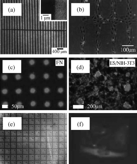
FIGURE 15.4 Surface patterning using various novel techniques. optical micrographs
of microcontact printed SAMs on gold having patterned regions with (a) fibronectin
(black in color) and (b) bovine capillary endothelial cells (reproduced from Reference
[56] with permission, from Academic Press). Layer-by-layer deposition using capillary
force lithography method; (c) fluorescence image of fibronectin adsorption on
hyaluronic acid patterned surface and (d) fluorescent image of patterned 1-day-old
cocultures of ES cells (red) with NIH-3T3 fibroblasts (green) (reproduced from
Reference [62] with permission, from Elsevier). (e) Fluorescence micrograph showing
selective protein adsorption to adhesive APS regions (line width of L =4mm) and (f)
reflectance image of green fluorescence protein-tubulin transfected HeLa cells adhering
to 100 mm
2
island (reproduced from Reference [64] with permission, from Nano Science
and Technology Institute.)
388
BIOMEDICAL NANOSTRUCTURES
demonstrated and is shown in Fig. 15.4c [62]. The ionic adsorption of PL onto
HA patterns was used to switch the HA surfaces from cell repulsive to adherent
for the purpose of patterned cocultures. Fibronectin- and PL-coated patterned
regions were selectively utilized for designing a coculture system with
hepatocytes, murine embryonic stem cells (ES), and NIH3T3 fibroblasts as
shown in Fig. 15.4d. The developed cocultures are independent of selective
adhesion of the cell type, the seeding order, and notably remained stable for 5
days. Subsequently, the cytotoxic PL is replaced by collagen, a component of the
natural ECM, and cationic in nature to form an ionic complex with HA [63].
Capillary force lithography is an inexpensive, simple, and powerful tool for
designing micropatterned coculture systems for probing cellcell and cellECM
interactions for designing and engineering complex tissues.
15.3.3 Biomolecular Patterning
Organization of biomolecules such as proteins, DNA, and cells into ordered
arrays on surfaces is called biomolecular patterning. This has been a subject of
considerable interest in many areas including biology and medicine. Recently,
photocatalytic lithography, scanning probe lithography, and computer-
controlled laser ablation methods have been used for organizing biomolecules
on silicon or glass surfaces. Photocatalytic lithography uses a specific
wavelength of light to chemically activate material on a stamp in contact
with an oxidizable thin film. In this method, a clean glass or silicon substrate is
modified with nonfouling polyethylene glycol (PEG) thiol coatings [64].
Selective removal of light-exposed regions leads to the formation of a bare
substrate and interpenetrating network of nonfouling regions. The bare glass
or silicon regions are then backfilled with adhesive aminopropylsilane (APS).
Subsequently, fluorescein isot hiocyanate (FITC)-labeled neutravidin is spread
on these surfaces and imaged using a fluorescence microscope. As shown in
Fig. 15.4e, the protein is attached to adhesive L regions (brigh t) and was
repelled by the PEG-based nonfouling regions (dark). Lipofectamine
transfected HeLa cells attached to the substrate and cell proliferation occurred
over time, as shown in Fig. 15.4f, which demonstrates that cell viability was
maintained on these patterned regions that were backfilled with APS.
Patterning using scanning tunneling microscopy or atomic force microscopy
(AFM) is known as scanning probe lithography. This pa rticular lithographic
approach is quite useful because the patterning, imaging, and pattern
characterization ca n be carried out sequentially within the microscope
chamber. This method allows making in situ observations of the material
immobilized on the patterns. Choi et al. demonstrated patterning streptavidin
on a PEG-modified Si surface using AFM. In this study, a silicon substrate was
uniformly coated with low molecular weight protein repellant methoxy PEG
silane [65]. Silane was converted into SiO
2
patterns by AFM anodic oxidation
by applying a voltage of 1020 V at the surface (anode) and the tip (cathode).
The square SiO
2
patterns were 23 nm thick when formed with an applied
NANOSTRUCTURES FOR TISSUE ENGINEERING/REGENERATIVE MEDICINE 389
voltage of 14 V and obs erved to be more reactive than nonpatterned regions.
These regions were chemically modified with functional groups having strong
affinity for selective protein patterning. Oxide pattern was first modified with a
self-assembled amine functional material. Protein patterning on these surfaces
was done by two ways. The first method immobilized Au-conjugated
streptavidin onto the amine -modified SiO
2
. The second approach used
gluteraldehyde to activate surface amino groups for protein attachment
through selective covalent bonding between aldehyde and amino groups on
streptavidin. These streptavidin patterns can be further utilized for the
patterning of biotinylated materials. Various groups around the globe are
working on creating features at 10100 nm length scales for producing
ultradense DNA and protein chips for various microarray and medical device
applications.
15.4 RELEVANCE OF NANOSTRUCTURED SCAFFOLDS
IN REGENERATIVE MEDICINE
In tissues and organs, cells are always surrounded and constantly in touch with
the ECM, which is a self-assembled nanofibrillar matrix consisting of various
proteins and polysaccharides. The ECM varies substantially in volume in
organs: abundant in cartilage and bone, and rare in brain and spinal cord. It
exhibits remarkable diversity and functional adaptation such as calcified in
bone and teeth, transparent in cornea, ropelike in tendon s, basal lamina
between epithelium and connective tissue. Due to its ubiquitous nature and
close association with cells, ECM controls cell functions and physical
properties of tissues. In addition, high surface area of the ECM nanofibrils
provides an opportunity to have proteins like proteoglycans, glycoaminogly-
cans, and integrins on the surface for better interaction with the cytoskeleton of
cells.
Cellular functions in vivo are regulated by cellcell communications and
cellsubstratum interactions and soluble factors. Ability to incorporate in vivo-
like features in synthetic scaffolds for tissue engineering may lead to
accelerated time regeneration. Cellsubstratum interactions are influenced
by topographical and chemical cues. Topographical cues dictate processes such
as cell adhesion, proliferation, differentiation, migration, and gene expression
based on the surface feature type and size. It is known that biological length
scale topography that ranges from 10 nm to 100 mm influen ces the cytoskeletal
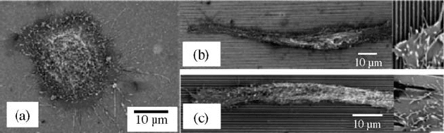
organization and eventually cellular behavior [66]. Karuri et al. observed SV40
human corneal endothelial cell rounded morphology on flat surface
(Fig. 15.5a), and elongated morphology on patterned surfaces (Fig. 15.5b).
The inset shows the extended filopodia and lamellopodia on the patterned
surfaces with grooves and ridges [67]. In a similar study with macrophage-like
cells on nanogrooves, cells aligned to the long axis of grooves with
390 BIOMEDICAL NANOSTRUCTURES
organization of cytoskeletal elements parallel to the grooves [68]. Actin and
microtubules were observed to align along the walls and edges of the grooves
while actin condensations appeared at topographic discontinuities [69]. Some
of the cells were also observed to have lamellae and filopodia bending around
the edges. There are also reports on the effect of groove depth on baby hamster
kidney (BHK) and Madin-Darby canine kidney (MDCK) cell orientation. For
example, BHK cell alignment increased with depth but decreased wi th width,
whereas width has no effect on MDCK cells [70]. Therefore, most of the cell
types including fibroblasts, endothelial, smooth muscle, macrophage, leuko-
cytes, osteoblasts, and mesenchymal stem cells respond to nanofeatured ridges,
steps, wells, nodes, pillars, pores, and spheres by showing good adhesion,
proliferation, migration, and differentiation. The cells have also shown to
exhibit a broad gene upregulation and increased production of ECM proteins
[71–74].
In vivo, cells are surrounded by the ECM and most of the cellular functi ons
inside the complex organs are mediated by the ECM in associ ation with
signaling molecules and growth factors. In addition to the chemical cues, the
ECM exhibits nanotopographical cues that influence various cellular functions
including adhesion and differentiation. Therefore, there is a great need and
demand for nanofeatured scaffolds that mimic and provide the same
environment as the in vivo ECM for tissue engineering/regenerative medicine.
15.5 ROLE OF NANOSTRUCTURED SCAFFOLD S
IN TISSUE ENGINEERING
Various in vitro and in vivo studies employed nanostructured scaffolds for
engineering bone, cartilage, vascular, neural, cardiac, and bladder tissue. Some
of the recent efforts where tissue regeneration was enhan ced by mimicking the
ECM topography via nanostructured scaffolds are discussed below.
FIGURE 15.5 SEM of adherent SV40 human corneal epithelial cells on (a) planar
surface and patterned surfaces with pitch size of (b) 400 nm and (c) 4000 nm. Inset
shows the tip of an elongated cell. (Reproduced from Reference [67] with permission,
from The Company of Biologists.)
NANOSTRUCTURES FOR TISSUE ENGINEERING/REGENERATIVE MEDICINE 391
15.5.1 Bone and Cartilage Tissue Engineering
Scaffold design and development for bone repair and regeneration has been a
priority for the tissue engineering community for many years. Traditional
methods like microsphere sintering, salt leaching, gas foaming, and freeze
drying are employed for fabricating 3D scaffolds for bone tissue engineering
applications [75]. Even though the fabricated scaffolds exhibit 3D pore
structure, they lack the nanoscale topography that cells experience in vivo.In
an attempt to mimic the ECM features, recently nanoscale features have been
incorporated in the design of scaffolds [76]. The fabricated nanofeatured
scaffolds were evaluated for their performance by culturing osteoblast and
mesenchymal stem cells in an effor t to engineer bone.
Li et al. used electrospinning for creating PLGA scaffolds for bone tissue
engineering applications [27]. Using the same techni que Vacanti et al.
developed 3D nanofibrous biodegradable PCL scaffolds for regenerating
bone [77]. The electrospun scaffold consists of fibers with diameters ranging
from 20 nm to 5 mm with the average diameter of 400 nm. Mesenchymal stem
cells (MSCs), derived from bone marrow of neo natal rats, were cultured with
osteogenic supplements under dynamic culture conditions. This study has
shown that nanofibrous scaffolds provide an environment that supports
mineralized tissue formation. The cellpolymer nanofiber constructs obtained
after the 4 weeks of in vitro dynamic culture were subsequently implanted in the
omenta of rats to assess the bone formation in vivo [78]. Histological
examination, on a specimen after 4 weeks of in vitro culture and 4 weeks of
implantation, revealed the presence of osteocyte-like cells and the formation of
mineralized bonelike tissue throughout the specimen confirming the suitability
of a nanofibrous construct for the treatment of bone de fects. A novel strategy
that combined stem cell-based gene therapy with nanostructured scaffolds was
recently developed for in vivo bone tissue engineering [79]. PLGA electrospun
scaffolds seeded with transfected MSCs to express BMP2 were implanted in the
thigh muscle of immunocompetent mice. After 4 weeks of implantation,
animals were sacrificed and the ectopic bone formation was assessed via
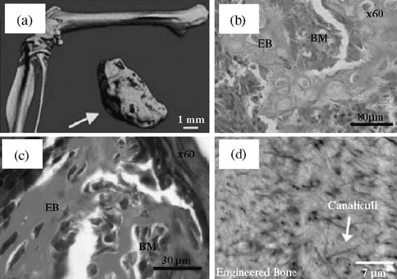
structural and nanoindentation studies. As shown in Fig. 15.6a, mCT
(computed tomography) clearly shows the growth of ectopic bone at the
transplantation site. Histological studies demonstrated the formation of bone
marrow (Fig. 15.6b) and collagen (Fig. 15.6c) regions within the new bone.
Ectopic bone further showed canaliculi structure as seen from Fig. 15.6d that is
similar to the femoral bone. The simila rity in mechanical properties (estimated
by nano-indentation), nanoscale topographies, overall microstructure, and
chemical composition between femoral and nanofiber-assisted engineered bone
further reveals the nanostructured scaffold’s potential in bone regeneration.
Self-assembly holds the reputation of creating 3D nanofibrous scaffolds with
the fiber diameters that fall in the range of natural collagen present in the ECM.
Self-assembled scaffolds have already been employed for regeneration of
various tissues. Hosseinkhani et al. employed self-assembled peptide-amphiphile
392 BIOMEDICAL NANOSTRUCTURES
nanofibrous scaffolds for osteogenic differentiation of mesenchymal stem cells
[80]. A 3D network of cellpolymer nanofiber construct prepared by mixing cell
suspension in media with dilute aqueous PA solution was used in this study. It
has clearly demonstrated that PA nanofiber scaffolds significantly enhanced in
vitro proliferation and osteogenic differentiation of MSC compared with a static
culture. The response was further improved by functionalizing nanofibers with
arginineglycineaspartic (RGD) sequence. Close resemblance of the in vivo
situation resulted in an increased alkaline phosphatase (ALP) activity and
osteocalcin expression. The authors further improved the mechanical strength by
employing hybrid scaffolds consisting of a hydrogel formed through self-
assembly of PA and the collagen sponge reinforced with PGA [81]. Hybrid
scaffolds were precultured using bioreactor perfusion culture system and 3-week
precultured scaffolds were used for the in vivo study. ALP activity and
osteocalcin content of the subcutaneous tissues around the implant site clearly
confirmed the ectopic bone-forming ability of these novel hybrid nanofibrous
scaffolds. Though this study has focused more on evaluating the effectiveness of
perfusion culturing system against the static culture, the observed in vitro and in
vivo osteogenic differentiation of MSCs clearly demonstrated the bone-forming
ability of nanofiber scaffolds.
FIGURE 15.6 (a) mCT image of a femoral and engineered bone. Histological
evolution of the engineered bone. (b) Hematoxilineosin staining for bone marrow
regions and (c) Masson’s trichrome staining for collagen regions (green in color).
(d) Backscattered electron microscope image of an engineered bone showing canaliculi
regions similar to the femoral bone (EB is engineered bone and BM is bone marrow).
(Reproduced from Reference [79] with permission, from Elsevier).
NANOSTRUCTURES FOR TISSUE ENGINEERING/REGENERATIVE MEDICINE 393
Phase separation process has been recently employed to create highly porous
(90% an d above) nanofibrous PLLA scaffolds for bone tissue engineering.
These scaffolds are three-dimensional with interconnected spheroid pores of
diameter 250420 mm and having pore walls with nanofibrous architecture with
fiber diameter ranging from 50 to 500 nm. Nanofibrous scaffolds further showed
improvement in protein adsorp tion with fibronectin and v itronectin that in turn
enhanced osteoblast attachment as compared to the scaffolds with solid pore
walls. Since the nanofibrous and nonfibrous scaffolds in this case are fabricated
using the same base material PLLA, the difference observed in protein
adsorption and cell attachment has been attributed to the nanoscale structure.
Nanohydroxyapatite (NHAP)/PLLA composite scaffolds were also developed
by the same group to better mimic the mineral component of the bone [82]. This
modification has greatly enhanced the mechanical properties, and also the
protein adsorption capacity. The similarity in microstructure and the bonelike
composition makes NHAP/PLLA composite scaffold a better candidate for
bone tissue regeneration. Li et al. in a related study investigated nanoscale
scaffolds for cartilage tissue engineering [83]. The authors examined in vitro
chondrogenesis of human MSCs cultured on electrospun PCL nanofibrous
scaffold with an average fiber diameter of 700 nm. The level of chondrogenesis
observed with nanofibrous scaffold was comparable to cell pellet cultures
traditionally used in the study of MSC chondrogenesis [84]. But the advantage
with nanofibrous scaffolds is that they are mechanically stable when compared
to cell pellet cultures and can find clinical applications.
In vitro and in vivo studies conducted thus far clearly indicated that
nanofibrous constructs are ideal candidates for bone and cartilage tissue
engineering applications for the follo wing reasons. Small fiber diameter
resulted in high surface area, which is beneficial for better cell attachment and
proliferation. Also, the high porosity associated with these constructs
facilitated cell migration and effective nutrient and waste transport.
15.5.2 Vascular Tissue Engineering
Layered architecture and the unique mechanical properties present an
enormous challenge to engineer blood vessels. The three types of blood vessels
are arteries, capillaries, and veins. Each blood vessel consists of three layers:
tunica intima, media, and adventitia that are built around endothelial, smooth
muscle, and fibroblast cell layers, respectively. Over the last 20 years, the
vascular tissue engineering communi ty has been looking at various biodegrad-
able vascular grafts that are biologically and mechanically sou nd, thrombosis
resistant, and immunologically safe. With the advent of biodegradable polymer
technology, various polymer candidates were developed with controllable
properties that are suitable as graft materials for vascular tissue engineering.
Initial studies indica ted the usefulness of biodegradable PGA scaffolds seeded
with vascular cells for vascular graft applications [85, 86]. Recently,
nanostructured scaffolds were being investigated to mimic ECM until the
394 BIOMEDICAL NANOSTRUCTURES

ECM secreted by the cells is capable of carrying out the required
biomechanical functionalities of a blood vessel.
For this purpose, Mo et al. electrospun poly(
L-lactide-co-e-caprolactone)
[P(LLA-CL)] (75:25) copolymer into randomly oriented nanofibers [87]. The
biocompatibility of the nanofiber mats has been confirmed by culturing
endothelial cells (ECs) and smooth muscle cells (SMCs). Both ECs and SMCs
proliferated well on these surfaces and maintained normal phenotypic shape on
the nanofibers. Simulation of the exact cell orientation present in tunica intima
and media layers of a native blood vessel has been further achieved through
oriented nanofiber matrices developed using a modified electrospinning process
[88]. An interesting observation in this study was that SMCs seeded on oriented
nanofiber mats proliferated along the longitudinal direction of the nanofiber
length. SMCs are found to adhere well (Fig. 15.7a) to the nanofibers and also
FIGURE 15.7 SEM micrographs showing the SMCs adhesion on the aligned P(LLA-
CL) nanofibers: (a) SMC alignment along the fiber orientation with focal contacts and
(b) close-up of SMC focal contacts that are attached to the surrounding fibers. Confocal
micrographs of SMCs after 1 day culture immunostained for (c) a-actin and (d) myosin
filaments. Arrow indicates the fiber orientation. (Reproduced from Reference [88] with
permission, Elsevier)
NANOSTRUCTURES FOR TISSUE ENGINEERING/REGENERATIVE MEDICINE 395