Molina-Par?s C., Lythe G. (editors) Mathematical Models and Immune Cell Biology
Подождите немного. Документ загружается.

1 Thymocyte Development 3
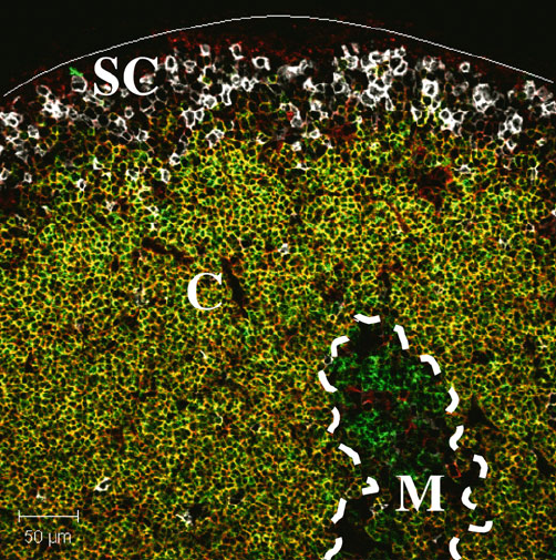
Fig. 1.1 The thymus displays a high degree of organization. Confocal analysis of mouse thy-
mus for CD4 (green), CD8 (red) and CD25 (white) reveals the distinct thymic compartments of
outer subcapsular region (SC), cortex (C) and inner medulla (M). Thymocytes at different stages
of development differentially express the cell surface markers CD4, CD8 and CD25 and specif-
ically localize to defined thymic regions. The subcapsular region is predominantly populated by
immature CD4
8
25
C
thymocytes (white), forming a discrete rim to the thymus. The cortex is
visualized as a densely packed region predominantly populated by CD4
8
double positive thy-
mocytes (appearing yellow), which undergo interactions with a network of interspersing cortical
thymic epithelial cells. The inner thymic medulla consists of a more sparsely populated region
formed by thymocytes at the single positive CD4
C
8
(green)andCD4
8
C
(red) undergoing
maturational interactions with a combination of medullary thymic epithelium and dendritic cells
In addition to thymic epithelium and thymocytes, a heterogeneous mixture of
additional cell types reside within the thymus, including stromal fibroblasts, thymic
macrophages and dendritic cells, all of which play an important role in thymus
biology.
During embryogenesis, the thymus develops as a bilateral outgrowth of endo-
derm, which buds off from a region of the foregut termed the third pharyngeal
pouch. A series of reciprocal signalling interactions with neural crest-derived mes-
enchyme ensheathing the thymic rudiment leads to a cascade of transcription factor
activity within the endoderm-derived thymus primordium, resulting in both out-
growth and differentiation of the thymus. Recent experimental data indicate that
endoderm-derived thymic epithelium producing cortex and medulla is not only gen-
erated from a single embryonic origin, but that both cortical and medullary thymic
4 W. Jenkinson et al.
epithelium derive from a bipotent progenitor population. Whether such a common
thymic epithelial progenitor population persists within the adult thymus and whether
such a population may represent a potential stem cell population with clinical ma-
nipulation implications remains an open area of research.
T cell development is not a cell autonomous process. The thymic microenviron-
ment nurtures the development of thymocytes, providing a wide range of signalling
interactions at defined developmental checkpoints in discrete anatomical locations.
Signals provided by thymic stromal cells regulate commitment to the T cell lin-
eage, regulation of proliferation, survival and importantly, selection of functional
T cells that are self-tolerant on the basis of thymocyte T cell receptor capability and
specificity. Due to the strict compartmentalization of thymic epithelial cells provid-
ing defined signals required at specific stages of thymocyte maturation, developing
T cells display a highly ordered pattern of migration within the thymus regulat-
ing stepwise interactions and sequential maturation. Amongst the important signals
provided by thymic epithelium to developing thymocytes, Delta-like 4 (Dl4) a lig-
and for the Notch signalling pathway is expressed by thymic epithelium. Signalling
through the Notch receptor, expressed by thymus colonizing thymocyte precursors,
is critical for commitment to the T cell lineage. Mice demonstrating defects in the
Notch signalling pathway lack the capacity to generate thymic T cells, instead B
cells are found to populate the thymus indicating that Notch plays an essential role
in the commitment of lymphoid precursors to the T cell lineage [2]. In addition,
thymic epithelium provides essential survival factors to developing thymocytes, in-
cluding IL-7 and stem cell factor [3]. Interestingly, the requirement for IL-7 appears
to be less important for thymocyte development within the fetal thymus compared to
the adult thymus, highlighting subtle differences in the developmental requirements
of T cells at defined temporal stages [4]. Of critical importance, thymic epithelium
also expresses high levels of both MHC class I and MHC class II. MHC expression
within the thymus plays an essential role in the selection of both CD4 and CD8
T cells through the capacity to test whether thymocytes with randomly generated
T cell receptors are able to recognise MHC presenting self-peptides with sufficient
affinity to ensure the TCR is functional but also ensure that those cells recognizing
MHC–self-peptide complexes with too high affinity are deleted to avoid potentially
autoreactive immune responses. Lack of MHC molecules on thymic epithelium re-
sults in a complete block in thymocyte development at the CD4
C
8
C
double positive
(DP) stage, as developing thymocytes are unable to test their randomly generated
receptors and fail to receive TCR transmitted survival signals.
It is clear that thymic epithelium provides essential signals to developing thymo-
cytes in a highly ordered stepwise manner. However, such signalling interactions
are not simply a one-way process. Analysis of the thymi of mice demonstrating
blocks in thymocyte developmentat defined stages demonstrates a clear correspond-
ing defect in differentiation, organization and maintenance of cortical and medullary
epithelium. Such findings highlight a complex reciprocal signalling mechanism op-
erating within the thymus whereby thymocytes provide important signals, such as
through the Lymphotoxin signalling pathway, that act to regulate the microenviron-
ments that regulate their own survival and maturation [5].
1 Thymocyte Development 5
The thymus is not an organ that is maintained at a constant size throughout life.
A programmed decrease in the proportion of functional thymic tissue ensues gen-
erally from the onset of puberty, resulting in a consequent decrease in T cell output
from the thymus of older individuals. The decrease in functional thymic tissue is
termed thymic involution or atrophy. The precise mechanics of thymus involution
remains unclear although several lines of evidence point to a pivotal role for sex
steroids in this process [6]. During thymocyte development, approximately 95% of
thymocytes are deleted due to production of either non-functional or auto-reactive
T cell receptors. Such huge wastage may provide an explanation as to the reason
for thymic involution, the necessity to maintain such a wasteful organ following
the establishment of a peripheral T cell pool of sufficient size and diversity as to
maintain immune protection may no longer be required. In support of this, thymec-
tomy of young individuals does not lead to significant immunodeficiency, indicating
that maintenance of a fully functional thymus with increasing age is not strictly
necessary. However, improving healthcare and associated increases in lifespan of in-
dividuals may present problems in the future as elderly individuals may demonstrate
a reduction in the T cell repertoire as a result of reduced thymic output. In addition,
the advent of clinical therapies resulting in depletion of lymphoid compartments
followed by bone marrow transplant presents a problem in terms of the reduced
presence of functional thymic microenvironments capable of supporting rapid re-
constitution of T cell compartments. Indeed, post-bone marrow transplant patients
demonstrate a high susceptibility to opportunistic infection due to a severely re-
duced capacity to produce T cells. Identification of potential thymic epithelial stem
cell populations and the precise signalling pathways involved in regulating thymic
epithelial development and expansion currently present intense areas of research
aimed at developing methods of regenerating functional thymic tissue and T cell
reconstitution.
Colonization and Export from the Thymus
The thymus lacks a resident population of self-renewing haematopoietic stem cells
(HSC). Unlike B cell development, where HSC exist within the same localized mi-
croenvironment as their B cell progeny, the remote location of the thymus therefore
requires the selected recruitment of T cell progenitors throughout life to ensure a
continuous supply of mature T cells. Recruitment of T cell progenitors to the thy-
mus, whilst continuous throughout life, does not occur in a steady stream but rather
entry is regulated in a periodically-gated manner [7]. The precise mechanisms reg-
ulating such stop-start thymic entry remains unclear, although evidence suggests
that this may at least in part be regulated by the availability of space within intra-
thymic niches capable of nurturing developing thymocytes [8]. Following entry to
the thymus, T cell precursors undergo a series of maturational events including
bursts of proliferation resulting in a single thymus colonizing cell generating up to
one million progeny [9]. The recruitment and specific entry of lymphoid progenitor
6 W. Jenkinson et al.
cells into the thymus therefore forms the first hurdle that potential thymocytes must
overcome in their journey to becoming a mature T cell.
Colonization of the thymus within the mouse appears to occur via two different
mechanisms operating within temporally defined windows. At approximately day
11.5 of gestation, recruitment of circulating fetal liver and AGM-region haematopoi-
etic cells to the embryonic thymus initiates and continues for a period of approx-
imately 2 days. During the initial window of thymus colonization, the thymus
remains an avascular structure, with entry of blood vessels into the thymus proper
occurring after day 14 of gestation. As a result, specific recruitment of haematopoi-
etic precursors occurs via the induction of haematopoietic precursor extravasa-
tion from capillaries in proximity to the thymus followed by migration through
perithymic mesenchyme and finally entry to the thymus through the presumptive
capsule. Attraction of haematopoietic cells to the early fetal thymus appears to be a
highly specific process regulated by the action of a restricted set of chemokines com-
prising CCL25, CCL21 and CXLC12. Absence of expression of CCR7 and CCR9,
the receptors for CCL21 and CCL25 respectively, on haematopoietic cells results
in a severe reduction in pre-vascularized thymus colonization and an associated ab-
sence of T cell subsets normally generated from these cells [10]. The importance
of CCL25 in mediating colonization of the fetal thymus is further highlighted in
Nude mice. Nude mice display a mutation in the transcription factor Foxn1. Lack of
functional Foxn1 expression results in a cell-autonomous defect in thymic epithelial
differentiation after day 11.5 of gestation and a corresponding absence of thymic
epithelial CCL25 expression. Whilst CCL25 expression is absent in Nude thymic
epithelium, CCL21 is expressed normally in a Foxn1-independent manner in cells
of the adjacent parathyroid. Importantly, whilst haematopoietic cells are attracted to
the proximity of the Nude thymus, no cells enter into the thymus itself. Such findings
suggest a differential two-step role for CCL21 and CCL25, whereby CCL21 attracts
haematopoietic cells into proximity of the thymus, whereupon CCL25 mediates en-
try of cells into the epithelial microenvironment of the thymus itself. Whilst the
diversity of chemokine expression within the embryonic thymus appears relatively
restricted, analysis of the chemokine receptors expressed by thymus colonizing cells
demonstrates a diverse heterogeneous mixture of cells, whether such cells demon-
strate differential developmental potential remains unclear [11].
Within the mature adult thymus a rich network of blood vessels penetrate into
the thymic tissue. Colonization of the thymus in vascularized adult stages occurs
via extravasation of thymus colonizing cells at post-capillary venules located pre-
dominantly at the junction between cortex and medulla. The role of chemokines in
the control of adult thymus colonization remains less clear than for the fetal thy-
mus. Recent evidence however, suggests that CCL25-CCR9 signalling may also be
involved in recruitment of cells to the adult thymus. In addition to chemokine action,
the protein P-selectin expressed on the endothelium of thymic post-capillary venules
binds the carbohydrate P-selectin ligand-1 (PSGL1) expressed by thymus coloniz-
ing cells, absence of either receptor or ligand results in a significantly reduced entry
of cells into the mature thymus highly implicating this pairing in thymus coloniza-
tion. Importantly, expression of P-selectin has been shown to be regulated by the
1 Thymocyte Development 7
availability of thymic stromal niches identifying a potential mechanism regulating
the gated entry of haematopoietic precursors into the thymus [12].
Until recently, the mechanisms regulating T cell exit from the thymus have re-
mained ill-defined. Again, chemokines have been implicated in the regulation of this
process. CCL19, an additional ligand for the chemokine receptor CCR7 expressed
by mature thymocytes, has been identified on blood vessels present within thymic
medulla. Whilst such chemokine expression is able to attract mature thymocytes
to exit thymic parenchyma into perivascular spaces within the thymus, additional
signals are required to facilitate full thymic egress. The cell surface receptor sphin-
gosine 1-phosphate type 1 (S1P1) appears to play an essential role in exit from the
thymus in response to a gradient of expression of the ligand S1P between thymus
and blood. As such, mice lacking the capacity to signal through the S1P1 receptor
expressed on haematopoietic cells demonstrate a traffic jam of mature thymocytes
within the thymus and perivascular spaces [13] as cells are unable to receive the cor-
rect signals to exit the thymus. Together, such data imply that thymic exit of mature
thymocytes occurs in a two step process driven firstly via initial chemokine attrac-
tion to blood vessels and into perivascular spaces followed secondarily by S1P1
receptor mediated exit into the bloodstream. Previous reports have also suggested
that exit from the thymus occurs in a lucky-dip haphazard manner whereby some
thymocytes leave early in maturation and some thymocytes leave late [14]. How-
ever, recent data now suggest that thymic emigration is a strictly ordered process
ensuring that only the most mature thymocytes, having completed the full develop-
mental program, have a preferential ability to leave the thymus. Such mechanisms
are thought to operate at least partly based on differential thymocyte expression of
the S1P1 receptor [15].
˛ˇ Versus ı T Cell Development
Two main lineages of T cells are produced within the thymus, a prevailing ˛ˇ T cell
lineage, and a minor ı T cells lineage. Whilst multiple aspects of ˛ˇ T cell de-
velopment and function have been defined, those relating to the ı T cell lineage
remain vague. Both ˛ˇ and ı T cells develop from a common haematopoietic
progenitor population colonizing the thymus. Branching of the ı T cell lineage
from ˛ˇ T cell lineage is though to occur within the window of the late DN2 to
DN3 stage [16]. During DN thymocyte development, Recombinase activating gene
(RAG) complex activity results in TCR gene rearrangement, in addition to ˇ-chain
rearrangements, DN thymocytes also undergo rearrangements of both -andı-TCR
chains. The fact that ˇ-chain rearrangements may be found within mature ı T cells
and conversely, ı-chain rearrangements within ˛ˇ T cells strongly supports the no-
tion of a common precursor origin for both T cell lineages [17, 18].
The precise developmental requirements of the ı T cell lineage within the thy-
mus remain uncertain. However, recent studies have indicated that a subset of ı
T cells bearing a canonical TCR of restricted diversity, generated exclusively during
8 W. Jenkinson et al.
fetal development, may undergo positive selection in response to interactions with
thymic epithelium. In contrast, the interactions of ı T cells, bearing diverse TCR
specificities, with thymic stromal elements are unclear. Within this chapter, we will
therefore concentrate on describing development of the ˛ˇ T cell lineage, which
form the majority of thymocytes developing within the thymus and also mature
T cells within the periphery.
Developmental Changes During Thymocyte Development
During thymocyte development, the linear progression of maturation can be charac-
terised by the differential expression of numerous cell surface molecules. Initially,
as thymic settling haematopoietic precursors colonize the thymus, the cells lack ex-
pression of both the T cell co-receptor molecules CD4 and CD8, as such these cells
are termed double negative CD4
8
(DN) and represent the most immature thy-
mocyte subset. The precise identity of thymus settling cells remains contentious.
While cells have been found within both bone marrow and blood with a lymphoid
bias, thymus-settling cells are capable of generating multiple different lineages, with
current data suggesting that multiple populations of haematopoietic cells colonize
the thymus, with each subset displaying different degrees of T cell potential. Upon
entry into the thymus, thymic settling progenitors undergo a series of interactions
with thymic stromal cells that regulate the proliferation, survival and progressive
differentiation of the haematopoietic cells. DN CD4
8
thymocytes subsequently
upregulate both CD4 and CD8 to become double positive CD4
C
8
C
(DP) thymo-
cytes, and finally mature to become single positive CD4
C
or CD8
C
(SP) cells
following stringent selection events. Development of thymocytes is strictly regu-
lated by sequential reciprocal signalling interactions with stromal cells of the thymic
microenvironment. Consequently, it is essential that developing thymocytes are lo-
cated in the right place at the right time to receive the right signals to drive efficient
maturational events.
Double negative thymocytes can be further subdivided into subpopulationson the
basis of CD25 and CD44 expression. The most immature thymocytes express CD44
but lack CD25 expression being termed CD44
C
25
DN1 cells. After approximately
10 days, thymocytes next upregulate CD25 to become CD44
C
25
C
DN2 cells. The
DN2 stage lasts 2 days, during which thymocytes begin the process of gene rear-
rangement within the TCRˇ chain locus. Downregulation of CD44 follows, marking
the transition to the CD44
25
C
DN3 stage, accompanied by ongoing V(D)J TCRˇ
gene rearrangement mediated by RAG1 and RAG2. Successful completion of TCRˇ
chain gene rearrangement leads to pairing of the TCRˇ chain with the invariant sur-
rogate pre-TCR˛ chain (pT˛) at the cell surface in conjunction with CD3 molecules.
Due to the imprecise nature of random TCRˇ gene rearrangement,a high proportion
of DN thymocytes are unable to generate a functional TCRˇ chain due to failed gene
rearrangement. The creation of the preTCR provides an early screening mechanism
to eliminate thymocytes bearing non-functional TCRs through the process termed
1 Thymocyte Development 9
ˇ-selection. Transient expression of the preTCR at the cell surface in conjunction
with CD3 elements results in the transmission of signal through the assembled re-
ceptor in what is currently thought to be a ligand-independent process. Successful
preTCR signals result in several important outcomes:
(a) Prevention of apoptosis
(b) Initiation of cell division
(c) Inhibition of TCR ˇ-chain rearrangement and
(d) Progression of differentiation through downregulation of CD25 to briefly
become CD44
25
DN4 thymocytes
The DN3 stage of development again lasts for approximately 2 days.
Following ˇ-selection and multiple rounds of cell replication, DN4 thymocytes
upregulate both CD4 and CD8 becoming double positive (DP) CD4
C
8
C
thymo-
cytes. During transit to the DP stage, cellular replication ends and rearrangement of
TCR˛ genes is initiated through re-expression of the RAG complexes responsible
for driving TCR gene rearrangement. During TCR ˛-chain rearrangement, thymo-
cytes are provided with multiple opportunities to express a functional TCR˛ chain.
Only thymocytes expressing a functional TCR of correct specificity receive survival
signals allowing escape from programmed cell death. During this period, thymo-
cytes will continue to rearrange ˛-chain genes until a functional ˛-chain paired with
the ˇ-chain allows for positive selection, or alternatively thymocytes run out of time
and are subsequently deleted as a result of failure to generate a functional TCR capa-
ble of interacting correctly with thymic stromal elements. The precise mechanisms
regulating DP thymocyte lifespan remain unclear. However, one potential mecha-
nism regulating the lifespan of DP thymocytes is thought to operate via the action
of the orphan nuclear receptor ROR regulating expression of the anti-apoptotic
factor Bcl-XL. Under steady-state conditions, it is thought that the pre-selection DP
stage of thymocyte development may last in the region of 2–3 days. Regulation of
DP thymocyte lifespan therefore plays an essential role in determining the diver-
sity of the T cell repertoire through the linked interplay with the amount of time
that thymocytes receive to continue TCR ˛-chain gene rearrangement and potential
for creating a functional TCR [19]. Once thymocytes are generated bearing a func-
tional TCR consisting of rearranged ˛-andˇ-chain subunits expressed at the cell
surface, positive selection of thymocytes ensues assuming appropriate recognition
of self-peptide:MHC complexes, resulting in downregulation of RAG and cessation
of further TCR˛ gene rearrangement.
Up until the DP stage of development, thymocytes are confined to and mature
within the cortex. Post-positive selection DP thymocytes upregulate CD69 and sub-
sequently down regulate either CD4 or CD8 becoming single positive (SP) CD4
C
8
or CD4-8
C
. In addition, SP CD4 and CD8 thymocytes demonstrate a redistribution
of compartmentalization, being primarily located within medullary areas. Within
the medulla , thymocytes undergo further maturational events thought to last up
to 12–14 days, although recent estimates suggest this may last as little as 4–5
days [15]. Within the medullary residency period, newly formed SP thymocytes
demonstrate a CD69
HI
CD62L
LOW
phenotype and exhibit relatively functionally
10 W. Jenkinson et al.
immature properties, following a period of maturation involving as yet poorly
defined events, SP thymocytes lose CD69 expression and increase CD62L
expression at which stage thymocytes become functionally mature and exhibit
a heightened responsiveness to thymic export signals.
Intrathymic Migration
After entry to the mature thymus at the cortico-medullary junction, thymocytes
demonstrate a clear pattern of migration. Such migration helps to establish compart-
mentalization of cells at different stages of maturation. This process acts to ensure
that cells are in the correct microenvironment at the right stage of development to
ensure access to the signals provided by stromal cells that dictate whether individual
thymocytes survive and differentiate or alternatively meet an early death through in-
duction of apoptosis (Fig. 1.2). The most immature thymocytes being CD4
8
DN
exhibit an outward pattern of migration moving from the cortico-medullary junction
towards a sub-capsular location. As such, DN1 thymocytes are located in relation
to the cortico-medullary junction, extending into the deep cortex, DN2 thymocytes
are present within mid-cortex and DN3 cells are present within the outer cortical
region [20]. Within the DN3 population, ˇ-selection induces proliferation and accu-
mulation of pre-DP thymocytes within the subcapsular region. Following transition
to the subcapsular region, progression to the CD4
C
8
C
DP stage is initiated resulting
in TCR ˛-chain rearrangement and cessation of proliferation. Subcapsular microen-
vironments may play a role in regulating the progression of DN thymocytes to the
DP stage through production of TGFˇ, which has been shown to negatively regulate
pre-DP thymocyte proliferation [21]. The outward migration of thymocytes across
the cortex is mediated by the action of several chemokine receptors inducing di-
rected cellular migration. Mice lacking the chemokine receptors CXCR4 or CCR7
both display an accumulation of early DN thymocytes near the cortico-medullary
junction and aberrant thymocyte development [22, 23]. In addition, mice lacking
CCR9, whilst demonstrating normal DN2 and DN3 localization within mid- and
outer-cortical regions display an inefficient localization of pre-DP thymocytes at
subcapsular sites [24]. However, CCR9-deficient mice do not appear to demonstrate
any gross defects in early thymocyte development suggesting that sub-capsular mi-
gration is not essential for thymocyte development.
In addition to directional cues provided by chemokine signalling gradients,
thymic stromal cells provide a defined matrix over which immature thymocytes are
able to crawl. Thymocytes express adhesion molecules differentially during their
maturational program, with studies demonstrating that expression of the adhesion
molecule VCAM1 by cortical stromal cells is necessary for normal thymocyte pre-
cursor migration [25].
As thymocytes enter the DP stage, they begin to rearrange the TCR ˛-chain and
test the function of newly generated TCR˛ˇ receptors to recognize peptide:MHC
complexes. DP thymocytes accumulate near the cortico-medullary junction but are
1 Thymocyte Development 11
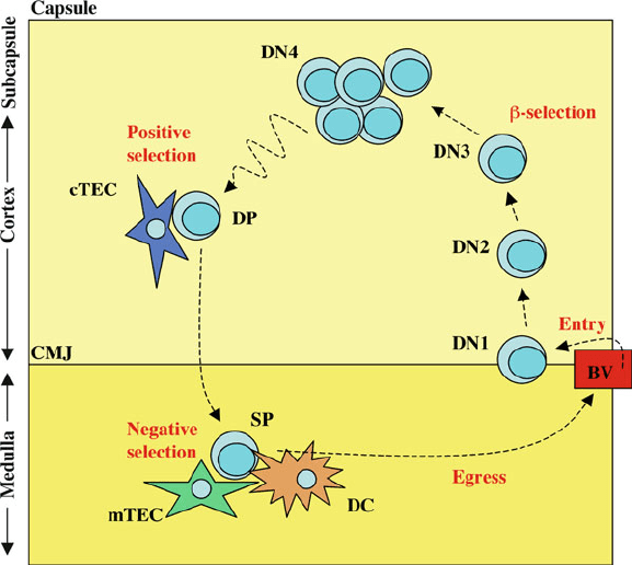
Fig. 1.2 Thymus settling cells enter the thymus through post-capillary venules located at the
cortico-medullary junction (CMJ) as DN1 (CD4
CD8
CD44
C
CD25
) cells. Differentiation
of thymocytes into DN2 (CD4
CD8
CD44
C
CD25
C
) and DN3 (CD4
CD8
CD44
CD25
C
)
subsets is accompanied by outwards-directed migration towards the subcapsular region. DN3
thymocytes passing the ˇ-selection checkpoint undergo both differentiation and extensive pro-
liferation whilst passing through the DN4 (CD4
CD8
CD44
CD25
) stage. Double positive
CD4
C
CD8
C
thymocytes subsequently undergo inwards-directed migration back through the cor-
tex, whilst undergoing a series of cognate interactions with cortical thymic epithelial cells in search
of positively selecting ligands. Positive selection of thymocytes results in rapid relocation to thymic
medulla where SP CD4
C
8
and CD4
8
C
thymocytes pass through further developmental check-
points prior to export from the thymus. Presentation of self-antigens by medullary thymic epithelial
cells (mTEC) and dendritic cells (DC) ensure that potentially auto-reactive thymocytes are deleted
via the mechanism of negative selection. Functional, self-tolerant SP T cells subsequently exit the
thymus through blood and lymphatic vessels via a combination of chemokine and S1P mediated
signals
unable to migrate into medullary areas. Thymocytes receiving positive signals con-
firming the generation of a functional TCR forming low affinity interactions with
self-peptide:MHC exhibit upregulation of the chemokine receptor CCR7 [26]and
begin rapid migration towards thymic medullary areas preferentially expressing the
CCR7 associated ligands CCL19 and CCL21. In support of this, mice lacking either
CCR7 or both ligands CCL19 and CCL21 exhibit a clear block in cortex to medulla
migration as a result of inhibited chemotaxis towards medullary regions. However,
SP thymocytes are still capable of entering medullary areas albeit in a seemingly
12 W. Jenkinson et al.
inefficient random manner occurring as a result of an absence of directed chemo-
taxis to medullary CCR7-ligands. Recent studies have further suggested that while
CCR7 is required for SP thymocyte chemotaxis towards medullary areas, a second
undefined G-protein coupled receptor mediated signal is required to enter thymic
medulla and migrate on medullary substrates possibly via activation of specific ad-
hesion molecules on SP thymocytes.
Positive Selection
The random nature of T cell receptor generation is accompanied by the intrinsic
drawback of the potential for T cells to be generated bearing receptors that either are
(a) Incapable of recognizing peptide presented by MHC, therefore being non-
functional or
(b) Capable of recognizing self-antigens to such an extent that they pose the dan-
gerous potential to generate autoimmune actions against the bodies own tissues
The quality control process operating within the thymus provides essential mech-
anisms acting to eliminate both non-functional and auto-reactive thymocytes yet
retain and facilitate the maturation of functional, self-tolerant T cells. Following
rearrangement and pairing of TCR˛ and ˇ-chains, three developmental outcomes
are open to DP thymocytes. Thymocytes generating TCR pairings incapable of
identifying self-peptide:MHC complexes with sufficient strength fail to generate
TCR transduced signals of sufficient level to cross the threshold required to induce
cell survival. In such cells, further TCR ˛-chain rearrangement provides a second-
chance for DP thymocytes via continued generation of alternative new TCR˛ˇ
pairings in order to try and generate a functional TCR. Alternatively thymocytes
may fail to win a reprieve and undergo deletion via neglect due to a lack of sur-
vival signal provision. On the flip-side to lack of sufficient TCR signalling inducing
death by neglect, an overly strong interaction between thymocytes bearing TCR and
self-peptide:MHC complexes presented by antigen presenting cells again results in
the induction of cell death in a process termed negative selection or clonal dele-
tion, thought to be an important factor in preventing the generation of potentially
autoreactive T cells. Interestingly, the threshold for TCR stimulation within DP thy-
mocytes undergoing selection events in the thymus appears to be lower than that
required by mature T cells responding to peptide in the periphery.
Sitting in a happy medium between these two negative outcomes of thymocyte
deletion lies the third potential fate of DP thymocytes, positive selection. Positive
selection occurs following engagement of DP thymocyte-expressed TCR with self-
peptide:MHC complexes with low affinity recognition remaining below the signal
strength threshold responsible for inducing negative selection. The three poten-
tial developmental outcomes open to developing thymocytes during selection has
been described as following the Goldilocks hypothesis. The Goldilocks conditions
command that positive selection and onwards development of thymocytes within