Scheffler Immo E. Mitochondria
Подождите немного. Документ загружается.

4.6 PROTEIN IMPORT INTO MITOCHONDRIA
4.6.1 Mitochondrial Targeting of Proteins
Mitochondria are distinct subcellular organelles. The existence of an outer and
an inner membrane therefore defi nes two compartments topologically sepa-
rated from the cytosol and from each other, with the matrix on the inside and
the intermembrane space between the two membranes. The identifi cation of
specifi c proteins in the intermembrane space occurred relatively more recently,
but from the time of the understanding of the morphology of mitochondria
the following question naturally arose: How do proteins get into the matrix?
This question was soon broadened to include the proteins found to be associ-
ated specifi cally with the inner and outer mitochondrial membranes.
The problem also became more urgent as soon as it was recognized that
the coding capacity of the mitochondrial genome was extremely limited and
that therefore the vast majority of the mitochondrial proteins were encoded
by the nuclear genome and synthesized in the cytosol. Pioneering and elegant
studies initiated in the laboratories of Schatz and Neupert with yeast, Saccha-
romyces cerevisiae , and many contributions by the disciples trained in these
laboratories and others have provided answers to increasingly complex and
detailed questions. Many complementary studies were also conducted with the
mold Neurospora crassa . Whenever it has been possible to test key features
or mechanisms with mammalian mitochondria, the similarities have been
striking, and it appears that the principal insights into mitochondrial protein
import derived from yeast will be applicable to mitochondria from many if
not all organisms. A question not often raised is the question about the evo-
lutionary origin of the import machinery (86) . With some exceptions, the pro-
teins involved are unique and unrelated to other proteins of eukaryotic cells.
From the point of view of the endosymbiont theory of the origin of mitochon-
dria, it is clear that the development of a protein import system must have
preceded the signifi cant transfer of genes from the endosymbiont cell to the
nucleus; that is, it constitutes an early event in the evolution of mitochondria.
There was no obvious need for protein import in the prokaryotic ancestors of
mitochondria, unless scavenging for “ loose ” proteins constituted a possible
source of nutrients. One might therefore expect closely related and homolo-
gous proteins in phylogenetically diverse organisms dedicated to this task. The
explosion of genomic sequence information from many organisms in the past
decade has provided a wealth of supportive data for the specialist (see Chapter
2 ). Plants present the additional challenge of understanding the biogenesis of
chloroplasts, where similar problems with protein import have to be solved.
In chloroplasts there is one additional compartment formed by the thylakoid
membranes. Comparative studies on these two organelles promise to be
revealing.
As always, the development of experimental approaches and model systems
was crucial for progress in this fi eld, and two breakthroughs can be singled
PROTEIN IMPORT INTO MITOCHONDRIA 141
142 BIOGENESIS OF MITOCHONDRIA
out. First, Schatz and his collaborators succeeded in using isolated mitochon-
dria from yeast in their import studies, with peptides to be imported also made
in vitro , using the reticulocyte lysate or wheat germ systems. This approach
became feasible when it was discovered that at least in vitro protein import
was post - translational, in contrast to the situation encountered with protein
synthesis and translocation into the endoplasmic reticulum. The second meth-
odological advance is becoming a refrain, or in more colorful language, “ d é j à
vu all over again ” : The combined approach of molecular genetics and cell
biology/biochemistry in yeast has facilitated the analysis in unprecedented
detail, because of (a) the ingenuity and power of selective methods and (b)
the effi cient way in which mutants can be analyzed in vivo and by in vitro
model systems.
There is still some uncertainty whether protein import is post - translational
or co - translational in vivo (86) . The absence of a prominent coating of mito-
chondria by polysomes (as in the rough endoplasmic reticulum) argues against
co - translational import, but it can occur, since ribosomes have been found to
be associated with mitochondria and specifi c cDNAs have been cloned from
mRNAs attached to mitochondria (264) . Polysome attachment was specifi cally
promoted in these experiments by slowing down protein synthesis with cyclo-
heximide. A problem that was solved in broad outline quite early in this fi eld
was concerned with the distinction between (a) proteins made in the cytosol
destined for import into mitochondria and (b) those which were unrelated to
any mitochondrial function and had to be discriminated against by the import
machinery. In the early, pioneering studies all proteins to be imported were
found to have a signal sequence (or targeting sequence) at the N - terminal. It
consists of 20 – 50 amino acid residues and is usually cleaved off upon import
on the matrix side of the inner membrane. The functional signifi cance of such
a signal sequence was demonstrated most convincingly by showing that almost
any protein — for example, the cytoplasmic protein dihydrofolate reductase
(DHFR) — could be imported into mitochondria when it was equipped with
such a signal at the N - terminal (265, 266) . Since most of the early studies were
performed with soluble proteins targeted to the matrix, the N - terminal
sequence in the present context will be referred to as the “ matrix targeting
signal. ” Many such sequences have been characterized to date in the hope of
defi ning criteria that would allow one to predict simply from inspection
whether a protein, known from its cloned and sequenced gene, was a mito-
chondrial protein (267 – 269) . The number of such sequences was surprisingly
diverse in length and composition. The major distinguishing feature appears
to be the ability to form an amphiphilic α - helix with positive charges on one
side when viewed down the long axis (268) . No obvious distinctions are found
between yeast and mammalian cells, for example. As the number of examples
increased, attempts were made to defi ne consensus sequences (or patterns),
and computer programs can now be found on the Internet that will identify
mitochondrial proteins from genomic sequence information ( www.123genomics.
com/fi les/analysis.html ). While useful, all of these programs fail to identify a
substantial fraction of mitochondrial proteins. There are several reasons. A
number of proteins are imported into mitochondria without further processing
by a peptidase, making it more problematical to defi ne the leader sequence.
Other proteins, especially the large number of proteins found as integral
membrane proteins in the inner membrane, have internal targeting sequences,
and such sequences are particularly diffi cult to identify and categorize (see
below).
The targeting sequence interacts with receptors on the mitochondrial
surface, but its removal on the matrix side is appropriately discussed here also,
since sequence recognition within or near the matrix targeting signal by one
or more highly specifi c endopeptidases is required. An exhaustive survey of
the literature is impossible, but a few examples will illustrate some major dis-
coveries and generalities. Human patients diagnosed with citrullinemia and a
defect in ornithine transcarbamylase (an enzyme in the urea cycle) prompted
efforts to understand this mitochondrial enzyme, leading Rosenberg and his
colleagues to perform some path breaking studies on mitochondrial protein
import in mammalian cells. It was discovered that the matrix targeting signal
of OTC was processed in two steps (270, 271) . A survey of proteolytic cleavage
sites identifi ed amino acid motifs recognized by the matrix proteases (269) . In
general, such studies have been helpful in identifying cleavage sites in the
maturation of mitochondrial proteins (the “ ARg2 rule ” ), but an absolutely
certain prediction is still not possible in all cases.
Finally, the cDNA for the β subunit of the mitochondrial processing pepti-
dase was cloned from rat liver, and sequence comparisons established relation-
ships with a proposed family of metallopeptidases (272) . A few years ago,
several of the prominent contributors to this fi eld established a uniform
nomenclature for the mitochondrial peptidases responsible for protein matu-
ration after these laboratories had independently purifi ed proteins, and they
characterized genes from rat liver, Saccharomyces cerevisiae, Neurospora
crassa , and potato (273, 274) . Three major activities are recognized at this time:
α - MPP and β - MPP are the subunits of the mitochondrial processing peptidase
responsible for the initial cleavage of the targeting sequence. MIP (mitochon-
drial intermediate peptidase) carries out the second cleavage required for
maturation of some matrix proteins, removing an additional eight amino acids
from the N - terminal. For proteins sorted into the intermembrane space (see
below), sorting signals have to be removed as well, and at least two integral
membrane proteins with peptidase activity have been identifi ed. They are
designated IMP I, and so on, and it appears that they are heterodimers. An
alternate approach to cloning the corresponding peptidases from yeast (origi-
nally designated MAS1 and MAS2 (275) ) will be discussed below.
The import of a protein into the matrix requires the traverse of two mem-
branes and hence the existence of a mechanism that allows a large, water -
soluble polypeptide to pass through the lipid bilayers. Before discussing the
proteins involved, and how they were identifi ed and assigned their specifi c
roles, it should be stated that this discussion will use the nomenclature adopted
PROTEIN IMPORT INTO MITOCHONDRIA 143
144 BIOGENESIS OF MITOCHONDRIA
by the leaders in the fi eld (276) . It therefore becomes possible to talk about
the “ TOM and TIM complexes ” (87) and remove at least one potential type
of confusion. Tom proteins are integral (or peripheral) membrane proteins of
the outer membrane participating in protein import. Tim proteins are integral
membrane proteins located in the inner membrane and participating in the
same process. Additional soluble proteins with distinct names are carrying out
important functions on the cytosolic side of the outer membrane; similarly, a
variety of soluble proteins on the matrix side have equally essential roles in
the overall process. More recently, a variety of Tim proteins in the intermem-
brane space have attracted attention. Tom and Tim proteins are distinguished
further by numbers corresponding to their molecular mass in kDa. For example,
Tom20, Tom22, Tom37, Tom70, and so on, represent outer membrane proteins
of the import machine.
It is diffi cult to do justice to the elegance and ingenious design of the many
experiments that have made the system less and less of a black box, or a few
rectangles symbolizing a pore through the membranes. But before losing our-
selves in details, an outline and some highlights will provide perspective and
allow the formulation of guiding questions.
A globular, soluble, cytoplasmic protein (for example, aquorin or the green
fl uorescent protein), when provided with a matrix targeting signal at the N -
terminal, was found to be imported into yeast and mammalian mitochondria
in vitro and in vivo . Therefore, the targeting signal had to be recognized by a
receptor on the mitochondrial outer surface. Simple notions of polypeptide
tails “ diving ” into lipid bilayers were entertained only temporarily. Since the
process was post - translational, one had to assume that the protein was in a
folded form; in fact the test protein, DHFR, was shown to be active before
import and after import. A simple but clever experiment provided evidence,
however, that the protein had to be unfolded to be imported: When the tertiary
structure of the protein was stabilized by the binding of a ligand (methotrex-
ate), import was prevented. From this and related experiments the notion was
generated that mitochondrial proteins are unfolded in the process of import
and threaded through an import pore as extended polypeptides. This reduces
the required pore size, but creates a problem at the beginning and at the end:
The protein has to be unfolded fi rst, and the protein has to be refolded once
it has arrived on the inside. More will be said about this later.
Since two membranes are involved, one could a priori imagine the import
process to occur in two stages, with the intermembrane space being an inter-
mediate, temporary location. However, matrix proteins were never found
there, even transiently, and no conditions were found which would have
arrested the process at such an intermediate stage. It could be shown that
proteins had their targeting signal removed before they were even completely
transferred to the matrix. For example, at lower temperatures, in vitro import/
translocation can be initiated, but the peptide remains “ stuck ” in the mem-
brane. The interpretation was that the N - terminal protruding into the matrix
was subject to proteolytic processing by the matrix peptidases while the
remainder of the peptide was still in transit. Another observation made with
the electron microscope was that the inner and outer membrane were fre-
quently in close contact, and it was hypothesized that these were locations
where a complex of proteins in the outer and inner membrane created a
channel traversing both membranes for protein import. Such a structure may
indeed exist, but only transiently (see below).
A key observation made relatively early in the course of these investiga-
tions was that the isolated mitochondria used in the in vitro experiments had
to be in a state where a membrane potential was maintained. The addition of
ionophores or uncouplers destroyed the membrane potential and also made
these mitochondria incapable of importing a peptide. This was and continues
to be a puzzling observation, although attempts to rationalize it have been
made. In general, current thinking attributes to the membrane potential a
driving force that pulls the positively charged matrix targeting signal across
the inner membrane, since the inside is negative relative to the outside (277) .
This problem is part of a larger question about the energetics of the process.
What are the thermodynamic parameters that make this process feasible,
effi cient, and essentially unidirectional. Electrophoresis of the targeting signal
across the inner membrane is part of, but not the complete, answer.
Mitochondrial protein import is more akin to active transport, fi rst because
of the recognized need for a membrane potential, and later because of the
involvement of the chaperones and their associated ATPase activities. The
conditions for manipulating external and/or internal ATP concentrations in
vitro in order to study import requirements have been described (278) , and
some general conclusions can be stated from such experiments. An obvious
one is that no internal ATP is required if a protein does not cross the inner
membrane. If matrix ATP is required, then the peptide or a segment of it will
have crossed the inner membrane on its path to its fi nal destination.
4.6.2 The Protein Import Machinery of Mitochondria
It now remains to understand each of these processes more detail and to
explain how the molecular architecture of the TOM and TIM complexes is
designed to execute translocation of a polypeptide across two membranes, or
to deliver them to other compartments. A genetic approach in yeast proved
invaluable. In pioneering studies, Yaffe and Schatz (85) were the fi rst to isolate
and characterize yeast mutants defective in general protein import. Recogniz-
ing that mutations affecting all mitochondrial functions might be lethal, a col-
lection of temperature sensitive mutants was screened for the inability to
import the β subunit of the F1 - ATPase at the nonpermissive temperature,
37
o
C. The assay was based on a clear distinction in electrophoretic mobility
between the precursor in the cytosol and the mature (processed) peptide after
import. More than 1000 mutagenized strains were screened; and in the fi rst
publication, two nonallelic, recessive mas mutants ( mas1 and mas2 , mitochon-
drial assembly) were characterized in more detail. As described expertly and
PROTEIN IMPORT INTO MITOCHONDRIA 145
146 BIOGENESIS OF MITOCHONDRIA
in some detail by Yaffe (279) , such mutants can be analyzed for import and
mitochondrial function in vivo , and isolated mitochondria can be further inves-
tigated in vitro . Specifi city, observed processing, dependence on a membrane
potential, and protection of the imported protein against exogenous proteases
were among the criteria defi ned for import experiments. A large collection of
reviews of methods can also be found in volume 260 of Methods of Enzymol-
ogy (1995). By one defi nition, protein import is defi ned as the process that
leads to the protection of the imported peptide against an exogenously added
protease. A simple test of this type will not distinguish between import into
the matrix, insertion into the inner membrane, or import into the intermem-
brane space. Insertion into the outer membrane may also leave a signifi cant
domain of a protein susceptible to proteolytic attack from the outside. A
further distinction can be achieved by a variety of schemes. Dilution of mito-
chondria in hypotonic medium causes swelling and rupturing of the outer
membrane, leading to the formation of mitoplasts. Outer membrane vesicles
can be made and purifi ed for functional analysis and biochemical/immuno-
chemical assays of composition. Well - characterized antibodies, especially
monoclonals, directed against specifi c epitopes can also give useful informa-
tion about the location and orientation of a protein of interest. Nowadays
the focus is often less on the mechanism of import, but on a determination
of the precise location of a mitochondrial protein that co - purifi es with
mitochondria.
Over the years, many more mas mutants were characterized and the corre-
sponding genes cloned. As described above, they were renamed Toms and
Tims, but can be found in the original literature under their original “ mas ”
designations. Not all Tom and Tim peptides were discovered from mutant
screens and cloning. A number of peptides, especially in the outer membrane,
were fi rst defi ned by cross - linking studies to already known components. Pro-
gress reports from this fi eld have been published on an almost annual basis
from the laboratories of Neupert, Pfanner, and others; only the most recent
reviews are cited here (280 – 286a) .
Multiple subunits of one kind may be found in the complexes being formed.
One of the major and uncontroversial conclusions about these complexes is
that they are not stable over time, but instead are transiently assembled from
the interaction of subcomplexes. An attractive and rational hypothesis is that
the assembly of one may promote or stabilize the assembly of the other in the
juxtaposed portion of the membrane, leading to the temporary alignment of
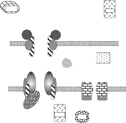
the two channels through the outer and inner membranes (Figure 4.10 ).
A logical start is with the accessory proteins in the cytosol. It was fi rst dis-
covered by Schekman ’ s laboratory (287) that a subset of stress proteins was
required for (a) the translocation of secretory proteins across the endoplasmic
reticulum membrane and (b) the translocation of mitochondrial proteins into
the matrix. It is now recognized that proteins such as those belonging to the
hsp70 heat - shock protein family bind to hydrophobic domains of denatured
proteins, thus stabilizing an unfolded state, or they may bind already to the
nascent peptide emerging from the ribosome during synthesis (288) . Matrix
proteins are also bound by these “ chaperones, ” so called because they are
believed to chaperone a peptide to its eventual destination, or to assist it in
folding into its functional tertiary structure. A mitochondrial protein is kept
in an unfolded state by association with hsp70 proteins and is thus able to
present the matrix targeting sequence to the receptor on the outer surface.
When import has been initiated, the chaperone proteins are postulated to be
sequentially released as more and more of the peptide is transferred into the
mitochondrion. The dissociation of the chaperone from the imported peptide
requires ATP hydrolysis. While hsp70 family members are participants in
protein translocation into several subcellular organelles, a factor that is exclu-
sively engaged in mitochondrial import has also been discovered. It is also an
ATP - dependent chaperone consisting of two subunits (30 and 32 kDa, respec-
tively) and is referred to as MSF (mitochondrial import stimulating factor).
The two chaperones, hsp70 and MSP, appear to be involved in distinct import
pathways that differ in the nature of the receptor on the outer surface and in
Figure 4.10 Highly schematized representation of the mitochondrial protein translo-
cation machinery. Not all peptides are shown. In this view, the complex in the outer
membrane (TOM) and the complex in the inner membrane (TIM23) are aligned to
form a continuous channel through both lipid bilayers for import of proteins into the
matrix. This association may be transient, as explained in the text. Proteins destined
for the inner membrane also pass thorough TOM, but they are guided to TIM22; they
may also interact with the small Tims in the IMS. Soluble factors on the cytosol side
(MSF and cHsp70), in the IMS (Mia40/Erv1), and on the matrix side (MPP and
mtHsp70) are also shown. Both TOM and TIM are made up of integral membrane
proteins and peripheral membrane proteins. See text for detailed discussion.
cytosol
matrix
inter-
membrane
space
TOM
OM
IM
MPP
mt Hsp70
c Hsp70
MSF
PAM
TIM23
TIM22
Mia40/
Erv1
small
Tims
PROTEIN IMPORT INTO MITOCHONDRIA 147
148 BIOGENESIS OF MITOCHONDRIA
the fi nal destination of the imported protein (288, 289) (see below). Their
behavior has also been contrasted in a review by Mihara and Omura (290) .
All precursors destined for the mitochondria are received by the outer -
membrane translocase (TOM complex); an important constituent of this
complex must be a receptor capable of recognizing the targeting signal. The
TOM complex consists of seven different subunits. Tom20, Tom22, and Tom70
are receptor subunits, Tom40 is the channel - forming subunit (291) , and Tom5,
Tom6, and Tom7 are small subunits of uncertain function, but appear to be
required for stabilizing the complex (292) . After the initial recognition the
protein is translocated through the β - barrel topology/structure of Tom 40, and
then the pathway of the imported protein depends on its structure and fi nal
destination: outer membrane (OM), intermembrane space (IMS), inner mem-
brane (IM), and matrix. One authoritative review characterizes six different
classes of proteins (281) .
The simplest OM proteins have a membrane anchor in the OM, with a large
C - terminal domain extending into the cytosol. The TOM complex assists in
membrane insertion and then releases the protein into the OM. It is still an
open question whether all OM proteins with a single transmembrane domain
follow this pathway. Another class of outer - membrane proteins has a more
complex topology, exemplifi ed by the β - barrel structure of porin (VDAC) and
of Tom40 itself. Such proteins enter into the IMS through the TOM complex,
where they become transiently associated with some “ small Tim ” proteins to
be delivered to the SAM complex (Sorting and Assembly Machinery) in the
outer membrane. The SAM complex consists of three subunits (Sam37 alias
Mas37, Sam50 (Omp85), and Sam35 alias Tom38). The need for the SAM
complex was demonstrated by genetic experiments; sam mutants fail to incor-
porate β - barrel proteins properly into the outer membrane. Sam50 is homolo-
gous to the bacterial Omp85 protein involved in β - barrel protein export from
the periplasm into the outer membrane and thus represents additional evi-
dence for the prokaryotic origin of mitochondria.
From genetic experiments in yeast (293) and with an impetus from the study
of the X - linked deafness – dystonia – optic atrophy syndrome in humans (294) ,
a group of “ small Tims ” (Tim8, Tim9, Tim10, . . . Tim13) was discovered; these
are small proteins localized in the IMS (295 – 297) . They form specifi c aggre-
gates (e.g., Tim9/Tim10) and act as chaperones for unfolded proteins in transit
through the IMS (see below). More recently, two other components of this
IMS import machinery have been identifi ed: Mia40 and Erv1. All of these IMS
proteins contain characteristic cysteine motifs, and it is speculated that the
oxidation of sulfhydryl groups with the formation of disulfi de bonds is an
essential part of the mechanism of action of these proteins (295) . Erv1 func-
tions as a sulfhydryl oxidase with Mia40 as its substrate. Proteins are imported
in their unfolded form, and upon folding and disulfi de bond formation they
are trapped in the IMS (298, 299) . Alternatively, it had been proposed that the
sulfhydryl groups form ligands for a metal ion (Zn
2+
) stabilizing the folded
structure of these proteins (296) . A subset of small Tims associates with β -
barrel proteins destined for the outer membrane via the SAM complex, while
another subset (Tim9/Tim10) chaperones multi - pass integral membrane pro-
teins (carriers) destined for the inner membrane.
The TIM complex has since differentiated into two distinct complexes in
the inner membrane. The fi rst one (TIM23) is identifi ed by the Tim23 subunit
in association with Tim50, Tim17, Tim21, and Tim44. It receives unfolded pro-
teins with an N - terminal targeting sequence (pre - sequence) that are to be
localized in the matrix. This ensemble of integral membrane proteins in the
IM has been referred to as the translocation unit (280) . Tim50 makes the initial
contact with the precursor emerging from TOM. Inserting the pre - sequence
through the membrane channel formed by the Tim23 subunits requires a
membrane potential ( ΔΨ ). Tim 23 has a most unusual topology. Fifty amino
acids at the N - terminal actually cross the outer membrane, followed by a
linker, and a C - terminal domain with four transmembrane helices in the inner
membrane forms the actual channel. The protein thus “ cross - links ” the OM
and IM, but it does not appear to interact specifi cally and strongly with the
TOM complex. The linker may serve as an additional receptor for the pre -
sequence. The C - terminal domain of Tim17 is homologous to that of Tim23;
it is required for the function of the translocation unit but cannot replace
Tim23.
A second essential component of TIM23 is the import motor (280) . Other
reviewers refer to this motor as the PAM complex (281) . Its constituents are
peripheral membrane proteins. In its fully assembled form, Tim44 acts as the
organizer binding to Tim17 and Tim23, it binds the Tim14 - Tim16 subcomplex,
and it recruits the mtHsp70 chaperone from the mitochondrial matrix. As the
unfolded precursor emerges from the Tim23 channel, it immediately becomes
engaged by mtHsp70, a well - studied protein that binds and releases unfolded
peptides as a result of conformational changes coupled to ATP hydrolysis. For
the operation of the motor, two models have been proposed. A Brownian
ratchet has been invoked to create a biased diffusion of the precursor into the
matrix. In this model a protein in transit is considered to diffuse back and forth
(oscillate), with chaperones on the outside and on the inside competing for
segments to bind to in a dynamic equilibrium. If the affi nity of the chaperones
on the inside is slightly higher than that of the outside chaperones, the diffu-
sion through the channel will be biased (300) . Alternatively, the ATPase of
mtHsp70 could execute a power stroke and actively pull the precursor through
the channel to which it is attached. The analogy with motors operating on
cytoskeletal elements (microfi laments) has been proposed; but in that case,
polymeric fi lamentous structures are involved with regularly spaced binding
sites, while in the case of mitochondrial import a single peptide chain with
almost any sequence can be imported. Can a chaperone creep along a peptide
chain? As noted by the authoritative review by Mokranjac and Neupert (280) ,
many more mysteries of the TIM23 translocase remain to be uncovered.
The presequence is cleaved by a matrix metallo - protease before the entire
protein has traversed the TIM23 complex. The mature protein is, however, not
PROTEIN IMPORT INTO MITOCHONDRIA 149
150 BIOGENESIS OF MITOCHONDRIA
released directly into the matrix, but is delivered to a large complex made of
hsp60 and cpn10 proteins and referred to as chaperonin. These proteins are
related to the bacterial chaperones GroEL and GroES. Further ATP hydro-
lysis leads to the eventual release into the matrix of a mature, biologically
functional, soluble enzyme. This is the simplest scenario.
Increasingly more complex situations arise when the protein, after import
into the matrix, becomes associated with other, heterologous subunits and
integral membrane protein domains on the inner membrane. A relatively
simple example is the assembly of the fl avoprotein and the iron – sulfur subunit
of succinate dehydrogenase with each other and with the two membrane
anchor proteins to form complex II, and a more complex problem is the
assembling of peptides of the F
1
oligomeric complex ( α
3
β
3
γδε ) and the F
0
oligomeric complex of the ATP synthase (complex V, see Section 5.5 ). Much
needs to be learned about the pathway of assembly of these complexes (see
Chapter 5 ). It is not surprising that the hsp60/cpn10 chaperonin complex is not
the only activity assisting with protein folding. For example, in some complexes
of the electron transport chain proteins have to become equipped with iron –
sulfur centers (see Section 6.8 ).
The second TIM complex is now referred to as TIM22, with subunits Tim22,
Tim54, Tim18, and a subset of small Tims. It is primarily responsible for the
insertion of polytopic integral membrane proteins into the IM. Early studies
focused on the ATP/ADP carrier (adenine nucleotide translocase, ANT), but
nowadays a very large number of metabolite carriers, shuttles, and uncoupling
proteins are recognized to have a similar topology in the IM and to follow a
common import mechanism. After traversing the TOM complex, such proteins
interact with an aggregate of small Tims that keeps them unfolded and soluble
in the IMS until delivered to the TIM22 complex. This complex then guides
these carrier proteins into the IM with the required topology for function (see
Section 6.9 ). A characteristic feature of these carrier proteins is the lack of an
N - terminal presequence. Instead, internal and still poorly defi ned targeting
signals serve initially in the recognition by the TOM complex, and later in their
interaction with TIM22.
Some special examples can also be considered. Cytochrome b2 is found in
the IMS. The precursor made in the cytosol has a matrix targeting sequence
at the N - terminal followed almost immediately by another short peptide
sequence for further targeting. Neither of them can be found on the mature
protein. Models for import differ in their view of the precise role of the second
targeting sequence. In the fi rst, the precursor is translocated into the matrix
following the path of a normal matrix peptide. Removal of the matrix targeting
signal exposes the second targeting sequence whose function is to initiate a
second translocation of the peptide in the opposite direction from the matrix
into the IMS. Presumably, a receptor and pore exist for this purpose. In the
alternative model the second peptide signal serves as a stop - transfer signal
similar to such signals found in protein translocation across the endoplasmic
reticulum. In this case, the matrix targeting signal initiates translocation and