Scheffler Immo E. Mitochondria
Подождите немного. Документ загружается.

. . . molecular oxygen reacts with divalent iron, whereby there results a higher
oxidation state of iron. The higher oxidation state reacts with the organic sub-
stance with the regeneration of divalent iron. . . . Molecular oxygen never reacts
with the organic substance.
In a remarkable paper he defi ned the respiratory enzyme ( Atmungsferment )
as “ . . . the sum of all the catalytically active iron compounds present in the
cell. ” The jump from the Atmungsferment to the defi nition of the mitochon-
drial electron transport complexes I – IV took almost another 40 years. Cyto-
chromes were fi rst identifi ed by MacMunn as histohematins and myohematins
in the 1880s. Their role in the respiratory chain was, however, not appreciated
until Keilin in 1925 placed them in the appropriate context, although Warburg
continued to have doubts about the intracellular function of cytochromes. The
objection was sustained on the one hand by Warburg ’ s attachment to the
notion of the Atmungsferment as a single enzyme. Another good reason for
Warburg ’ s resistance was that he had found that oxygen uptake by yeast was
inhibited by carbon monoxide, and therefore cytochrome could not be the
Atmungsferment, since cytochromes had not been discovered to react with
carbon monoxide (1927). It took another 12 years for Keilin and Hartree to
demonstrate the existence of a CO - sensitive cytochrome absorbing near
600 nm, and its spectroscopic properties were identical to those of Warburg ’ s
Atmungsferment complexed to CO. A detailed personal history of the evolu-
tion of ideas about respiration and the role of the cytochromes has been
written by Keilin (1) , and the reader is referred to this highly readable
account.
We should remind ourselves that all of these studies were carried out with
whole - animal tissues by spectroscopic techniques, since mitochondria had not
been isolated, and the solubilization of the cytochromes with the exception of
cytochrome c had not been achieved.
There was a widespread assumption for some decades that all the oxygen
consumed during respiration was accounted for in the carbon dioxide pro-
duced, and the hydrogens of carbohydrates appear to have been temporarily
ignored by many. Again to quote C. Bernard (1876):
Later, more precise studies having shown that all of the oxygen was not repre-
sented in the carbonic acid, it was supposed that the surplus was used to burn
hydrogen and form water. But, as noted by M. M. Regnault and Reiset, that is a
gratuitous hypothesis. It has no reason for existence except to explain a defi cit,
itself hypothetical.
Experiments started primarily by Wieland in around 1912 drew attention to
the activation of hydrogen by palladium black, and biochemists were made
aware that the conversion of ethanol to acetic acid (in Acetobacter ) was also
an oxidation which could, however, proceed anaerobically — that is, in the
absence of oxygen. Similarly, observations on putrefaction of organic materials
had shown that hydrogen - rich compounds could be formed (methane,
HISTORICAL INTRODUCTION 171
172 MITOCHONDRIAL ELECTRON TRANSFER
hydrogen sulfi de), and it began to dawn on some that organic substances can
be changed and cleaved by the action of water. From a number of pathbreak-
ing discoveries on the interconversion of organic substances in various cell
extracts, enzymology was born. From a more limited perspective it also became
clear that oxidative processes can also be considered dehydrogenations. This
was shown elegantly by T. Thunberg (1917 – 1920), who prepared frog muscle
as a suspension in an anaerobic environment and demonstrated that several
organic acids (succinate, citric acid, glutamic acid, etc.) could be oxidized with
the simultaneous reduction of methylene blue. These experiments and ideas
culminated around 1924 – 1926 with the hypothesis that
hydrogen is transported, not directly from primary donators to oxygen, but by
stages. It would appear that the path may in a given case be smoothened, and so
the velocity of transport increased, by the intervention of a substance which can
alternately act as an intermediate acceptor and donator. Such a substance acts
therefore catalytically, as a carrier of hydrogen (Hopkins, 1926; Szent - Gyorgyi,
1924) (2) .
We can begin to see the glimmer of the idea of an electron transport chain,
and the reader is invited to pursue the detailed lineage of ideas on this fasci-
nating subject in the wonderful and scholarly account written by H. J. Fruton
(2) . Some updated information may be found in a more recent book by the
same author (3) . Some further milestones in the history of biochemistry and
bioenergetics will be presented in Chapter 6 . We have reached the stage where
it is clearly understood that organic acids and ultimately NADH are the hydro-
gen donors and that a “ carrier ” exists which separates these substances to be
oxidized from molecular oxygen.
The discussion so far has focused on only one aspect of respiration as the
driving force behind biological work, namely, the nature of the redox reactions
and the participation of oxygen as the ultimate electron acceptor. Thermody-
namic theory predicted that part of the free energy change resulting from
these reactions would be in the form of the release of heat, as in ordinary
combustions, but it became clear that many biochemical phenomena and reac-
tions are endergonic and therefore required an input of energy in some form.
What was the nature of the chemical free energy driving the biosynthesis of
macromolecules, muscular contraction, or conduction of signals in the nervous
system?
“ Energy - rich ” compounds were fi rst isolated from muscle, leading to the
characterization of ATP by Lohman, and of creatine phosphate, but the uni-
versal signifi cance of ATP was not immediately recognized. In 1934, ATP was
still considered a “ co - enzyme ” in glycolysis. On the other hand, when in 1939
Engelhardt and Lyubimova identifi ed myosin in muscle as an ATPase, and
they and others linked the contractile process directly to the energy derived
from ATP hydrolysis, the need for synthesizing ATP (or recycling it from
ADP) became obvious. Many illustrious names in the history of biochemistry
THE ELECTRON TRANSPORT CHAIN 173
are linked to the clarifi cation of ATP synthesis in the process of glycolysis, and
the details of “ substrate - level phosphorylation ” were beginning to be eluci-
dated, culminating in the most infl uential articles by Kalckar and Lipman in
1941. These authors defi ned the concept of “ high - energy phosphate bonds ”
and clarifi ed the thermodynamics of phosphoryl - transfer reactions. From this
initial focus on muscle the general notion of ATP as the “ energy currency ” of
all cells evolved, and within the next two decades the role of ATP in the syn-
thesis of DNA, RNA, and proteins and other macromolecules was rationalized
and explained. It took signifi cantly longer to fully understand how ATP was
essential for the conduction of nerve impulses. Transmission of electrical
signals required a membrane potential, and how such a membrane potential
could be created could be understood only after the role of ATP in primary
active transport was defi ned. It will become obvious from the discussion of the
chemiosmotic hypothesis in the section below how the problem of ATP
synthesis in mitochondria is conceptually related to active transport. The role
of ATP in force generation and motility is appreciated now not only for
muscle, but also for the dynein - based movement of cilia and fl agella. The dis-
covery of whole families of molecular motors (myosin, dynein, kinesin) using
ATP has expanded the horizon even further. ATP is now recognized to be
essential for protein targeting to subcellular compartments, membrane fusions
in the secretory pathway, protein degradation by the proteasome, and last but
not least: What would signal transduction be without ATP, kinases, and
phosphatases?
However, it is necessary to step back and dwell on another problem.
Glycolysis and ATP production in the course of glycolysis are only part of
the story, and every undergraduate is taught how much energy is “ wasted ”
when lactate (or ethanol) is the end product of glycolysis (or fermentation)
under anaerobic conditions. Converting pyruvate to CO
2
, along with trap-
ping the released chemical free energy in the form of ATP, became the
next great challenge. It was decomposed into several problems: (1) the path-
ways of the breakdown of carbon compounds, solved brilliantly by H. Krebs
with his formulation of the tricarboxylic acid cycle (Krebs cycle); (2)
the oxidation of NADH and succinate by the mitochondrial electron trans-
port chain; and (3) the coupling of electron transport to ATP synthesis
(oxidative phosphorylation), culminating in the Chemiosmotic Hypothesis
of P. Mitchell.
5.2 THE ELECTRON TRANSPORT CHAIN
5.2.1 The Biochemical Components
The only component of the electron transport chain which was successfully
purifi ed early on (1930s and 1940s) was cytochrome c, since it could be easily
solubilized. The purifi ed protein was further characterized as consisting of a

174 MITOCHONDRIAL ELECTRON TRANSFER
peptide ( ∼ 13 kDa), the apo - protein, and a heme linked covalently to two cys-
teine side chains in the peptide (Figures 5.1 and 5.2 ). Because of the ease of
its purifi cation, it could be obtained readily from many different species. Fur-
thermore, its small size invited efforts to determine the amino acid sequence
long before cloning and DNA sequencing made it a routine. In turn, sequence
Figure 5.1 The basic structure of a heme prosthetic group.
N
N
N
N
CH
3
CH
3
CH
3
Fe
3+
CH
2
CH
2
COO
-
CH
2
CH
2
COO
-
R
1
R
2
R
3
Figure 5.2 Structure of cytochrome c showing the association of the heme with the
polypeptide chain. See color plates.

THE ELECTRON TRANSPORT CHAIN 175
comparisons and other characteristics, as well as its ubiquitous distribution in
living organisms, made it a favorite for molecular evolutionists. It is sobering
to learn that already in 1871 Lankester expressed the idea that “ The chemical
differences among the various species and genera of animals and plants are
certainly as signifi cant for the history of their origins as the differences in form
reference ” (2) , p. 314). The fi rst phylogenetic trees constructed from amino
acid sequence comparisons were made by Margoliash and Dayhoff, and those
of cytochrome c were very prominent among them.
When mitochondria were fi nally identifi ed as the “ powerhouse of the cell, ”
it became easier to look for the “ carrier ” between organic hydrogen and
oxygen, but almost another 15 years elapsed before the “ carrier ” was resolved
into a series of complexes forming the electron transport chain, with ubiqui-
none (coenzyme Q) and cytochrome c acting as electron carriers between the
complexes. It is diffi cult to do justice to all the discoveries and to defi ne in a
simple, linear progression the chain of ideas which contributed to the picture
of the electron transport chain as presently found in elementary Biology text-
books. As noted earlier, cytochromes were characterized at fi rst mainly by
spectroscopic methods, and eventually their chromophore was established as
a heme, bound to proteins, and thus localized in a microenvironment that
determined its spectroscopic properties and its redox potential. Type c cyto-
chromes have covalently bound heme, while type a and b cytochromes have
hemes in a noncovalent attachment. Parallel studies of heme in soluble hemo-
globin were, of course, most valuable.
Another major breakthrough belonging to the earlier phase was Warburg ’ s
discovery of the “ old yellow enzyme ” (1932 – 1933), the identifi cation of the
coferment (cofactor) as a fl avin, and the concept of a fl avoprotein. The struc-
tures of ribofl avin phosphate and fl avin - adenine dinucleotide (FAD) are now
familiar to most students (at least at one time in their careers) (Figure 5.3 ).
The discovery of ubiquinone (coenzyme Q) in 1957 contributed a key, low -
molecular - weight lipid constituent with a central role in electron transport (4,
5) . The use of ubiquinone as an acceptor, and of ubiquinol as a donor of
hydrogen, proved to be invaluable in the assays devised by Hatefi to fraction-
ate the complexes of the electron transport chain.
Ingenious spectroscopic studies with relatively intact systems, in combina-
tions with highly specifi c inhibitors (rotenone, antimycin, CO and cyanide
to name a few), were able to defi ne a pathway which can be outlined as
follows:
NADH Æ FMN
CoQ Æ cytb Æ cytc
1
Æ cytc Æ cyta/cyta
3
Æ O
2
Succinate Æ FAD
176 MITOCHONDRIAL ELECTRON TRANSFER
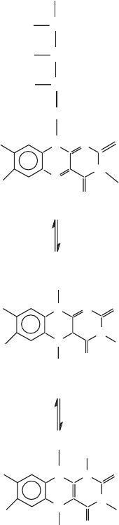
Figure 5.3 The structure of fl avin mononucleotide (FMN) showing the fl avin in the
fully oxidized form (top) and in the fully reduced form (bottom).
CH
2
OPO
3
C
C
C
CH
N
N
N
NCH
3
CH
3
O
O
H
H
H
HO
HO
HO
O
O
CH
3
CH
3
N
N
N
N
O
O
CH
3
CH
3 N
N
N
N
H
R
R
H
H
H
H
H
2
Many individuals have contributed to the precise defi nition of this electron
transport chain, and the reader is referred to the monograph of W. Wainio (6)
for an authoritative and detailed review of the literature up to about 1970.
Key contributions were many, but the experiments from the B. Chance labora-
tory using dual - beam, dual - wavelength spectrophotometers stand out. They
contributed to a further defi nition of the spectroscopic properties of the cyto-
chromes, and kinetic and inhibitor studies resolved the direction of electron
THE ELECTRON TRANSPORT CHAIN 177
fl ow, defi ned coupling sites, and further supported earlier thermodynamic cal-
culations by Ball and Lipman showing that the free energy change associated
with the oxidation of NADH is suffi cient for the generation of three ATPs.
Oxidation of succinate was predicted to yield only two ATPs. The enzyme
succinate dehydrogenase (SDH) was found to be the only enzyme of the Krebs
cycle which was insoluble and attached to the inner mitochondrial membrane,
coupling the Krebs cycle to the electron transport chain. Thus, succinate can
reduce the fl avin in SDH directly. The recognition of several distinct fl avopro-
teins was important, because the above simple scheme emphasizes the distinc-
tion between FMN and FAD.
The advent and application of more sophisticated spectroscopic techniques
in the 1950s and 1960s brought a fi nal major discovery about electron transport
with proteins. Electron spin resonance (ESR), now referred to as electron
paramagnetic resonance (EPR), which detects the spins of unpaired electrons
in a transition metal ion, was applied to whole heart tissue or mitochondria in
pioneering studies by H. Beinert (7) , leading to the discovery of a novel kind
of metalloprotein containing iron in a nonheme form. The iron is found in so -
called iron – sulfur clusters, and their diversity and abundance in the ETC was
at fi rst quite a surprise (7) . They had escaped detection because the optical
properties, measurable by spectrophotometry, were obscured by the strong
absorption of the cytochromes. The structures of many such clusters have since
been characterized (Figure 5.4 ). In addition to their role in electron transfer,
their structural versatility has been adapted in a large variety of proteins to
“ accept, donate, shift, and store electrons. ” Cluster construction as well as
cluster interconversions by ligand exchanges and oxidative degradation con-
stitute interesting biological reactions (8) . The past decade has seen a particu-
larly dramatic progress in our understanding of the biosynthesis of iron – sulfur
clusters (9 – 11) , and this subject will be reviewed in a separate section ( 6.8 ).
The iron is coordinated with two to four cysteine side chains from the
polypeptide, and in addition the cluster generally contains an equal number
of sulfi de ions (S
2 −
); these sulfi des are acid - labile and released at low pH as
hydrogen sulfi de (H
2
S), a biochemical diagnostic for the presence of such
clusters. In most clusters, each iron is coordinated to a total of four S atoms
in a roughly tetrahedral arrangement. The [Fe – S] cluster, with a single Fe
linked to four cysteine residues (and no sulfi de), has been found only in bac-
teria. Eukaryotic proteins have been found to contain the clusters [2Fe – 2S],
[3Fe – 4S], and [4Fe – 4S]. The iron atoms in each cluster form a conjugated
system, and instead of a single iron forming a Fe
2+
/Fe
3+
redox couple, the entire
cluster can lose or gain electrons. For example, the [4Fe – 4S] cluster contains
one Fe(II) and three Fe(III) ’ s in the oxidized form and two each of Fe(II) and
Fe(III) in the reduced form. In general the cluster can be represented by
[m Fe – n S]
x −
Each cluster has its characteristic redox potential, and with
unpaired electrons each cluster also has a characteristic EPR spectrum (when
x = 1 or x = 3). EPR can thus detect the gain or loss of electrons at each
cluster.
178 MITOCHONDRIAL ELECTRON TRANSFER
The presence of such clusters in the mitochondrial ETC provided another
mechanism for the passage of an electron through a protein. It should be
mentioned that Fe – S clusters were eventually also found in many proteins
unrelated to the mitochondrial electron transport chain. Not surprisingly, they
were found in proteins involved in photosynthesis and nitrogen fi xation, but
also in soluble proteins such as xanthine oxidase, ferredoxins and numerous
hydrogenases in bacteria. A particularly interesting example is the protein
binding to the iron - responsive element (IRE) in certain mRNAs. This protein
has been identifi ed as the cytosolic aconitase, and iron binding involves the
formation of an Fe – S center accompanied by a conformational change in the
protein. Disassembly of the center in the absence of iron leads to a conforma-
tion with high affi nity for the IRE. Not all Fe – S centers are devoted to electron
transfer, but they may also serve in the stabilization of the tertiary structure
of proteins. The literature on the subject has become vast, with whole books
on the subject appearing periodically. A very insightful and comprehensive
review on recent developments has been written by Beinert et al. (8) . In mito-
chondria, the next challenge was to localize them and place them in a position
relative to the fl avins, ubiquinones, and cytochromes. The original discoveries
(reviewed by Beinert (7) ) were made in the NADH and succinate dehydro-
genases (see below), and a third such center was found in what is now known
as Rieske ’ s protein in the cytochrome b - c
1
complex (complex III), named after
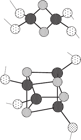
Figure 5.4 Structure of the [2Fe – 2S] and [4Fe – 4S] iron - sulfur clusters.
Fe
S
2-
S
2-
Fe
Cys
Cys
Cys
Cys
Cys
S
2-
S
2-
S
2-
S
2-
Fe
Fe
Fe
Fe
Cys
Cys
Cys
THE ELECTRON TRANSPORT CHAIN 179
the original discoverer (12) . More centers in mitochondria were discovered
when EPR spectroscopy was carried out at very low temperatures with a sig-
nifi cant gain in resolution, but their specifi c position within the overall ETC
could not be determined without further purifi cation.
The assembly of iron – sulfur centers in proteins was originally thought to
be a simple process, but research within the last decade has shown that it
depends on many proteins. These will be discussed in more detail in a separate
section (13) .
5.2.2 Physical Separation of the Complexes of the ETC
5.2.2.1 Biochemical Fractionations A hotbed of biochemical research on
the electron transport chain was the laboratory of D. Green at the Enzyme
Institute in Madison, from which many of the pioneers in the fi eld emerged in
the late 1950s and early 60s. The stage was set for the pioneering break-
throughs achieved by Y. Hatefi and his collaborators in the late 1960s (14 – 16) .
Starting with enormous quantities of beef heart, mitochondria were isolated
and purifi ed in correspondingly large amounts and subjected to systematic
solubilizations and fractionations (Figure 5.5 ). It should be recalled that our
concepts of membrane structure and membrane proteins were rudimentary at
that time. While other laboratories had attempted to isolate individual cyto-
chromes, Hatefi sought to isolate proteins or protein complexes which would
be functionally intact and capable of carrying out individual redox reactions
that had been associated with respiration. He exploited the observation that
coenzyme Q (ubiquinone; Figure 5.6 ) and cytochrome c could be easily and
reversibly removed from mitochondria by an appropriate solvent or salt
extraction, respectively, and the suggestion that these two compounds were
mobile electron carriers: Ubiquinone was found to act between complexes I
or II and III, and cytochrome c was found to act between complexes III and
IV. Based on these insights, assays for the function of each individual complex
could be developed. Complex I oxidized NADH with ubiquinone as electron
acceptor; the same electron acceptor was used in the oxidation of succinate
by complex II; a reduced complex III was able to donate electrons to cyto-
chrome c; reduced cytochrome c could be oxidized by complex IV with oxygen
as electron acceptor. Finally, complex V was assayed as an ATPase. The devised
scheme allowed the isolation of all fi ve complexes from the same batch of
mitochondria. For the fi rst time it became apparent that each complex con-
sisted of multiple peptides and required the presence of lipids for solubiliza-
tion and stabilization.
These isolation procedures provided relatively pure complexes with which
more detailed biochemical and biophysical analyses could be performed. The
earliest data were confi rmed and refi ned over the years, and there is no reason
to dwell on some of the uncertainties that were resolved with time. The most
challenging problem was to establish very precise stoichiometries in the various
complexes with respect to fl avin content (complexes I and II), iron content,
180 MITOCHONDRIAL ELECTRON TRANSFER
and acid - labile sulfi de (complexes I, II, and III). Another issue was to deter-
mine the number of polypeptides that were specifi cally associated with each
complex, and while this number was settled quite quickly for complexes II to
V, the peptide composition of complex I continued to be a signifi cant challenge
over the years. Initial estimates of over 10 polypeptides (14) were revised to
approximately 25 polypeptides based on two - dimensional gel electrophoresis
(17) , and the most recent analysis by high - resolution chromatography and
sequencing yielded at least 45 peptides (18, 19) . As discussed elsewhere in this
book, all complexes except complex II were shown to contain peptides made
in the mitochondrial matrix, and their number and identity were settled
after the complete human mtDNA sequence had been published and analyzed
(20, 21) .
In the early 1970s the mitochondrial electron transport chain in mammalian
mitochondria had taken on its present form which is schematically represented
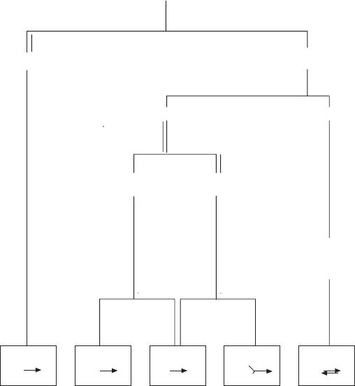
Figure 5.5 An outline of the purifi cation scheme devised by Hatefi to obtain all fi ve
complexes from the inner mitochondrial membrane of bovine heart. The complexes
are assayed and defi ned by the reactions shown in the boxes at the bottom.
IV
ATP Pi
NADH
Q
NADPH
Succ Q QH cyt c
2
cyt c O
2
MITOCHONDRIA
DOCA, KCl
DOCA, KCL,
ammonium
sulfate
DOCA, ammonium
acetate
cholate
ammonium
sulfate
Red
Green
S1
S1 sup
II - III
cholate
ammonium
sulfate
cholate
ammonium
sulfate
dialyze
spin
II III I V
I - III