Scheffler Immo E. Mitochondria
Подождите немного. Документ загружается.


MOLECULAR GENETICS OF HUMAN MITOCHONDRIAL DISEASES 361
tion immediately raises two questions, one less challenging, the other diffi cult
to answer at this time. How can such individuals survive? And how does
homoplasmy for the mutation come about?
The fi rst question can be answered theoretically and experimentally simply
by showing that the point mutation leads to a “ leaky ” phenotype, a protein or
a tRNA that is still partially functional. Another possibility is that the mutation
in a tRNA affects the rate or effi ciency of maturation of individual mRNAs
from the polycistronic transcript with no effect on tRNA function. While this
answer may satisfy temporarily, it immediately generates more questions,
especially in the case of homoplasmy. First, one of the outstanding and most
challenging questions in the whole fi eld is to explain why only certain tissues
are affected; in the most extreme case, why, for example, do LHON patients
suffer the central vision loss (although other symptoms have also been
observed)? Second, the optic atrophy (similar to other syndromes discussed
here) is delayed in onset that is variable among affected individuals in a pedi-
gree. Third, the severity of the vision loss is also variable. Finally, pedigrees
harboring the more severe mutations in a homoplasmic state can include
family members that are not affected at all.
The distinction between severe and mild mutations is in part based on an
assessment of the amino acid substitution and the effect it might have on the
residual activity of the peptide. A Leu - to - Pro change observed in one pedigree
with homoplasmy is expressed in 80% of the individuals of the family, presum-
ably because of a drastic alteration in the conformation of the peptide. Where
amino acids are conserved in evolution, functional or spatial restraints are
TABLE 7.1 Mitochondrial Diseases Resulting from Mutations in mtDNA
Clinical Description Inheritance
Ragged
Red
Fibers Defect in mtDNA
Kearns – Sayre
syndrome (KSS)
Sporadic + Single deletion or insertion
Incomplete KSS Sporadic + Single deletion
Pearson syndrome Sporadic + Single deletion or insertion
Leber ’ s hereditary
optic neuropathy
(LHON)
Maternal − Point mutation in ND4,
ND1, CYTB, COI
Neuropathy, ataxia,
retinitis pigmentosa
(NARP)
Maternal − Point mutation in ATPase 6
MELAS Maternal + Point mutation tRNA
Leu(UUR)
MERRF Maternal + Point mutation tRNA
Lys
MIMyCa Maternal + Point mutation tRNA
Leu(UUR)
Sensorineural hearing
loss
Maternal 12 S rRNA, tRNA
362 MITOCHONDRIAL MUTATIONS AND DISEASE
indicated, and changes at such positions are expected to be more serious.
Changes found in nonconserved regions of the peptide are diffi cult to evaluate
in the absence of a complete structure of the complex.
In more formal genetic terminology, one can speak of the expressivity of
the mutation in individuals within affected families and the penetrance of the
mutation in the general population. Penetrance is defi ned as the probability
of detecting a particular combination of genes (mutations) when they are
present (in the population or in a pedigree). This may depend on the criteria
and means of observation (measurement). In other words, the degree of pen-
etrance of a gene is a measure of the fraction of individuals possessing the
mutation and showing manifestations of the mutation. Expressivity refers to
the degree to which a particular phenotype due to a particular mutation mani-
fests its presence. “ Expressivity refers to the type of manifestation of a gene
that is penetrant ” ( 82a , page 290) . Both parameters are most likely dependent
on other genes, and in the present discussion the expression of mitochondrial
mutations will depend on a combination of nuclear genes, often referred to as
modifi er genes.
While the possibility of the existence of homoplasmy in viable individuals
can be explained on the basis of a leaky mutation, the development of homo-
plasmy for a mutation in a rare pedigree in the human population is more
diffi cult to explain. In almost all normal individuals, ∼ 99.9% of mtDNAs are
identical with no harmful mutations. In its simplest form the problem can be
stated as follows: A rare point mutation occurs in a mtDNA in germ - line cells
of a female. At the time of origin the mutated mtDNA is one among hundreds,
if not thousands, of normal mtDNAs. How many generations does it take to
accumulate a signifi cant fraction of mutated mtDNAs (heteroplasmy)? How
quickly is homoplasmy achieved?
There are indications that a signifi cant change in the mitochondrial geno-
type can occur within a few generations, or even a single generation, in
mammals. Hauswirth, Laipis, and colleagues (83, 84) have taken advantage of
a common polymorphism in a herd of Holstein cows to demonstrate that a
switch between alleles differing by point mutations was frequently almost
complete from one generation to the next. In other words, homoplasmy with
respect to a specifi c base defi ning the alleles was achieved in the change and
transmission from mother to offspring.
7.3.5 Mitochondria and Oogenesis
To escape from this conundrum, investigators have proposed the idea of a
“ bottleneck ” : a very small number of mtDNAs, or even a single mtDNA, serve
as templates for replication to populate the oocytes from which the mtDNA
is transmitted to the offspring (85 – 87) . A critical review has challenged the use
of the term “ bottleneck ” because it can be confusing in its meaning and has
been used inconsistently by different authors (88) . Instead, the term “ sampling
and amplifi cation ” has been suggested by these authors. They also point out
MOLECULAR GENETICS OF HUMAN MITOCHONDRIAL DISEASES 363
that while either random drift or a complex sampling process can readily
explain a rapid segregation of mitochondrial genomes, the real puzzle is the
stable heteroplasmy observed in some pedigrees. A female with a relatively
small fraction of mutated mtDNAs can have offspring with a signifi cantly
higher proportion of mutated mtDNAs, if the mutated DNA is preferentially
amplifi ed. But unless there is bias in the sampling, in the majority of her off-
spring the heteroplasmy should be reduced.
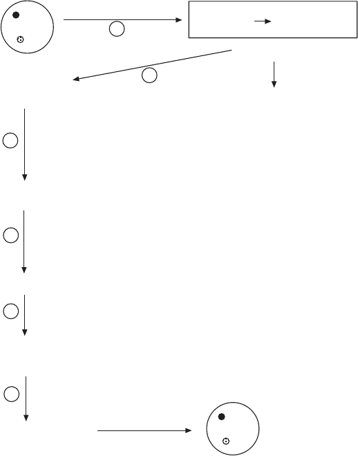
At what stage in gametogenesis or embryogenesis is the sampling and
amplifi cation mechanism operational? A priori , several stages could be con-
sidered (Figure 7.1 ). One should be aware that the human egg has an estimated
200,000 mtDNAs (77) and that as much as one - third of the total DNA of an
oocyte is mtDNA. There is certainly no “ bottleneck ” at this level, in the sense
that only a small number of mitochondria are transmitted by the oocyte. After
fertilization and prior to implantation, a number of rapid zygotic (mitotic)
divisions occur (step 1 in Figure 8.1 ), apparently without any further mtDNA
replication, in which the pool of oocyte mitochondria is distributed between
the daughter cells and the number of mtDNAs per cell is reduced to the
normal range of ∼ 1000 or less (89) . At a relatively early stage in development
a primordial germ cell lineage is set aside (step 2) to migrate to the ovary, and
within the ovary a subpopulation of diploid cells (oogonia) is formed by
repeated mitotic divisions (step 3). The oogonia increase signifi cantly in size,
and a multiplication in the number of mitochondria could accompany this
growth (step 4). Meiosis and maturation leads to one ovum (and polar bodies)
from one oogonium. (It should be noted that in mammals, meiosis I is arrested,
possibly for years, and completed only at periodic intervals when eggs mature.
Furthermore, meiosis II is subsequently arrested and completed only if fertil-
ization by a mature sperm takes place.) It has also been suggested that a series
of zygotic divisions with nuclear but no mtDNA replication could reduce the
number of mtDNAs to a much smaller number ( “ the bottleneck ” ), accompa-
nied by large stochastic variations in mitochondrial genotype between differ-
ent cells. In this hypothesis, segregation is a postzygotic event. Thus, after 12 – 15
zygotic divisions without mtDNA replication, oogonia could have mtDNAs
numbering in the range of 50 – 56 . If the subsequent amplifi cation of mtDNA
molecules during the maturation of the primary oocyte from an oogonial
precursor cell started from such a small population, or it involved only a small
subpopulation of a larger number of mtDNA templates ( “ sampling and ampli-
fi cation ” ), shifts in the distribution of “ alleles ” could be achieved quite
rapidly.
To restate the two plausible hypotheses, sampling and amplifi cation could
occur in the process of formation of an oocyte from an oogonial cell, or a
reduction in mtDNA copy number could occur by simple dilution during the
formation of the germ cell lineage in the early embryo. A third hypothesis has
proposed that the sampling may occur during a series of mitotic divisions
leading to a large population of oogonia (step 3). Finally, it is conceivable that
the partitioning of the embryonic cells between the inner cell mass and the
364 MITOCHONDRIAL MUTATIONS AND DISEASE
zygotic divisions
cleavage
blastula
gastrulation
somatic cells
primordial germ cells
migration to the
ovary
oogonia
primary oocyte
secondary oocyte
+ 1st polar body
ovum
+ 2nd polar body
multiple mitotic
divisions
growth of oogonia
large increase
in cell volume
increase in number
of mitochondria
meiosis I
unequal cell division
meiosis II
5
6
4
3
2
1
Fertilization
2 Pro-nuclei
Fertilized
Egg
A
Figure 7.1 (A) Mitochondrial DNA amplifi cation and relationship to oogenesis. The
fertilized egg contains an estimated 100,000 mtDNAs. These may be distributed during
the early zygotic divisions (step 1) to yield cells with considerably smaller populations
of mtDNA. Early in development, germ cells are differentiated from somatic cells (step
2). A very limited number of mtDNAs could be replicated during step 3 (expansion of
the precursor pool of oogonia by mitotic divisions), or in step 4 when oogonia increase
in size and prepare for meiosis. (B) A model for the origin of the bottle neck based on
the assumption that the fi rst n zygotic divisions occur without any mtDNA replication,
diluting the population of mtDNA molecules to a small number per cell at the time
when germ line cells are set aside. This small number then expands again during meiosis
and formation of the mature oocyte.
MOLECULAR GENETICS OF HUMAN MITOCHONDRIAL DISEASES 365
extraembryonic tissues is accompanied by an unequal partitioning of mtDNAs
which could be responsible for the segregation and reduction of mtDNA
genotypes in the ICM. A review by Chinnery et al. (90) presents these hypoth-
eses with more references to the original literature, and it also illuminates
some interesting differences in oogenesis between mice and humans. They
illustrate that precise quantitative predictions for genetic counseling based on
mouse models may be diffi cult.
Several experimental systems have been developed in recent years to
achieve “ cracks in the bottleneck ” (85) Heteroplasmic mouse strains have now
been created to be used to understand the mechanisms for segregation of
genetically marked mtDNAs (48) (49, 91, 92) . Advances in the manipulation
of mouse embryos have been instrumental in creating the kind of experimental
conditions allowing a direct observation of the segregation of mtDNAs in
heteroplasmic mice. Jenuth et al. (93) produced mouse embryos by the fusion
of cytoplasts containing mitochondria of one haplotype with one - cell embryos
of a strain with another mitochondrial haplotype. For simplicity the mtDNA
genotypes will be referred to as NZB and BALB, respectively. The cytoplasts
were obtained from one cell embryos by using a glass pipette to draw up and
pinch off cytoplasm surrounded by plasma membrane. These were injected
under the zona pellucida of recipient one - cell embryos and electrofused.
Transplantation into pseudopregnant females produced female and male
fertilized egg (zygote)
100-500,000 mtDNA
(
of maternal origin)
n zygotic divisions
with no
mtDNA replication
1 - 5 x 10
5
2
n
cells
= m = mtDNAs/cell
somatic cell
lineages
early germ line cells
large pool of oogonia
oocyte
1 - 5 x 10
5
mtDNAs/cell
expansion of mtDNA
pool from m to > 100,000
B
Figure 7.1 (Continued)
366 MITOCHONDRIAL MUTATIONS AND DISEASE
animals whose mtDNA could be analyzed from tail biopsies. Five founder
females were produced with 3.1 – 7.1% of the donor mtDNA genotype. When
a female with 5% donor (NZB) mtDNA was crossed to a BALB male, 28
offspring were analyzed and found to contain, on average, 6.7% NZB mtDNA.
However, in individual offspring the proportion of donor (NZB) mtDNA
varied from 0% to 29.6%. A rigorous statistical analysis of 191 F
1
offspring
from all fi ve crosses, along with 176 offspring from several more back - crosses
with F
1
females, indicated a large variance of haplotype frequencies. It was
concluded that the results supported the idea that germ - line segregation is a
stochastic process capable of producing signifi cant fl uctuations from an origi-
nal distribution of genotypes.
In mice approximately 50 primordial germ cells can be identifi ed in 7.5 - day
mouse embryos. When several (3 – 12) such primordial germ cells from a given
embryo were analyzed, the coeffi cient of variation was very small, suggesting
that a given proportion of donor mtDNAs received in the zygote was main-
tained during the earliest stages up to the establishment of the germ cell
lineage. These primordial germ cells, when properly located on the genital
ridge, divide and give rise to oogonial cells, from which approximately 25,000
primary oocytes are derived in a series of synchronous divisions. Based on
these experimental results, Jenuth et al. (93) propose that segregation of
mtDNA variants must occur during the mitotic cell divisions that precede
meiosis in the ovary.
At this point, one can only speculate about details of the process (86, 93) .
Several aspects of mtDNA replication must be considered (see Chapter 4 ).
Unlike the case of the nuclear genome, mtDNA replication is relaxed; that is,
in a given population of mtDNAs, some may replicate multiple times during
the cell cycle, while others do not replicate at all. This still leaves the question,
How many tDNA molecules are selected for replication, and is there any kind
of selection for a subpopulation? There is evidence that localization within a
cell can infl uence mtDNA replication, with proximity to the nucleus being a
determining parameter (94) . Employing mathematical modeling using popula-
tion genetic theory, along with assuming that segregation is the result of
genetic drift, one can calculate the effective number of segregating units, N ,
from the observed variance in the nth generation, if the initial distribution of
genomes is known. With an estimated 15 cell divisions required to produce the
total population of primary oocytes in the mouse ovary, Jenuth et al. (93) cal-
culated that N is in the range of 200.
7.3.6 Clinical Aspects of Mitochondrial DNA Mutations
In humans, the distinction between variants with point mutations and variants
with large deletions is an important one. The fact that changes are occurring
post - fertilization is clearly indicated by the differences in heteroplasmy
observed between muscle tissue and blood cells in patients with mtDNA dele-
tions. Homoplasmic cells with deleted mtDNA would not be expected to be
MOLECULAR GENETICS OF HUMAN MITOCHONDRIAL DISEASES 367
viable. Whatever the starting position in the fertilized egg (zygote), an unre-
solved issue is whether there is genetic selection in somatic cells in specifi c
tissues, or simply random genetic drift due to relaxed mtDNA replication.
7.3.6.1 MtDNA Deletions: Kearns –Sayre Syndrome and Pearson Syn-
drome It need not be stressed that homoplasmic null mutations would be
lethal. Two distinct mechanisms can lead to partial defi ciency. In heteroplas-
mic cells the most severe mutation, a complete deletion of one or more genes,
can be partially compensated by the presence of normal mitochondrial
genomes. In homoplasmic cells a mutation must be less severe — for example,
by causing a missense substitution in a protein which reduces its activity. In
either case, one expects the tissue to suffer from an inadequate production of
ATP by oxidative phosphorylation; and one may now ask, What is it that dis-
tinguishes the various “ mitochondrial diseases ” from each other. A priori , one
would expect the severity of the disease to depend on the degree to which a
particular function is compromised. In the same vein, one would expect the
defi ciency to be most deleterious in tissues such as the brain and muscle which
are energetically the most demanding. In practice, the situation is much more
complicated in ways that continue to challenge our understanding of the basic
phenomena.
Large - scale rearrangements or deletions of mtDNA are associated with two
clinical conditions that share overlapping symptoms. The fi rst, recognized by
Kearns and Sayre in 1958 (95) includes among the symptoms retinitis pigmen-
tosa, external ophthalmoplegia, a complete heart block. Morphological altera-
tions in the brain were also noted at autopsy, and these have since been
confi rmed as the rule rather than an exception or artifact. The disease is char-
acterized by a childhood onset, progressive ophthalmoparesis, pigmentary
retinopathy, increased propensity of complete heart block (which could lead
to sudden death), and neurological symptoms including incoordination, mental
retardation, and episodic coma. Growth retardation and hearing loss are
common, and ragged red fi bers are found in almost all affected patients. An
unbalanced and insuffi cient energy metabolism is indicated by elevated lactate
and pyruvate levels in plasma. Named after its original discoverers, the syn-
drome is now referred to as the Kearns – Sayre syndrome (KSS). As explained
earlier, the occurrence of KSS is sporadic not familial, tissues are heteroplas-
mic with respect to wild type and rearranged mtDNA, and both sexes are
affected at about equal frequencies.
A second syndrome named after its discoverer is Pearson syndrome (96) .
The affected infants had pancytopenia and altered pancreatic exocrine func-
tion, and it was fatal in some cases, but others survived into adulthood and
developed clinical features of KSS. When molecular genetic analyses became
possible, lesions in mtDNA were found very similar to those found in KSS.
How is this syndrome survivable, only to be converted into the second severe
condition? Speculation centers around cell population dynamics in the bone
marrow, where cells with the greater proportion of mutated mtDNAs have a
368 MITOCHONDRIAL MUTATIONS AND DISEASE
disadvantage during rapid cell divisions and may be eliminated; that is, in this
tissue there may be a selection for cells with the highest proportion of normal
mtDNAs. At the same time, in tissues with nondividing cells like muscle or the
brain there may be a selection at the level of mtDNA replication which favors
the deleted mtDNAs, and ultimately an oxidative phosphorylation defi ciency
is developed concomitant with encephalomyopathy. Patients diagnosed in
infancy with Pearson syndrome do not have ragged red fi bers, whereas muscle
biopsies showing an abundance of ragged red fi bers also show deleted mtDNAs
in excess.
Patients with Pearson syndrome and those KSS patients with the most
severe symptoms have detectable heteroplasmy not only in muscle, but also
in fi broblasts and leukocytes, tissues easily available from biopsies. At autopsy,
brain and liver tissue (and other tissues) become available, with the brain
showing pronounced heteroplasmy or an abundance of mutated mtDNAs,
while liver may have a smaller proportion. On the one hand, the ubiquitous
presence of mutated mtDNA is an indication that the mutation was present
in the oocyte before fertilization, but the variability in the ratio of normal to
mutated DNA in different tissues is indicative of perhaps two selection mecha-
nisms based on (1) the fi tness of individual cells in rapidly dividing or renewing
tissue (blood, liver), and the replicative advantage of mutated mtDNA mole-
cules in nondividing tissue. As a result, these distributions may vary between
patients and, most signifi cantly, may shift with age. Delayed onset and variable
expressivity may be explained quite satisfactorily in general terms, but more
defi nite predictions at the time of the fi rst diagnosis may not be possible. Even
a determination of the proportion of mutated DNA in a muscle biopsy (which
muscle?) may not have any predictive value, since there is no correlation
between this fraction and the clinical severity.
It is commonly assumed that there are several mtDNAs per mitochondrion,
and that intra - mitochondrial complementation occurs. Continuous fi ssion and
fusion of mitochondria would be expected to contribute to a mixing of the two
genomes. However, such a picture, derived from observations in tissue culture,
may be misleading in tissues such as muscle, where the highly structured cyto-
plasm and the cytoskeleton (sarcomeres) may prevent extensive diffusion and
mixing of mitochondria.
The severity and the precise combination of symptoms may also be depen-
dent on the nature of the rearrangement and the specifi c segment of mtDNA
missing in heteroplasmic cells. Nevertheless, for reasons explained above,
patients with identical deletions may exhibit different clinical abnormalities.
Almost any deletion of some length will cause the loss of several genes, includ-
ing structural genes encoding essential subunits for complexes I, II, and IV,
alone or in combination, and almost always the loss of at least one tRNA gene.
Thus, very specifi c predictable biochemical activities will be compromised, but
the loss of one or more tRNA genes will also diminish the capacity for mito-
chondrial protein synthesis, and hence the level of all complexes of the elec-
tron transport chain may be affected. A more detailed discussion of several
MOLECULAR GENETICS OF HUMAN MITOCHONDRIAL DISEASES 369
specifi c cases can be found in the review of Harding (73) . Superimposed on
the effects of the specifi c deletions are (a) mechanisms of post - transcriptional
regulation such as mRNA stability and the control of steady - state levels of
individual mRNAs and (b) translational control mechanisms (initiation?) that
may infl uence the half - life of an mRNA. If such mechanisms are tissue - specifi c,
a simple consideration of heteroplasmy and gene dose is defi nitely not suffi -
cient to predict the full scope of clinical symptoms and their severity.
7.3.6.2 Familial Mitochondrial DNA Depletion A rare disorder with
an inevitably fatal outcome in early childhood has been studied in several
families. Healthy parents have produced children suffering a severe depletion
of mtDNA in specifi c tissues, most frequently liver or muscle. First recognized
by Moraes et al. (97) as a novel genetic abnormality, the defect appears to
reside in a nuclear gene, since no mitochondrial mutations could be
detected.
7.3.6.3 Point Mutations Table 7.1 lists fi ve distinct clinical conditions
resulting from point mutations in mtDNA which are maternally inherited.
They are: LHON, NARP, MELAS, MERRF, MIMyCa, and sensorineural
hearing loss. A genetically interesting and challenging feature of these diseases
is the frequent fi nding of homoplasmy (see discussion in Section 7.3.5 ); and a
perhaps oversimplifi ed aspect of each mutation is that it is “ leaky, ” which in
biochemical terms implies that some function is retained. A detailed con-
sideration of each condition and patients having the mutation leave many
other questions still unanswered.
7.3.6.2.1 Leber ’ s Hereditary Optical Neuropathy (LHON) Leber ’ s heredi-
tary optic neuropathy (LHON) has as the defi ning feature the sudden onset
of bilateral central vision loss. The condition was fi rst described by Leber in
1871. Onset is usually delayed until young adulthood (20 – 24 years of age), but
may occur as early as age 5. Although bilateral, vision may be affected fi rst in
one eye, followed by involvement of the other eye weeks or months later.
Individuals are generally healthy, although the mutation is found in all tissues.
Detailed histological and anatomical studies have established the specifi c
degeneration of the ganglion cell layer and the optic nerve. Although vision
loss is permanent in most cases, a number of LHON patients have shown
improvement, sometimes relatively soon after the diagnosis, sometimes after
several years. Disedema and subtle alterations in retinal vessels are part of the
diagnosis, but curiously, the clinical condition is not followed rapidly by the
appearance of other neurologic abnormalities, although patients (and family
members) have exhibited some or all of the following: hyperrefl exia, Babinski
signs, incoordination, peripheral neuropathy, and cardiac conduction abnor-
malities. How this highly restricted pattern of degeneration of the optic nerve
is initiated is still a matter of debate, which has recently been summarized by
Howell (98) . The pathogenesis of LHON is not yet well understood. A detailed
370 MITOCHONDRIAL MUTATIONS AND DISEASE
discussion requires the description of the microanatomy of the optic nerve
head, the region around the lamina cribosa, and the possibility of a “ choke-
point ” in the optic nerve where axoplasmic transport of mitochondria may be
impaired. The resulting swelling may accentuate ischemia and ultimately neu-
rodegeneration. One plausible model is that increased or altered mitochon-
drial ROS production renders the retinal ganglion cells vulnerable to apoptotic
cell death (99) . Patients do not have ragged red fi bers in muscle, and lactate
levels in blood and cerebrospinal fl uid are normal.
Members of some families, predominantly females, have been reported to
be at risk for developing symptoms resembling multiple sclerosis (MS). The
risk of LHON is inherited from the mother, typical of mitochondrial diseases,
and the mother may or may not have been affected. The primary mutation
(see below) in most LHON patients is homoplasmic, but patients with hetero-
plasmy have also been found. There is incomplete penetrance even in the
homoplasmic population, and the current view is that the primary mutation
predisposes toward LHON, but secondary genetic and environmental etiologi-
cal factors contribute to the development of the disease (98, 100) . Eighty - fi ve
percent or more of the patients are male (100) . Historically, this fact was
thought to indicate X - linkage, with the appearance in a larger - than - expected
fraction of females requiring an explanation. Today the maternal inheritance
is fi rmly established, challenging us to explain what genetic factor(s) contrib-
ute to the biased expression of this phenotype. An X - linked susceptibility locus
has been proposed to play a role. However, while the issue has not been
defi nitively resolved, recent studies have failed to obtain evidence for such a
locus on the X chromosome. On the other hand, three X - linked genes required
for complex I biogenesis have been identifi ed (36) .
In their review, Brown and Wallace (101) have described 16 point mutations
associated with LHON, in subunits ND1, ND2, ND4, and ND6 of complex I,
in CYT b, and in the CO1 subunit of complex IV. These authors express the
view is that mtDNA LHON mutations “ represent a continuum of severity of
OXPHOS defects with each imparting a proportional increased risk for
LHON. ” The issue is controversial, and Howell (100) emphasizes that the vast
majority of LHON families have one of three primary pathogenic mutations
in mtDNA at nucleotides 3460 (ND1 gene), 11778 (ND4 gene), and 14484
(ND6 gene). The most abundant mutation in the ND4 gene at nucleotide
11778 converts a highly conserved arginine to a histidine. Other, less common
mitochondrial mutations may have a secondary etiological role, or they may
appear in rare “ nonclassical ” LHON families suffering from optic neuropathy
and additional neurological abnormalities. Another perspective is that mild
“ LHON mutations ” may act synergistically. This is deduced from the fi nding
that at least four “ LHON mutations ” by themselves do not cause loss of vision,
but in combination with other “ LHON mutations ” the phenotype is expressed
in almost 100% of individuals carrying the two mutations. The probability of
such double mutants would at fi rst seem extremely small, until it is realized
that some of these mutations are present in a substantial fraction of the