Scheffler Immo E. Mitochondria
Подождите немного. Документ загружается.

yielding highly purifi ed, intact mitochondria was an important contribution.
Such isolated organelles could be viewed by electron microscopy and shown
to have a morphology similar to those viewed in situ .
A plausible explanation made already at the time was that this topology
vastly increased the surface area of the inner membrane. The full signifi cance
of the compartmentalization into an intermembrane space and a matrix could
not be fully appreciated until later, but in this chapter, attention will be largely
confi ned to a discussion of observable morphology of individual mitochondria.
It is evident that the size and shape of mitochondria seen in the electron
microscope depends on the plane of sectioning relative to their long axis.
As more examples of mitochondria in different tissues were examined, dis-
tinguishing features soon became apparent. The matrix was not homogeneous
and exhibited a “ fi ne granularity, ” and frequently there were distinct crystalline
inclusions or small particles of high electron density (Figure 3.3 ). Their number
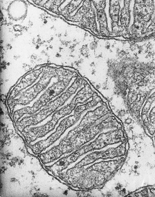
Figure 3.1 Mitochondrion from centroacinar cell of pancreas ( × 190,000). (Courtesy
of Dr. G. Palade.)
STRUCTURE AND MORPHOLOGY 21
22 STRUCTURE AND MORPHOLOGY. INTEGRATION INTO THE CELL
could in some instances be shown depend on the metabolic state of the tissue
observed. The high electron density may be the result of a sequestration and
storage of calcium. Other studies have suggested that the electron density is the
consequence of the retention of osmium tetroxide by a phospholipoprotein
that may bind calcium in vitro , but may not have such a role in vivo .
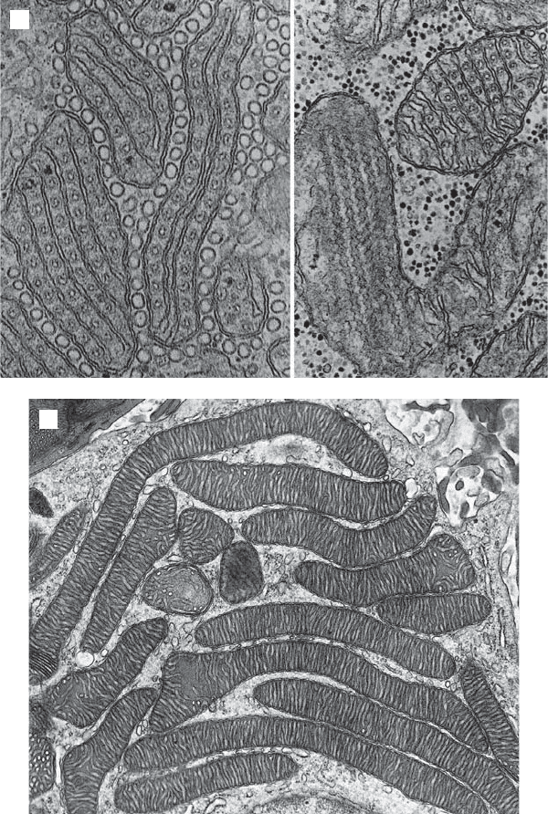
Most notable was the variable appearance of the cristae (Figure 3.4 ). They
were often lamellar in appearance, but also tube - like in other cells, and a
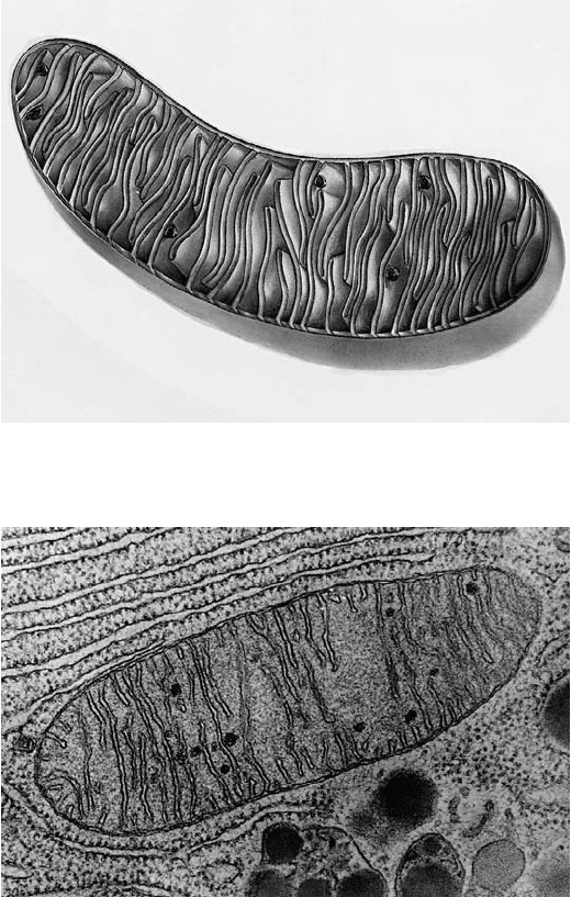
Figure 3.2 Schematic view of mitochondrion showing the outer and inner membranes
and the folded cristae. (Drawing provided by Dr. D. Fawcett.)
Figure 3.3 Mitochondrion showing electron - dense inclusions. (Original photograph
of K. Porter; from The Cell , by D. W. Fawcett, 1981, W. B. Saunders Co.)
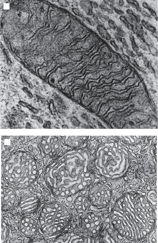
Figure 3.4 (A) Mitochondria with angular cristae from ventricular cardiac muscle.
(Original photograph of J. P. Revel; from The Cell , by D. W. Fawcett, 1981, W. B. Saun-
ders Co.) (B) Mitochondria with tubular cristae in amoeba. (Original photograph of
T. Pollard; from The Cell , by D. W. Fawcett, 1981, W. B. Saunders Co.) (C) Mitochondria
from adrenal cortex. (Original photograph of D. Friend; from The Cell , by D. W.
Fawcett, 1981, W. B. Saunders Co.) (D) Mitochondrion from astrocyte. (Original pho-
tograph of K. Blinziger; from The Cell , by D. W. Fawcett, 1981, W. B. Saunders Co.) (E)
Mitochondria from fi sh pseudobranch. (Original photograph of J. Harb and I. Cope-
land; from The Cell , by D. W. Fawcett, 1981, W. B. Saunders Co.) (F) Mitochondria from
adrenal cortex. (Steroid secreting cells). (Original photograph of D. Fawcett; from The
Cell , by D. W. Fawcett, 1981, W. B. Saunders Co.)
A
B
STRUCTURE AND MORPHOLOGY
23
24 STRUCTURE AND MORPHOLOGY. INTEGRATION INTO THE CELL
D
C
Figure 3.4 (Continued)
complete description required many images, and ideally, serial sections. Thin
sections in the electron microscope still require an interpretation in three
dimensions, and frequently the continuity of a membrane is not seen in a given
section. Thus, tubes become “ free ” circular (vesicular) membranes in cross
section, and sheet - like foldings of the inner membrane in some sections may
appear as fl attened sacs fl oating in the matrix. In the current view the inner
membrane is continuous, and there appears to be no functional reason to have
membrane vesicles within the matrix analogous to the thylakoid membranes
(grana) of chloroplasts.
The interpretation of cristae topology from thin - section electron micro-
scopy by Palade and Sjostrand postulated that the cristae are sheet - like
invaginations of the inner membrane into the matrix of the mitochondrion
(Figure 3.2 ). A maximum number of cristae can be accommodated if the sheet -
like membranes are arranged in parallel, but it seems that the models fudge
E
F
Figure 3.4 (Continued)
STRUCTURE AND MORPHOLOGY 25
26 STRUCTURE AND MORPHOLOGY. INTEGRATION INTO THE CELL
the issue of what happens to the image if a different plane of sectioning were
presented. The T - shaped junction cannot be continuous around the entire cir-
cumference of the mitochondrion. Recent advances in electron tomography
have made it possible to view complex 3 - D objects like mitochondria in thick
sections and reconstruct a high - resolution 3 - D image. Data are collected by
recording 2 - D images over a wide range of tilt angles, and 3 - D reconstructions
are achieved by computer - assisted imaging. A 3 - D resolution in the range of
5 – 10 nm can be achieved, hence images of the outer and inner membranes of
mitochondria can be viewed at a hitherto unobtainable resolution (5, 6) . Only
a limited number of tissues have been examined by this powerful methodology
(Figure 3.5A,B ).
Of particular interest and signifi cance is the conclusion by Perkins et al. (5)
that cristae consist of a lamellar portion similar to that envisioned before, but
the continuity with the inner boundary membrane (which lies close to the
outer membrane) is achieved by a relatively small number of narrow tube - like
connections. In other words, though continuous, the inner membrane juxta-
posed to the outer membrane and the inner membrane of the lamellar struc-
tures are connected by a narrow “ bottleneck ” referred to as a cristae junction.
Topologically, the intermembrane space is still continuous with the “ inside ” of
the lamellae, but the authors raise the question whether the bottleneck can
constitute a barrier and hence serve to separate two compartments: intracristal
and intermembrane. It may also serve to segregate patches of inner membrane
with differing protein compositions. Studies exploring the implications of these
morphological observations have been challenging (7 – 12) . The tubular struc-
ture of the cristae junctions requires a mechanistic explanation. On the one
hand, theoretical models have considered the local composition of the lipid
bilayer and the packaging of lipids to create the required curvature (13, 14) .
Other speculations have centered on specialized proteins required for main-
taining cristae junctions. Attempts to purify cristae junctions have not yet met
with success. More recently, several proteins have been identifi ed and impli-
cated in the control of cristae formation, but their relationship to cristae junc-
tions is unclear. The inner - membrane protein mitofi lin is a candidate requiring
serious attention (15) . Mitofi lin is localized in the space between the inner
boundary membrane and the outer membranes, where it can assemble into a
large multimeric protein complex. A perhaps more surprising development is
the discovery in yeast that the ATP synthase (complex V), and specifi cally the
subunits e and g play a role in generating mitochondrial cristae morphology.
The dimerization or oligomerization of the ATP synthase appears to be a
critical step in this mechanism (16 – 18) . The appearance of the cristae and
cristae junctions has been shown to depend on the conditions under which the
mitochondria are prepared for tomography (12) . They are likely to be highly
dynamic in vivo , responding to changes in the metabolic state, ADP/ATP
ratios, ion fl uxes, and matrix swelling. There is evidence that in some but not
all cells, inner membranes are being remodeled during apoptosis, possibly as
part of a mechanism for mobilizing cytochrome c release (19, 20) .
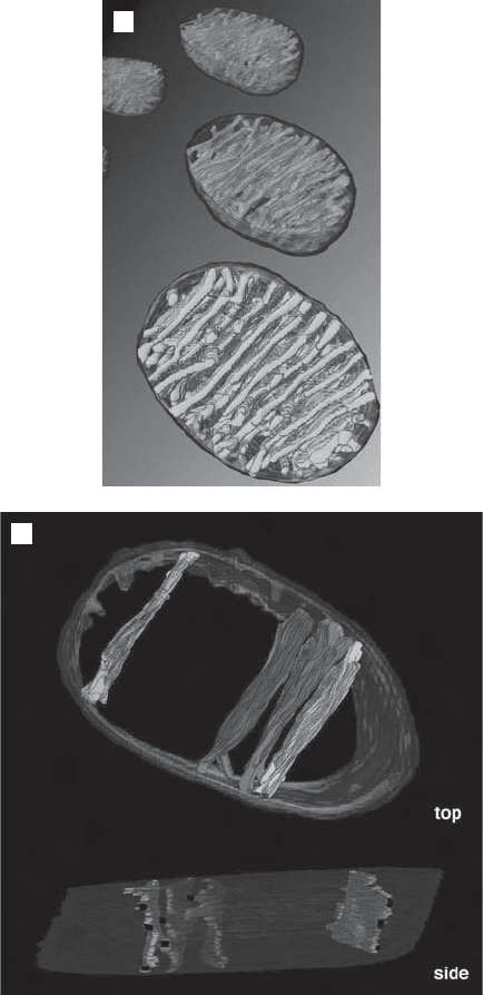
Figure 3.5 (A) Mitochondria seen by electron microscope tomography. Cells were
derived from chick CNS. [The original fi gure was provided by G. Perkins, University
of California, San Diego; with permission from Academic Press (Perkins et al. (5) ).]
(B) The 3 - D reconstruction was achieved after the volume was segmented manually
contouring into the regions bounded by the outer, inner, and cristal membranes. [For
details the reader is referred to the original publication (Perkins et al. (5) .] In the
bottom of the fi gure, individual lamellar sections of crista are shown, together with
the tubular portions which connect the disks to the inner boundary membrane. (The
photographs were generously provided by Dr. G. Perkins, University of California, San
Diego; with permission by Academic Press.) See color plates.
A
B
STRUCTURE AND MORPHOLOGY
27
28 STRUCTURE AND MORPHOLOGY. INTEGRATION INTO THE CELL
The suggestion has been made that cristae junctions represent diffusional
barriers between the IMS and the cristal compartment. If the barrier were
complete, a great similarity to chloroplasts would emerge, where the thylakoid
membranes enclose a compartment distinct from the space between the inner
and outer membranes. Such a view might also imply a difference in the
biochemical composition of different domains of the inner membrane. At the
moment, such speculations have limited experimental support, but a promising
beginning has been made by Vogel et al. (21) . These authors show that proteins
are distributed in an uneven, yet not exclusive, manner between the inner
boundary membrane and cristae membranes.
The number and morphology of cristae are likely to refl ect the response of
the mitochondria to the energy demands of the cell. Highly folded, lamellar
cristae with a large surface area are typically found in muscle cells and neurons,
where the respiratory rate is the greatest. These observations clearly raise
questions about the control of cristae formation and the expression of both
nuclear and mitochondrial genes encoding mitochondrial proteins. A telling
example, although perhaps an extreme one, comes from the study of mito-
chondrial morphology in the yeast Saccharomyces cerevisiae . When grown in
the presence of a nonfermentable substrate such as glycerol or ethanol, the
cells are dependent on oxidative phosphorylation, and hence they have mito-
chondria easily recognizable even by the non - expert. When grown in the pres-
ence of glucose, the biogenesis of the inner membrane is severely repressed;
and the morphology, but not the number of the mitochondria, changes dra-
matically. The details of the mechanism of “ glucose repression ” of respiration
will be presented in a later chapter. A similar change in the abundance and
morphology of an intracellular organelle can be observed for peroxisomes —
for example, in the yeast Piccia pastoris , where growth on methanol or fatty
acids induces the appearance of large peroxisomes. Free - living microorgan-
isms are exquisitely attuned to their environment and capable of adjusting and
optimizing their metabolism to the ever - changing conditions.
The question about the number and shape of mitochondria in yeast,
Saccharomyces cerevisiae , took a different turn when a series of serial sections
were analyzed to reveal that in the extreme there may be only one mitochon-
drion per cell, existing as a highly branched, continuous structure. A single
section alone will show what appear to be discrete mitochondria with spherical
or elongated shapes (22, 23) . (See Section 3.2 for further details.)
In multicellular organisms, mitochondria are likely to have a more invari-
able morphology in a given tissue, but different tissues have distinct energy
needs. It may well be that an altered mitochondrial morphology is also related
to functions of mitochondria not strictly linked to respiration. Steroid secreting
cells such as Leydig cells have mitochondria with an unusually wide range of
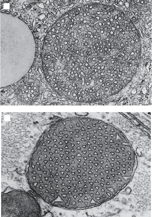
sizes and often a very distinct array of tubular cristae (24) . Perhaps the most
puzzling display of cristae morphology is one that occurs sporadically (?) in
many tissues of diverse species. These cristae appear to be made of parallel,
hexagonal arrays of tubules with a triangular cross section. This may be an
extreme manifestation of cristae with sharp angulations regularly spaced
along their length, which are found in mitochondria of cardiac cells, among
others (24) .
It is evident that the more subtle aspects of the variations in cristae mor-
phology can generally not be explained in terms of some sweeping generaliza-
tion about surface area and respiratory activity. The inner mitochondrial
membrane is an exceptional membrane with respect to the protein : lipid ratio.
While in most membranes this ratio is close to 50 : 50, conducive to the image
of “ proteins fl oating in a sea of lipids, ” the inner mitochondrial membrane has
a ratio of ∼ 75 : 25, suggestive of relatively densely packed proteins. Researchers
focused on the inner mitochondrial membrane were among the most reluctant
in their acceptance of the “ fl uid mosaic model ” as a model for all biological
membranes (J. Singer, personal communication). However, numerous studies
have provided strong experimental support for the view that the inner mem-
brane is in a fl uid state rather than a solid state (for a review see references
25 and 26 ). Thus, the proteins of this membrane cannot be exclusively respon-
sible for its folding. Note, however, the role of ATP synthase oligomerization
mentioned above (17) . The discovery of supercomplexes formed by complexes
of the electron transport chain (see Section 5.2 ) also requires considerable
rethinking about the complexes fl oating in a sea of lipids.
If the outer membrane has a fi xed surface area and hence can confi ne the
contents to a fi xed volume, the inner membrane with a larger surface area
must necessarily fold to fi t into the available volume. Some order to this
folding may be provided by the periodic attachment of the inner membrane
to the outer membrane. Such rivet - like attachment points have been observed,
and there is now agreement that such attachment points may be made up of
the components that are responsible for the import of peptides made in the
cytosol. They may be formed transiently by the interaction of integral mem-
brane proteins in the outer membrane with others in the inner membrane, thus
aligning two channels through both membranes. Further details on such a
proposed structure and its function in protein import will be presented in a
later chapter.
Formation and maintenance of tubular cristae or cristae with a triangular
cross section presents a greater challenge to the imagination. Theoretical
studies on the conformation of membranes can lead to stimulating specula-
tions (27) , but experimental verifi cation is still a major unachieved goal. The
morphology of membranes such as the plasma membrane and the Golgi mem-
branes is demonstrably determined by interactions with cytoskeletal elements,
but there is no convincing evidence for the existence of such structures within
mitochondria. Nevertheless, our view of the mitochondrial matrix as an amor-
phous soup of soluble enzymes is most likely too simple - minded. Mitofi lin in
the IMS may represent the fi rst in a number of structural proteins required
for organizing the cristae (15) .
In summary, the variation in mitochondrial morphology has been abun-
dantly documented. Three - dimensional reconstructions from serial sections
STRUCTURE AND MORPHOLOGY 29
30 STRUCTURE AND MORPHOLOGY. INTEGRATION INTO THE CELL
and more recently by confocal microscopy (even in live yeast cells) have
removed some of the last ambiguities of interpretation of thin sections used
in electron microscopy. A general correlation of the total area of the inner
membrane (density of cristae) with the capacity for oxidative phosphorylation
is apparent, but some of the more detailed aspects of cristae morphology in
relation to function and activity are still relatively obscure. Even less is known
about the control of this morphology by nuclear genes in the course of devel-
opment and differentiation. As will be discussed in the following section,
mitochondrial shapes and distributions within a cell may be controlled by
extra - mitochondrial, cytoskeletal elements, but the establishment of the par-
ticular, cell - specifi c interior morphology remains mysterious and therefore a
challenge for the future.
3.2 INTEGRATION INTO THE CELL
3.2.1 Distribution in the Cytosol
So far in the discussion it has suffi ced to consider mitochondria fl oating around
in a cell, with highly diffusible molecules entering and exiting the organelle.
This view is certainly too simplistic, and there are now numerous observations
indicating that mitochondria have a nonrandom distribution in cells; they can
either be restrained in preferred positions, or move or be moved to sites where
their output is needed. They are not only not static in their positions within a
cell, but they are also quite changeable in their shape. Individual mitochondria
can undergo shape changes, or shapes and sizes can be altered by processes
of fusion or fi ssion. Thus, some of the dynamics may be due to internal or
intrinsic driving forces. Other movements or dislocations are quite clearly due
to interactions of mitochondria with the cytoskeleton and the possible involve-
ment of molecular motors.
Studies on the dynamics of shape and position of mitochondria began early
in this century before their identity had even been established. Stains were
used, but the effect of these stains on the “ viability ” of these organelles was
often not clear. Later (1950s) the phase contrast microscope opened up new
ways of exploring live cells and the visible subcellular structures whose identity
was, however, not always fully established. When immunofl uorescence tech-
niques were perfected and specifi c antisera against various identifi ed organ-
elles and cytoskeletal elements became available, another signifi cant expansion
of opportunities for the study of mitochondria was achieved. However, the
need for permeabilization of cells also imposed a limitation: Static images had
to be interpreted that could provide only hints of the dynamics in living cells.
The fi rst indications that mitochondria interact with the cytoskeleton were
made in immunofl uorescence studies with distinct labels for mitochondria and
microtubules (28, 29) . The most powerful technology became available in the
1980s when fl uorescence microscopy and the discovery of new specifi c dyes