Scheffler Immo E. Mitochondria
Подождите немного. Документ загружается.

with minimal harmful effects on mitochondria were combined to study mito-
chondrial distributions and dynamics in living cells. A landmark paper describ-
ing the laser dye rhodamine 123 was published by Chen ’ s group in 1980 (30) ,
illustrating the distribution of mitochondria in a variety of mammalian cell
types. It also already gave descriptions of alterations observed in cell transfor-
mation, and as the consequence of the disruption of microtubules by colchi-
cine. Many studies followed and have been expertly evaluated and summarized
at various times (31 – 34) . A following discussion (Chapter 5 ) will address in
more detail the use of lipophilic cationic dyes to measure the membrane
potential and the possibility of heterogeneity within a mitochondrial popula-
tion of a cell with respect to staining, hence membrane potential, refl ecting a
difference in metabolic activity. The present discussion will deal with the dis-
tribution of mitochondria within cells and the mechanisms by which charac-
teristic or functionally meaningful distributions are achieved or maintained.
The review by Bereiter - Hahn (34) , in particular, includes many historical refer-
ences and is comprehensive in its coverage of all aspects of the dynamics of
mitochondria in living cells.
Like in so many other areas of cell biology, the green fl uorescent protein
(GFP) has been used as a versatile reporter to observe the dynamic behavior
and fl uctuating distribution of mitochondria in various cell types. When
produced as a chimeric protein with a mitochondrial targeting sequence, it
has been localized into the mitochondrial matrix of yeast cells (35) and HeLa
cells (36) and in the intermembrane space (37) . Combined with powerful
new imaging technology and computers, high - resolution 3 - D images can be
recorded almost continuously with live cells. The conclusions on the dynamic
aspects of mitochondria will be presented below. In studies of HeLa cells with
differential labeling of mitochondria and the endoplasmic reticulum (ER), the
authors reported close appositions between mitochondria and the ER, and
they estimated that about 5 – 20% of the mitochondrial surface was involved
in such contacts (38) . A possible signifi cance is that at such sites, microdomains
are created where Ca
2+
is released from stores in the ER mediated by IP
3
.
However, caution is in order, because both mitochondria and the ER network
can become attached to microtubules via kinesin or dynein - like motors, and
thus their physical proximity may be coincidental. Furthermore, the density of
these subcellular structures also makes apparent contacts quite likely. A
broader discussion of the interaction of the smooth endoplasmic reticulum
with mitochondria has been presented recently (39) .
It is clear that a consideration of a single generic cell does not suffi ce in
discussions of the broad range of observations made on mitochondrial distri-
butions in cells, and the reader is referred to specialized reviews on the subject
(33, 34) . Some general principles will be emphasized.
Mammalian cells in tissue culture (fi broblasts, cancer cells, etc.) have been
popular for study because of ease of access, and important deductions have
been made. When certain highly differentiated vertebrate or invertebrate cells
are investigated, special considerations have to be included. Unique situations
INTEGRATION INTO THE CELL 31
32 STRUCTURE AND MORPHOLOGY. INTEGRATION INTO THE CELL
may be encountered in cells of lower eukaryotes, with the kinetoplast of try-
panosomes being one specifi c example. The discussion of events associated
with cell divisions and the cell cycle usually ignores mitochondria, but it is
obvious that there must be some mechanism assuring that each daughter cell
obtains half of the mitochondria of the dividing cell. This creates a special
problem for the budding yeast, Saccharomyces cerevisiae , to give one example,
which will be addressed below.
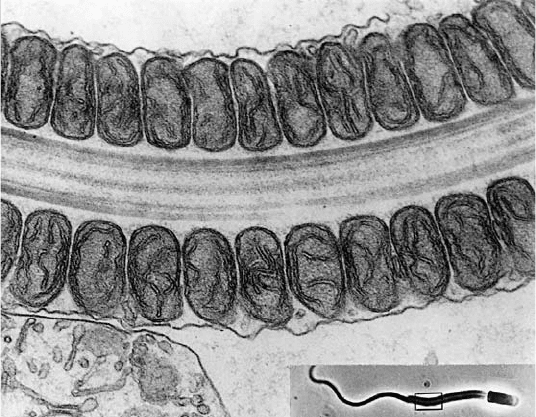
What dictates the distribution of mitochondria within a cell? In large and
highly asymmetrical cells, mitochondria may have to be localized in regions
where the ATP supply has to be high. Thus, mitochondria wrapped around
or packed along the axonemes of sperm tails can be easily rationalized.
(Figure 3.6 ).
In neurons, mitochondria have to be moved and distributed along the often
considerable length of axons and into the growth cone (40, 41) . Skeletal muscle
cells require a very uniform distribution of mitochondria in the myofi brils
along their entire length to provide a uniform supply of ATP to all the sarco-
meres (Figure 3.7 ). A particularly impressive example of a highly regular
arrangement of mitochondria in striated muscle cells is found in insect fl ight
muscles.
Rod cells in the retina appear to have their mitochondria concentrated in
the cell body closest to the outer segment. In round yeast cells or in a gamete
of Chlamydomonas the mitochondria are distributed around the cell peri-
phery, perhaps to maximize their access to oxygen. Thus it has been speculated
Figure 3.6 Mitochondria aligned along an axoneme of a sperm tail. (From The Cell ,
by D. W. Fawcett, 1981, W. B. Saunders Co.)
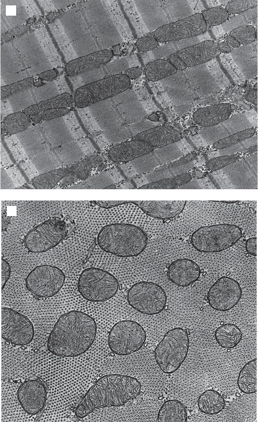
Figure 3.7 (A) Mitochondria distributed in skeletal muscle between sarcomeres.
(From The Cell , by D. W. Fawcett, 1981, W. B. Saunders Co.) (B) Cross section of cardiac
muscle showing uniform distribution of mitochondria. (From The Cell , by D. W. Fawcett,
1981, W. B. Saunders Co.)
B
A
INTEGRATION INTO THE CELL
33
34 STRUCTURE AND MORPHOLOGY. INTEGRATION INTO THE CELL
that ATP (and/or ADP) gradients might be responsible for infl uencing mito-
chondrial distributions. Alternatively, the clustering of mitochondria within a
cell might create gradients of other metabolites and ions such as calcium and
cause transient, highly localized perturbations, including pH gradients.
3.2.2 Interaction with Cytoskeleton
Mitochondria often tend to have an elongated, thread - like appearance, and
when viewed in live cells they appear to be roughly aligned along their long
axes. When they were colocalized with cytoskeletal structures, their association
with microtubules became immediately apparent in several cell types. In fact,
mitochondria surrounding the meiotic spindle in insect cells were noted before
the composition of the mitotic and meiotic spindles was known. Observations
in the laboratory of Singer (28, 29) (Figure 3.8 ) and of Chen (30) provided
defi nite proof, and many more observations in diverse cell types have followed
these early studies.
One interpretation is that mitochondria are simply trapped and partially
aligned within this cytoskeletal array of microtubules and are further dis-
persed by their own “ intrinsic motility ” (shape changes, fi ssion, and fusion; see
below). At the other end of the spectrum of interpretations, it has been argued
that molecular motors attached to the outer membrane of mitochondria can
become engaged with microtubules. In other words, dynein - and kinesin - like
molecules have been looked for, initially based on the differential sensitivity
of these motors to specifi c inhibitors. The evidence clearly points to an ATPase
activity bridging the outer mitochondrial membrane and microtubules (34) .
Before discussing the locomotion of mitochondria along cytoskeletal struc-
tures, it is relevant to consider whether the elongated shape of mitochondria
may be related to or even dependent on their attachment to fi lamentous or
long tubular structures, in the same way as the fl attening of cells on a substrate
is dependent on interactions between integral membrane proteins in the
plasma membrane and extracellular matrix elements. In other words, is the
sausage shape of mitochondria observed in vivo retained when mitochondria
are isolated and highly purifi ed in vitro ? Unfortunately, the isolation of mito-
chondria tends to break up mitochondria, and the resulting mitochondrial
preparations typically consist of smaller, spherical or only slightly elongated
vesicles. A more gentle test of the dependence of mitochondrial morphology
on intact microtubules can be made by treating intact cells with colchicine, and
one of the earliest experiments of this type showed clearly that the distribution
or orientation of mitochondria was perturbed, but their threadlike appearance
was not altered by such a treatment. Therefore, some internal mechanism must
be active that controls the shape. In the absence of an internal mitochondrial
skeleton, the answer has to be found in understanding the maintenance of the
varying curvature of the membranes. Under some conditions, however, defi ned
by specifi c mutations in extra - mitochondrial proteins, giant spherical mito-
chondria can be observed in yeast (see below).
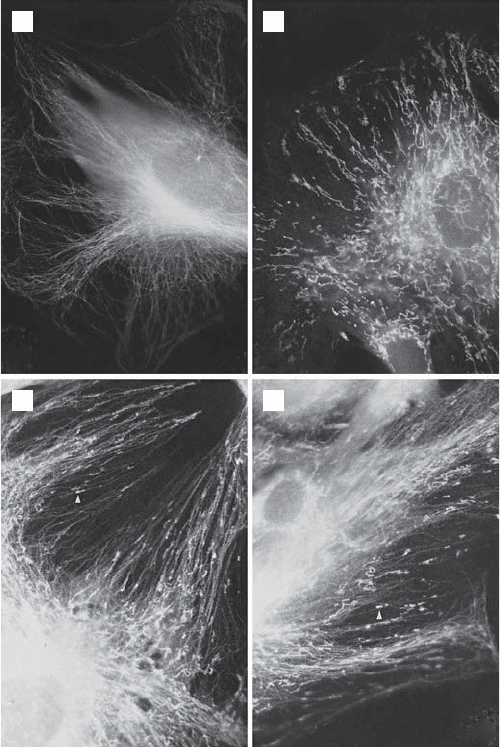
Figure 3.8 Correlation of microtubule alignment and mitochondria (27, 28) . Anti-
bodies against beef heart cytochrome oxidase and against chick tubulin were used in
indirect immunofl uorescence microscopy to localize microtubules and mitochondria
in permeabilized cells. (A) Normal rat kidney (NRK) cell stained with antitubulin
antibody and rabbit anti - chick tubulin IgG. (B) NRK cell stained with rabbit anti -
cytochrome oxidase antibody and goat anti - rabbit IgG. (C, D) NRK cells double -
stained with anti - tubulin and anti - cytochrome oxidase antibodies. The axis of elongated
mitochondria ( arrow ) is always parallel to the microtubule axis.
A B
C D
Neurons, axons, and axonal transport have received the maximum of atten-
tion, since both retrograde and anterograde movement of mitochondria
in axons has been observed by video - enhanced microscopy in intact axons
as well as in extruded axoplasms (41) . Closely related studies with systems
INTEGRATION INTO THE CELL 35
36 STRUCTURE AND MORPHOLOGY. INTEGRATION INTO THE CELL
of highly aligned microtubules of uniform polarity have been possible in
unsheathed nutritive tubes of insect ovaria and in permeabilized reticulopodia
of the giant amoeba Reticulomyxa . (The primary references to these studies
are found in reference 34 .) A characteristic aspect of such movements is the
saltatory nature of the displacements typical for motor - driven movements on
cytoskeletal elements, and not compatible with simple Brownian motion.
Unresolved problems are related to the bidirectional displacements, the spe-
cifi c speeds observed, the effectiveness (or lack of it) of known inhibitors such
as vanadate (a dynein inhibitor in vertebrates), and the possible contribution
of additional mechanisms (e.g., an actin - based motility).
A series of reports from the laboratory of Hollenbeck serves to illustrate
the experimental approaches and the conclusions drawn from the observations
(42 – 48) . In one study, chick sympathetic neurons were cultured in the continu-
ous presence of either cytochalasin E to eliminate microfi laments (F - actin,
MF), or in the presence of vinblastine or nocodazole to eliminate microtubules
(MTs). Mitochondrial movements were followed by computer - aided video
analysis after staining the organelle with rhodamine 123. In such neurons,
mitochondria continued to move with saltatory movements in both directions
in neurites with MTs, but no MFs, but failed even to enter the neurites in the
absence of MTs but presence of MFs. The dependence of mitochondrial move-
ment on MTs was confi rmed, and it appeared that MFs by themselves were
insuffi cient to support the movement of mitochondria into the neurites. Inter-
esting and partially contrasting results were obtained when the neurons were
fi rst cultured in the absence of any drug to allow for the normal development
of axons. Subsequently, the mature neurons were treated with either cytocha-
lasin E or with vinblastine. The drugs disrupted the cytoskeletal structures as
before, but in this case the axons already contained mitochondria. Now the
average mitochondrial velocity increased in both directions in axons lacking
only MFs, but net directional transport decreased. Surprisingly, in axons with
MFs but no MTs, mitochondria also moved in both directions at a reduced
average velocity and excursion length; net retrograde transport was favored
under these conditions. When both drugs were added to eliminate both MTs
and MFs, but leaving neurofi laments, no movement was observed. The studies
clearly support an actin - based motility of mitochondria over shorter distances
and a faster MT - based motility over long distances. They suggest that mito-
chondrial movement in neurites is a complex process involving several motors
(kinesins, dyneins, and myosins), each with characteristic intrinsic properties
and presumably able to fi ne - tune mitochondrial distributions for optimal func-
tioning of the axon (47) . The discussion must also include the nature of various
linkers, adaptors, or anchors on the surface of the organelle as reviewed by
Hollenbeck (46) . Finally, attention has now shifted to cellular signals modulat-
ing the activity of the motors and their attachment to mitochondria. An assort-
ment of kinases, a GTPase (Miro in Drosophila ), G proteins, and growth
factors (NGF) have been shown in a variety of model systems to infl uence
axonal transport of mitochondria (see reference 47 for a recent collection of
THE DYNAMICS OF MITOCHONDRIAL MORPHOLOGY 37
references). Miller and colleagues have made the very interesting observation
that mitochondria with high membrane potentials are transported primarily
toward the growth cone, while mitochondria with low potential were trans-
ported toward the cell body (49) . This may refl ect an aspect of quality control
by turnover (mitophagy; see below).
There have been numerous investigations aimed at establishing an interac-
tion between mitochondria and intermediate fi laments. A co - distribution has
been observed in a few instances (see reference 34 for references), but since
there are few systems with highly organized, regular arrays of such fi laments,
model systems and especially cell - free systems are lacking. Some observations
point to a role of desmin in anchoring mitochondria in skeletal muscle cells
to preserve their relative positions during repeated contraction – relaxation
cycles, but a more defi nite proof is lacking. In non - muscle cells deliberate
attempts to demonstrate an alignment of mitochondria with intermediate fi la-
ments have failed.
Further impetus to relate mitochondria and intermediate fi laments has
come from pionering studies in Yaffe ’ s laboratory (50) with the yeast Saccha-
romyces cerevisiae . Taking a genetic approach, this laboratory has isolated
mutants defective in mitochondrial inheritance; that is, these mutants fail to
transfer mitochondria into daughter buds. A gene for an essential protein was
thus identifi ed ( MDM1 ), and the Mdm1 protein was found to have modest
sequence similarity to animal intermediate fi lament proteins. In vitro this
protein can form intermediate fi lament - like (10 nm) structures, while in vivo
immunochemical staining reveals a punctate distribution of the antigen. A
further discussion will be deferred to one of the following sections.
3.3. THE DYNAMICS OF MITOCHONDRIAL MORPHOLOGY
3.3.1 Mitochondrial Shape Changes
The phenomenon referred to as “ intrinsic mitochondrial motility ” (34) has
been observed in animal cells, in fungal hyphae, in algal cells, and in the cyto-
plasms of plant cells where cytoplasmic streaming is absent to allow longer -
term observations. A mitochondrion appears to be able to slither, creep, or
slide at rates ranging from 2 to 30 μ m/min in the direction of its long axis. The
displacement is accompanied by subtle shape changes: slight thickening or
contractions along the axis, possibly refl ecting localized changes in the internal
arrangement of the cristae. Microinjection studies with ATP and/or ADP have
shown that these reagents can immobilize mitochondria; and in general, the
infl uence of metabolic conditions on mitochondrial motility have been inter-
preted in favor of intrinsic activities driving their displacements (34) .
Intrinsic dynamic changes in mitochondria are most strongly indicated by
the well - documented studies on mitochondrial fusions and fi ssions. Such events
can be followed in the phase - contrast microscope by time - lapse photography
38 STRUCTURE AND MORPHOLOGY. INTEGRATION INTO THE CELL
(34) ; and in some cases, observations on live cells have been elegantly related
to follow - up examinations with the electron microscope (see Figure 3.9 ) .
As a result of the combination of the power of genetics with the in vivo
visualization of mitochondria in the yeast Saccharomyces cerevisiae , an explo-
sion of papers has occurred in the past few years, contributing to an under-
standing of mitochondrial shape changes (fusion, fi ssion, and tubulation). A
variety of novel proteins were identifi ed, and their characterization was fre-
quently extended to mammalian cells by “ homology cloning. ” In other words,
genetic studies in yeast have generally provided guidance for studies in
mammalian cells. Many, but not all, proteins characterized in yeast have been
identifi ed in mammalian cells, either because they cannot yet be recognized
or because there are mechanistic differences between organisms. The growth
of the relevant literature dictates that reference is made primarily to review
articles (51 – 58) . The recent reviews by Okamoto and Shaw ( 59 ) and by Hoppins
et al. ( 59a ) are noteworthy for being up-to-date, authoritative, comprehensive,
and illustrated with highly informative drawings. Table 3.1 and Figures 3.11
and 3.12 represent summaries of information from these reviews.
Historically, fusion - defective mitochondria were fi rst discovered in sterile
Drosophila males (see below). Subsequently, the discovery and observations
of yeast mutants with defective mitochondrial morphologies defi ned three
major mechanisms/pathways in genetic and biochemical terms. When fusion
is interrupted, ongoing fi ssion produces a large number of mitochondrial frag-
ments. When fi ssion is defective, ongoing fusion leads to extensive intercon-
nected nets. A third pathway that so far is unique to fungi has been called
tubulation; defects in this pathway cause tubular mitochondria to form large
spheres.
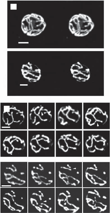
Serial sections and image reconstructions have revealed that in the yeast
Saccharomyces cerevisiae a cell contains a small number of mitochondria exist-
ing in a highly branched, reticulated shape, such that a section viewed in the
electron microscope gives the appearance of multiple mitochondria (Figure
3.10 ) . One to ten such reticular structures are expanded below the cortex of
the cell (22, 23) . A state of the art reexamination of this feature and its dynamic
aspects in live cells was pioneered by Nunnari and colleagues (35) , who
modifi ed a green fl uorescent protein (GFP) for import into mitochondria to
be able to use wide - fi eld fl uorescence microscopy for the acquisition of three -
dimensional images over long time intervals. The use of GFP avoids the prob-
lems of photobleaching. Beautiful 3 - D images were obtained, but more
signifi cantly, time - resolved images from single optical sections yielded quanti-
tative data on fusion and fi ssion events. While the authors are cautious to state
that the resolution does not unambiguously allow the distinction between
fusion and close juxtaposition, many events leading to stable connections were
documented. During vegetative growth of wild - type cells, 0.39 ± 0.14 fi ssion
events per minute and 0.40 ± 0.08 fusion events per minute were observed,
indicative of a highly dynamic situation. The matching fi gures indicate a steady
state in which a continuous network undergoes constant fragmentation and
THE DYNAMICS OF MITOCHONDRIAL MORPHOLOGY 39
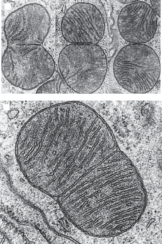
Figure 3.9 (A) Dividing mitochondrion. (Original photograph of T. Kanaseki; from
The Cell , by D. W. Fawcett, 1981, W. B. Saunders Co.) (B) Successive stages in a dividing
mitochondrion. (Original photograph of T. Kanaseki; from The Cell , by D. W. Fawcett,
1981, W. B. Saunders Co.)
B
A
re - assembly. They confi rmed previous observations that at least one tip (end)
was involved, where the putative fusion machinery may be located.
A similar study with GFP in HeLa mitochondria (36) also obtained high -
resolution 3 - D images at 30 - s intervals, and it was concluded that these mito-
chondria also formed a connected, continuous, and highly dynamic network
40 STRUCTURE AND MORPHOLOGY. INTEGRATION INTO THE CELL
exhibiting growth and retraction, as well as fusions to other portions of the
network. Photobleaching of a small area within a cell was followed by a rapid
recovery (5 – 20 min) of GFP fl uorescence within that area.
3.3.1.1 Fission Fission is the postulated mechanism for mitochondrial
proliferation. The insertion of new components into the matrix and all the
Figure 3.10 Mitochondria in living yeast cells (35) . Green fl uorescent protein was
modifi ed to be imported into mitochondria. Wide - fi eld fl uorescence microscopy was
used for the acquisition of three - dimensional images over long time intervals. (A)
Three - dimensional images of yeast mitochondria in wild - type cells grown on galactose.
(B) Fission and fusion of mitochondria. Time - resolved images from a single optical
section of 0.2 μ m. Time intervals from left to right in 3 - min intervals.
A
B