Vij D.R. Handbook of Applied Solid State Spectroscopy
Подождите немного. Документ загружается.


15. Laser Raman Spectroscopy
infrared guiding configurations. The availability of these glasses in substantial
quantities and the capability we have to fabricate good optical quality thin
films by thermal evaporation and other deposition techniques enables us to
realize relatively low cost As-S-(Se) integrated optical devices. Another
attractive feature is our capability to create integrated components with one-
and two-photon laser writing [48, 49].
Figure 15.10 Raman spectra of a series of bulk chalcogenide glasses obtained with near-
infrared excitation.
674
Figure 15.10 illustrates the near-infrared Raman spectra (incident and
scattered polarization resolved along the z-axis) for a series of binary and
ternary compounds. The spectra were obtained at a spectral resolution of 1.5
cm
–1
. The bulk spectra clearly show that each of the dominant bands consists
of several overlapping components [50, 51]. The dominant feature in the
binary sulfide and selenide compounds are bands at 345 cm
–1
(As
40
S
60
) and
230 cm
–1
(As
40
Se
60
), respectively. These spectra are in good agreement with
other studies [52, 53], and the strong, broad band is attributed to an
antisymmetric As-(S,Se)-As stretching vibration in the As (S,Se)
3
pyramids.
According to the analysis of Lucovsky and Martin [54], the normal modes of
the bulk glasses (e.g., clusters of As(S,Se)
3
modes (As(S,Se)
3
) and bridging chain modes (As-(S,Se)-As) independently.
molecules with weak inter-
molecular coupling) are obtained by treating the molecular pyramid
In the ternary compounds with S/Se = 1 molar ratio and decreasing As
content, a progressive decrease of these broad bands is observed, indicative of
a decrease in the number of As-containing pyramidal sites. New bands
appearing around 255 cm
–1
and 440–480 cm
–1
form in the now chalcogen-rich
glasses and are attributed to Se-Se and S-S homo-polar bonds. These may be
correlated with the enhancement of nonlinear optical properties (n
2
) in ternary
compounds with S/Se = 1 molar ratio and decreasing as content [10]. These
units serve as chalcogen chains connecting the remaining pyramidal units.
The small number of S-S bonds indicated by a weak band near 495 cm
–1
for
equal concentrations of S and Se suggests that the S stays with the remaining
pyramids, and that it is the Se that dominates the connecting chain units.
15.4.3 Chalcogenide Thin Films
-
Waveguide Raman
Waveguide Raman spectroscopy (WRS) using guided mode excitation has
been applied to thin organic and polymeric films to probe spontaneous [55,
16] and coherent [56] scattering, and very recently, to sol-gel-derived planar
germano-silicate [57] waveguides and lead titanate [58] films. The relative
low refractive index n of these organic and oxide materials allows the use of
glass prisms such as LaSF5 (n ~ 1.8) for coupling a range of propagation
vectors into the waveguide structure. In spite of its sensitivity WRS has not
been applied to the structural characterization of chalcogenide glasses until
recently [17], most likely due to their high index (n ~ 2.45), the lack of
suitable prism couplers, and difficulties associated with working in the near-
infrared.
Cleaved silicon substrates were employed for high efficiency endface
coupling of the near-infrared laser beam into a single-layer channel
waveguide structure [17]. The waveguides were nominally 1.75–2 Pm thick.
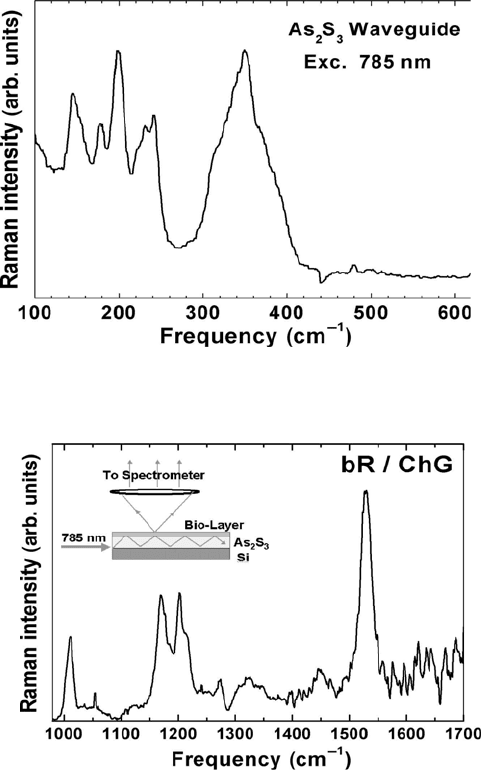
Figure 15.11 displays the Raman spectrum of As
2
S
3
and Figure 15.12 the
spectrum obtained from bacteriorhodopsin layered on the waveguide substrate
[15]. As shown in Figure 15.13, As
2
S
3
has Raman active vibrational bands
below 500 cm
–1
. The vibrational frequencies below 300 cm
–1
are attributed to
S-S interactions and the vibrational frequencies between 300–400 cm
–1
are
attributed to AsS
3
pyramidal units and their interactions [52].
For the integration of waveguide structures with photosensitive proteins,
bacteriorhodopsin (bR) is of particular interest, since it has applications in
molecular electronic devices and optical switching [59, 60]. As with all
proteins and organic assemblies, the characteristic vibrational frequencies of
bR are in the 1000–4000 cm
–1
range. This allows for no interference in the
protein’s signal from that of the waveguide. The bR spectrum in Figure 15.12
is indicative of the light-adapted state [61]. The light-adapted form of bR is
the initial state for the proton pumping cycle. At 785 nm the Raman spectrum
is still associated with the vibrations of the atoms that make up the
15.4 Applications
675
15. Laser Raman Spectroscopy
chromophore. Resonance Raman, nuclear magnetic resonance (NMR), and
chemical extraction studies have established that the chromophore in bR
568
is
a C
13
=C
14
trans, C
15
=NHR trans protonated Schiff base of retinal. As shown
in Figure 15.13, bR has several vibrational frequencies that are identifiable.
The band at 1012 cm
–1
is assigned to the rocking vibrations of a C-CH
3
group
of the bR molecule. The 1100–1300 cm
–1
region of the Raman spectrum is the
fingerprint region for C-C bonds and is very sensitive to isomerization. The
intense band at 1528 cm
–1
is attributed to the ethylenic stretching of C=C
bonds [61]. The data demonstrate that evanescently excited near-infrared
Raman spectra can be measured with high signal-to-noise ratio providing an
in situ probe of the native state of the protein.
Figure 15.11 Raman spectrum of a chalcogenide thin film obtained with waveguide excitation
and a power of 20 mW.
Figure 15.12 Evanescent wave-excited Raman spectrum of a bacteriorhodopsin layer on a
As
2
S
3
waveguide.
676
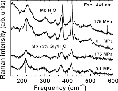
Figure 15.13 Low frequency region of the Raman spectra of deoxygenated myoglobin at
ambient and high pressure. Samples are in aqueous solution or glycerol-water mixtures at pH
of 7. Note the shift of the band near 220 cm
–1
to higher frequency with increasing pressure.
ChG waveguides are optimized for near-infrared excitation allowing one to
obtain the Raman spectra of biological compounds at minimal background
fluorescence. This feature of the substrates can be a useful tool in the study of
cells and microorganisms. Obtaining the Raman spectra at wavelengths less
than 1 Pm allows for the resolution of small spatial features compared to mid-
infrared absorption wavelengths of 5–10 Pm. Integrated optical components
fabricated with ChG can be combined with proteins such as bR. Evanescent
wave excitation may be employed for optical switching and spectroscopy of
bio-assemblies on patterned ChG semiconductors. There are also potential
applications involving biomolecular sensors.
15.4.3 High Pressure Raman Spectroscopy of Proteins
Resonance Raman spectroscopy is one of the few techniques that can probe
the local environment of the active site inside a large biological system. The
laser excitation wavelength is chosen near an electronic transition of the
chromophore, and the Raman scattering cross-sections for vibrational modes
that couple to this transition are selectively enhanced [29]. Raman spectra of
heme proteins reveal a number of bands that have been well characterized and
yield information on the spin state, coordination, and environment of the
heme [29]. The small, globular heme protein myoglobin has served as a
model system for extensive experimental and theoretical studies of protein
15.4 Applications
677
15. Laser Raman Spectroscopy
dynamics. During the process of reversibly binding small ligands such as O
2
,
CO, or NO to the heme iron, both the chromophore and the protein undergo
conformational changes. The bond between the iron and the proximal
histidine imidazole nitrogen is the only covalent linkage between the heme
group and protein. Resonance Raman studies at ambient pressure support the
view that modulation of the iron by the protein through the proximal histidine
exerts control at the level of reactivity [62, 63, 64].
In vivo, functional properties of proteins are affected by environmental
parameters such as viscosity, pH, temperature, and pressure. For instance, sea
animals survive over a wide range of pressure, from sea level to extreme
depths. On the other hand, pressure can deactivate enzymes and kill bacteria
[65]. For a description on a molecular level the effect of high pressure on
prototype reactions of isolated proteins must be understood [66, 67]. The
approach to combine high pressure and vibrational spectroscopy is motivated
by the following observations: Spectral band parameters (frequencies,
intensities, lineshapes and linewidths) are sensitive to dynamic and structural
changes of biomolecules [68] at the sub-Angstrom level, a length scale where
small, yet significant conformational changes for enzyme activity occur. From
changes in the Raman spectra, pressure effects on protein function can be
correlated with structural changes, for instance at the chromophore-protein
interface [69] and compared with theoretical models [70]. Deoxymyoglobin is
used as a reference structure since the reaction process is absent, and pressure-
induced changes of the conformation can be separated from those along the
reaction coordinate.
Resonance Raman scattering was excited with the frequency doubled
output of a Ti:sapphire laser tunable from 441 to 425 nm or by the 457.9-nm
line of an Ar ion laser. Detection of the backscattered Raman radiation is
accomplished using a thin back-illuminated, charge-coupled in conjunction
with a single-grating spectrograph and a Rayleigh line rejection filter. The
pressure cell is constructed of beryllium-copper that combines the ability to
resist high pressure (up to 400 MPa) with good thermal conductivity.
Sapphire windows allow measurements from the near UV to the near infrared
region. The high pressure Raman setup has been described in more detail in
[71, 72].
The resonance Raman spectra of horse deoxy myoglobin (Mb) in the
frequency range from 150 to 600 cm
–1
are shown for ambient and high
pressure in Figure 15.13. The band at 220 cm
–1
has been assigned to the iron-
histidine (Fe-His) stretching mode [73, 74, 75]. The other lines have been
classified as follows [75]: The band near 241 cm
–1
is a pyrole ring tilting
mode. The modes from 250 to 420 cm
–1
all involve peripheral substituents,
and those from 420 to 520 cm
–1
are attributed to out-of-plane distortions of
the pyrole rings [76, 75].
The most significant spectral change with pressure is a shift of the peak
frequency Q
Fe-His
of the iron histidine mode. Q
Fe-His
shifts to higher
wavenumber by ~3 cm
–1
between 0.1 and 175 MPa. The shift of the Fe-His
678
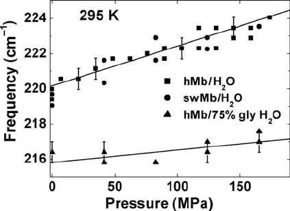
mode has been observed in different solvents (75% gly/H
2
O) and in Mb from
sperm whale. The peak position of Q
Fe-His
as a function of pressure is plotted in
–1
.
A smaller shift is apparent in the band near 343 cm
–1
. Choi and Spiro [76]
assigned this band to out-of-plane modes of propionate porphyrin macrocycle
substituent groups. Since the hydrogen bonding partner of the carboxyl group
of the propionic acid attached to the D pyrole [77] is Arg 45 (CD3), changes
in the 343 cm
–1
band may indicate motion of the protein helices.
Figure 15.14 Pressure dependence of the peak frequency of the iron-histidine mode in
myoglobin in aqueous solution and 75% gly/H
2
O.
We attribute the observed frequency shift to a conformational change,
which alters the tilt angle between the heme plane and the proximal histidine
and the out-of-plane iron position. The geometry of the proximal histidine
influences both the frequency and the intensity of the Fe-His stretch band
[74]. An important factor affecting the global protein conformation, the heme
pocket structure, and the iron-histidine mode is water activity [78]. The
altered water activity in a glycerol/H
2
O mixture causes a 2.6 cm
–1
downshift
of n
Fe-His
as compared to aqueous solution. Apparently glycerol influences the
protein-water interaction with a possible release of bound water molecules
from the surface, though this perturbation of the Fe-His frequency is opposite
to the perturbation from pressure.
The main conclusion from the shift of the iron-histidine mode is that
pressure causes global conformational changes in the protein as well as
rearrangements of the active site environment. Indeed, a very recent high-
pressure crystallographic study [79] of myoglobin confirms that the change in
protein structure due to pressure is not purely compressive but involves
conformational changes. Large collective displacements are observed in six
regions including sliding of the F-helix towards the E-helix [79].
In the case of ligand bound myoglobin (MbCO, MbO
2
), photolysis by the
laser beam during the acquisition of a Raman spectrum creates a stationary
mixture of bound and photolyzed molecules. Photostationary experiments
15.4 Applications
Figure 15.14. The error bars correspond to a precision of 0.6 cm
679
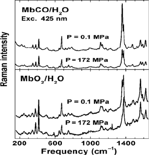
15. Laser Raman Spectroscopy
demonstrate a significant pressure-dependence of the ligand rebinding rate in
myoglobin [80]. The photolysis of ligated myoglobin by the laser beam
during the acquisition of a Raman spectrum (100 seconds) creates a stationary
mixture of bound and photolyzed molecules (Figure 15.15).
Figure 15.15 Resonance Raman spectra of MbCO (top) and MbO
2
(bottom) in aqueous
solution as a function of pressure at 295 K. The sample is in photostationary equilibrium. As
the pressure is increased the intensity of the Q
2
band of MbCO (1374 cm
–1
) increases relative to
that of Mb (1356 cm
–1
).
This is evident from the oxidation state marker band (n
4
), which appears at
1354 cm
–1
for deligated Mb and 1372 cm
–1
for MbCO [62]. The spectrum at
high pressure (keeping all other parameters fixed) shows a significant increase
in intensity of the peak at 1372 cm
–1
relative to that at 1354 cm,
–1
reflecting an
increase in population of the bound state [71]. This population increase is due
to a speedup of the overall rebinding. Consistent changes between bound and
unbound states are also seen in the core size marker band n
2
(1560 and 1582
cm
–1
) and the vinyl modes (1618 and 1632 cm
–1
). The lower amount of
photolysis at high pressure is also indicated by the reduced intensity of the
iron-histidine mode at 220 cm
–1
[71].
15.4.4 Micro-Raman Spectroscopy
For the measurement of a minuscule amount of sample or when spatial
resolution is required (in addition to spectral resolution) various combinations
of a Raman spectrometer with an optical microscope have been developed
[81, 18]. This is known as Raman microscopy or micro-Raman spectroscopy.
680

A typical setup is shown in Figure 15.16. The excitation laser is reflected
by a beam splitter, goes through a microscope objective, and is focused on the
sample. The backscattered Raman is then collected with the same objective.
The major part of the collimated beam is reflected from the beam splitter and
is focused on the entrance slit of the monochromator. The small sample area,
which is at the focusing point, is thus imaged through the entrance slit. It is
important to understand that a narrow slit width will determine a well-defined
sharp image, and thus result in a high resolution.
Figure 15.16 Schematics of micro-Raman spectrometer. The sample is located on an
dimensional positioning stage.
Applications in an industrial environment or for routine testing of samples
require an experimental technique that requires no specialized sample
preparation. Raman spectroscopy is a nondestructive probe and spectra can be
measured on materials in solid or liquid form. Using a Raman microscope the
sample can simply be placed on a slide and the area of interest is selected
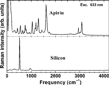
optically by using a viewing system. Figure 15.17 shows the Raman spectrum
of a piece of silicon and an aspirin sample obtained with a commercial micro-
be measured from micron-sized particles, which makes the technique well
suited as an analytical tool in chemistry and biotechnology.
Raman spectroscopy has proven to be an informative and nondestructive
technique in III-V material characterization including local structure
determination and stress analysis [82]. One of the key issues for the
performance of wide band gap semiconductor-based device structures is the
control of growth-induced defects and their impact on optoelectronic and
transport properties. Despite the impressive progress in device applications, a
deeper understanding of defect and impurity issues is necessary for continued
rapid development in the areas of LEDs, laser diodes, UV detectors, and high
voltage unipolar and bipolar [83].
We present an example where micro-Raman spectroscopy was used to
probe optical phonons in gallium nitride close to the GaN/sapphire substrate
15.4 Applications
3-
Raman instrument. With micro-Raman, spectroscopy vibrational spectra can
681
15. Laser Raman Spectroscopy
interface [84]. Frequency shifts in vibrational modes correlate with
independently obtained data on the dislocation density and are connected to
the strain due to lattice mismatch at the interface. Micro-Raman spectra were
measured in a backscattering geometry using the 514.53-nm line of an Ar
+
ion
laser. An infinity-corrected microscope objective focused the laser beam to a
spot size of about 1 Pm in diameter. A very narrow slit width (5 Pm) in
combination with binning in the vertical direction of the CCD chip acts as a
confocal aperture which leads to a 1–2 Pm depth resolution. A translation
stage with submicron sensitivity was used for laser beam positioning at the
predetermined distance from the GaN/sapphire interface. The excitation beam
was parallel to the surface of the interface and perpendicular to grows
direction of the GaN layers.
Figure 15.17 Raman spectra of aspirin and silicon obtained with a commercial micro-Raman
instrument (LabRam HRUV). Integration time is 1 sec, HeNe laser power 10 mW, and slit
width 100 Pm.
Figure 15.18 depicts the Raman spectra at various distances from the
GaN/sapphire interface over a range of 60 Pm. The data were measured with a
backscattering geometry corresponding to x(..)x configuration in Porto
notation. The propagation direction of the laser beam was perpendicular to the
c-axis but not along one of the principal axes so the A
1
(TO), E
1
(TO), E
1
(LO),
and E
2
(high) modes [85] are observed at 535 cm
–1
, 562 cm
–1
, 745 cm
–1
and
569 cm
–1
, respectively. Due to limitations of the Rayleigh filter the E
2
(low)
mode (144 cm
–1
) was not investigated. The spectral resolution of 1 cm
–1
was
achieved by recording the Raman spectrum in second order. The raw data
presented in Figure 15.18 show a shift to lower frequency with increasing
distance from the sapphire substrate. The precision in the determination of the
frequency shifts could be further increased by fitting Lorentzian lines to the
Raman peaks. Since the lineshape remains constant we estimate that spectral
shifts of 0.1 cm
–1
can be reliably detected [86].
682

Figure 15.18 Raman spectra of a 64-mm thick GaN film measured with 514.5-nm excitation.
Insert shows E
1
(TO) and E
2
(high) modes for various distances from the GaN interface.
The peak frequency of the Raman modes are displayed as a function of
the distance from the GaN/sapphire interface in Figure 15.19. To verify, the
reproducibility measurements were made in both directions: from
the interface to the surface and then backwards. For the A
1
(TO) mode no shift
could be observed. Also there were no significant changes in full width at half
maximum (FWHM) or intensity of the spectral bands. The E
2
Raman mode is
known to be shifted by stress. In GaN films large compressive stresses
causing frequency shifts of +4.5 cm
–1
compared to single crystals have been
observed [87, 88]. The small shifts in the data presented here indicate the
good quality of our films. The absence of a shift in the A
1
phonon can be
explained by the direction of the optical phonon eigenvectors with respect to
the c-axis. Among the Raman active modes A
1
is the only one with
displacements along the c-axis as opposed to the E
2
(high) and E
1
(TO)
vibrations [82, 85]. Thus it appears that the stresses are perpendicular to the c-
axis.
Raman microscopy enables measurement of spectra from a single “point”
defined by the optical resolution limit of the microscope. It thus allows point-
to-point mapping whereby the spectroscopic information from different points
of a relatively large sample can be detected and compared. Raman imaging
allows one to noninvasively visualize chemical heterogeneity through the
integration of microscopy and spectroscopy. It provides information that is
useful in the fabrication of new materials, evaluation of the performance of
existing materials, and control of product quality.
15.4 Applications
683