Berg J.M., Tymoczko J.L., Stryer L. Biochemistry
Подождите немного. Документ загружается.


26.3.4. The LDL Receptor Is a Transmembrane Protein Having Five Different
Functional Regions
The amino acid sequence of the human LDL receptor reveals the mosaic structure of this 115-kd protein, which is
composed of six different types of domain (Figure 26.18). The amino-terminal region of the mature receptor consists of a
cysteine-rich sequence of about 40 residues that is repeated, with some variation, seven times to form the LDL-binding
domain (Figure 26.19). A set of conserved acidic side chains in this domain bind calcium ion; this metal ion lies at the
center of each domain and, along with disulfide bonds formed from the conserved cysteine residues, stabilizes the three-
dimensional structure. Protonation of these glutamate and aspartate side chains of the receptor in lysosomes leads to the
release of calcium and hence to structural disruption and the release of LDL from its receptor. A second region of the
LDL receptor includes two types of recognizable domains, three domains homologous to epidermal growth factor and
six repeats that are similar to the blades of the transducin β subunit (Section 15.2.2). The six repeats form a propeller-like
structure that packs against one of the EGF-like domains (Figure 26.20). An aspartate residue forms hydrogen bonds that
hold each blade to the rest of the structure. These interactions, too, would most likely be disrupted at the low pH in the
lysosome.
The third region contains a single domain that is very rich in serine and threonine residues and contains O-linked sugars.
These oligosaccharides may function as struts to keep the receptor extended from the membrane so that the LDL-binding
domain is accessible to LDL. The fourth region contains the fifth type of domain, which consists of 22 hydrophobic
residues that span the plasma membrane. The final region contains the sixth type of domain; it consists of 50 residues
and emerges on the cytosolic side of the membrane, where it controls the interaction of the receptor with coated pits and
participates in endocytosis. The gene for the LDL receptor consists of 18 exons, which correspond closely to the
structural units of the protein. The LDL receptor is a striking example of a mosaic protein encoded by a gene that was
assembled by exon shuffling.
26.3.5. The Absence of the LDL Receptor Leads to Hypercholesteremia and
Atherosclerosis
The results of Brown and Goldstein's pioneering studies of familial hypercholesterolemia revealed the physiologic
importance of the LDL receptor. The total concentration of cholesterol and LDL in the plasma is markedly
elevated in this genetic disorder, which results from a mutation at a single autosomal locus. The cholesterol level in the
plasma of homozygotes is typically 680 mg dl
-1
, compared with 300 mg dl
-1
in heterozygotes (clinical assay results are
often expressed in milligrams per deciliter, which is equal to milligrams per 100 milliliters). A value of < 200 mg dl
-1
is
regarded as desirable, but many people have higher levels. In familial hypercholesterolemia, cholesterol is deposited in
various tissues because of the high concentration of LDL cholesterol in the plasma. Nodules of cholesterol called
xanthomas are prominent in skin and tendons. Of particular concern is the oxidation of the excess blood LDL to form
oxidized LDL (oxLDL). The oxLDL is taken up by immune-system cells called macrophages, which become engorged
to form foam cells. These foam cells become trapped in the walls of the blood vessels and contribute to the formation of
atherosclerotic plaques that cause arterial narrowing and lead to heart attacks (Figure 26.21). In fact, most homozygotes
die of coronary artery disease in childhood. The disease in heterozygotes (1 in 500 people) has a milder and more
variable clinical course. A serum esterase that degrades oxidized lipids is found in association with HDL. Possibly, the
HDL-associated protein destroys the oxLDL, accounting for HDL's ability to protect against coronary disease.
The molecular defect in most cases of familial hypercholesterolemia is an absence or deficiency of functional receptors
for LDL. Receptor mutations that disrupt each of the stages in the endocytotic pathway have been identified.
Homozygotes have almost no functional receptors for LDL, whereas heterozygotes have about half the normal number.
Consequently, the entry of LDL into liver and other cells is impaired, leading to an increased plasma level of LDL.
Furthermore, less IDL enters liver cells because IDL entry, too, is mediated by the LDL receptor. Consequently, IDL
stays in the blood longer in familial hypercholesterolemia, and more of it is converted into LDL than in normal people.
All deleterious consequences of an absence or deficiency of the LDL receptor can be attributed to the ensuing elevated
level of LDL cholesterol in the blood.
26.3.6. The Clinical Management of Cholesterol Levels Can Be Understood at a
Biochemical Level
Homozygous familial hypercholesterolemia can be treated only by a liver transplant. A more generally applicable
therapy is available for heterozygotes and others with high levels of cholesterol. The goal is to reduce the amount
of cholesterol in the blood by stimulating the single normal gene to produce more than the customary number of LDL
receptors. We have already observed that the production of LDL receptors is controlled by the cell's need for cholesterol.
Therefore, in essence, the strategy is to deprive the cell of ready sources of cholesterol. When cholesterol is required, the
amount of mRNA for the LDL receptor rises and more receptor is found on the cell surface. This state can be induced by
a two-pronged approach. First, the intestinal reabsorption of bile salts is inhibited. Bile salts are cholesterol derivatives
that promote the absorption of dietary cholesterol and dietary fats (Section 22.1.1). Second, de novo synthesis of
cholesterol is blocked.
The reabsorption of bile is impeded by oral administration of positively charged polymers, such as cholestyramine, that
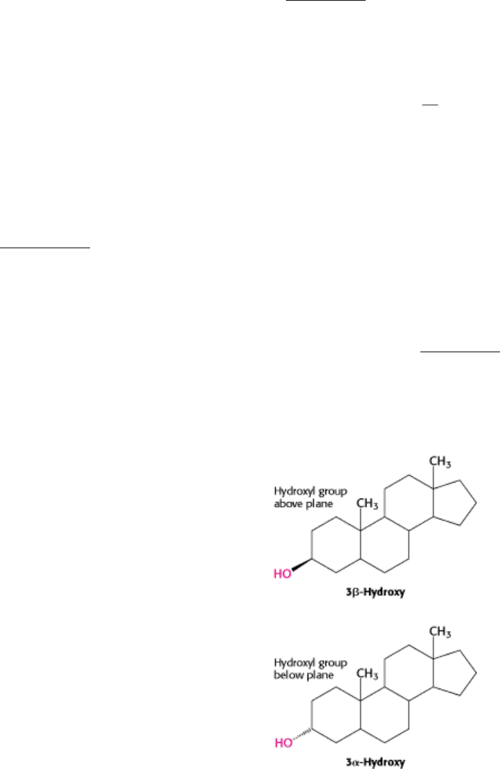
bind negatively charged bile salts and are not themselves absorbed. Cholesterol synthesis can be effectively blocked by a
class of compounds called statins (e.g., lovastatin, which is also called mevacor; Figure 26.22). These compounds are
potent competitive inhibitors (K
i
< 1 nM) of HMG-CoA reductase, the essential control point in the biosynthetic
pathway. Plasma cholesterol levels decrease by 50% in many patients given both lovastatin and inhibitors of bile-salt
reabsorption. Lovastatin and other inhibitors of HMG-CoA reductase are widely used to lower the plasma cholesterol
level in people who have atherosclerosis, which is the leading cause of death in industrialized societies.
III. Synthesizing the Molecules of Life 26. The Biosynthesis of Membrane Lipids and Steroids 26.3. The Complex Regulation of Cholesterol Biosynthesis Takes Place at Several Levels
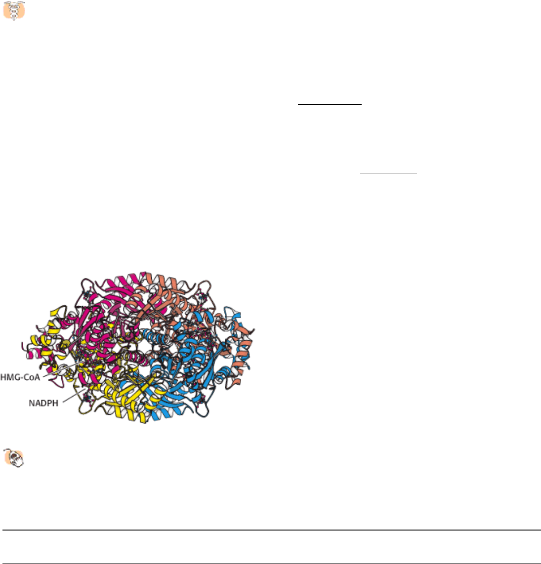
Figure 26.14. HMG-CoA Reductase.
The structure of a portion of the tetrameric enzyme is shown.
III. Synthesizing the Molecules of Life 26. The Biosynthesis of Membrane Lipids and Steroids 26.3. The Complex Regulation of Cholesterol Biosynthesis Takes Place at Several Levels
Table 26.1. Properties of plasma lipoproteins
Lipoproteins Major core lipids Apoproteins Mechanism of lipid delivery
Chylomicron Dietary triacylglycerols B-48, C, E Hydrolysis by lipoprotein
lipase
Chylomicron remnant Dietary cholesterol esters B-48, E Receptor-mediated
endocytosis by liver
Very low density lipoprotein
(VLDL)
Endogenous triacylglycerols B-100, C, E Hydrolysis by lipoprotein
lipase
Intermediate-density lipoprotein
(IDL)
Endogenous cholesterol esters B-100, E Receptor-mediated
endocytosis by liver and
conversion into LDL
Low-density lipoprotein (LDL) Endogenous cholesterol esters B-100 Receptor-mediated
endocytosis by liver and other
tissues
High-density lipoprotein (HDL) Endogenous cholesterol esters A Transfer of cholesterol esters
to IDL and LDL
Source: After M. S. Brown and J. L. Goldstein, The Pharmacological Basis of Therapeutics. 7th ed., A. G. Gilman, L. S.
Goodman, T. W. Rall, and F. Murad, Eds. (Macmillan, 1985), p. 828.
III. Synthesizing the Molecules of Life 26. The Biosynthesis of Membrane Lipids and Steroids 26.3. The Complex Regulation of Cholesterol Biosynthesis Takes Place at Several Levels
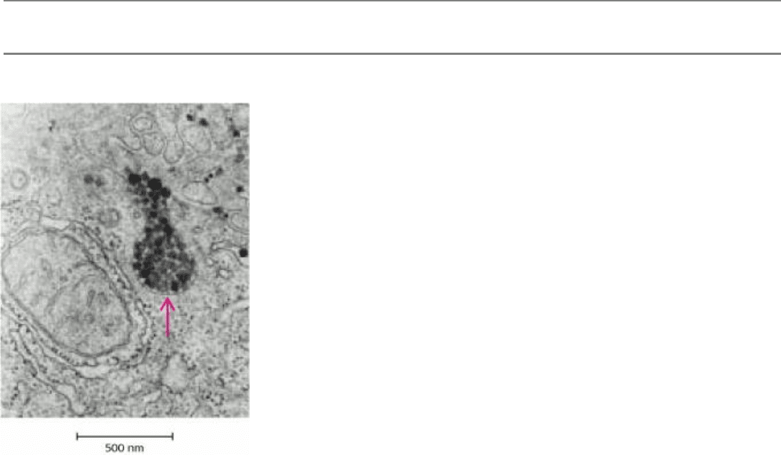
Figure 26.15. Site of Cholesterol Synthesis. Electron micrograph of a part of a liver cell actively engaged in the
synthesis and secretion of very low density lipoprotein (VLDL). The arrow points to a vesicle that is releasing its content
of VLDL particles. [Courtesy of Dr. George Palade.]
III. Synthesizing the Molecules of Life 26. The Biosynthesis of Membrane Lipids and Steroids 26.3. The Complex Regulation of Cholesterol Biosynthesis Takes Place at Several Levels
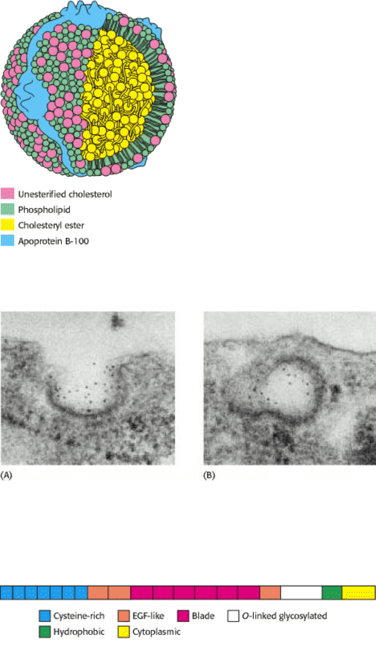
Figure 26.16. Schematic Model of Low-Density Lipoprotein. The LDL particle is approximately 22 nm (220 Å) in
diameter.
III. Synthesizing the Molecules of Life 26. The Biosynthesis of Membrane Lipids and Steroids 26.3. The Complex Regulation of Cholesterol Biosynthesis Takes Place at Several Levels
Figure 26.17. Endocytosis of LDL Bound to Its Receptor. (A) Electron micrograph showing LDL (conjugated to
ferritin for visualization, dark spots) bound to a coated-pit region on the surface of a cultured human fibroblast cell. (B)
Micrograph showing this region invaginating and fusing to form an endocytic vesicle [From R. G. W. Anderson, M. S.
Brown, and J. L. Goldstein. Cell 10 (1977): 351.]
III. Synthesizing the Molecules of Life 26. The Biosynthesis of Membrane Lipids and Steroids 26.3. The Complex Regulation of Cholesterol Biosynthesis Takes Place at Several Levels
Figure 26.18. LDL Receptor Domains. A schematic representation of the amino acid sequence of the LDL receptor
showing six types of domain.
III. Synthesizing the Molecules of Life 26. The Biosynthesis of Membrane Lipids and Steroids 26.3. The Complex Regulation of Cholesterol Biosynthesis Takes Place at Several Levels
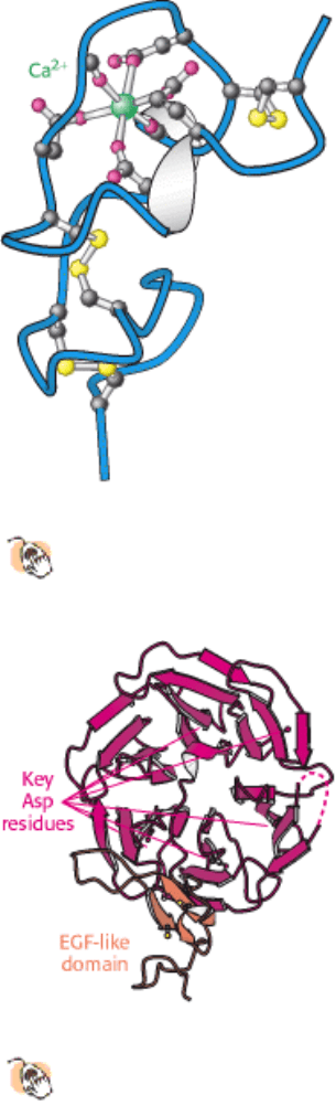
Figure 26.19. Structure of Cysteine-Rich Domain.
This calcium-binding cysteine-rich domain is repeated seven times
at the amino terminus of the LDL receptor.
III. Synthesizing the Molecules of Life 26. The Biosynthesis of Membrane Lipids and Steroids 26.3. The Complex Regulation of Cholesterol Biosynthesis Takes Place at Several Levels
Figure 26.20. Structure of Propeller Domain.
The six-bladed propeller domain and an adjacent EGF-like domain of
the LDL receptor.
III. Synthesizing the Molecules of Life 26. The Biosynthesis of Membrane Lipids and Steroids 26.3. The Complex Regulation of Cholesterol Biosynthesis Takes Place at Several Levels
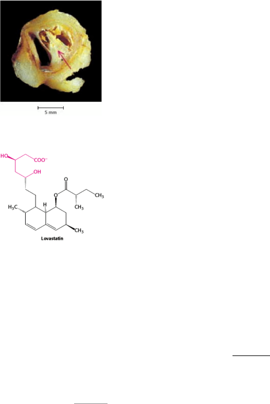
Figure 26.21. An Atherosclerotic Plaque. A plaque (marked by an arrow) blocks most of the lumen of this blood
vessel. The plaque is rich in cholesterol. [Courtesy of Dr. Jeffrey Sklar.]
III. Synthesizing the Molecules of Life 26. The Biosynthesis of Membrane Lipids and Steroids 26.3. The Complex Regulation of Cholesterol Biosynthesis Takes Place at Several Levels
Figure 26.22. Lovastatin, a Competitive Inhibitor of HMG-CoA Reductase. The part of the structure that resembles
the 3-hydroxy-3-methylglutaryl moiety is shown in red.
III. Synthesizing the Molecules of Life 26. The Biosynthesis of Membrane Lipids and Steroids
26.4. Important Derivatives of Cholesterol Include Bile Salts and Steroid Hormones
Cholesterol is a precursor for other important steroid molecules: the bile salts, steroid hormones, and vitamin D.
Bile Salts.
As polar derivatives of cholesterol, bile salts are highly effective detergents because they contain both polar and
nonpolar regions. Bile salts are synthesized in the liver, stored and concentrated in the gall bladder, and then released
into the small intestine. Bile salts, the major constituent of bile, solubilize dietary lipids (Section 22.1.1). Solubilization
increases in the effective surface area of lipids with two consequences: more surface area is exposed to the digestive
action of lipases and lipids are more readily absorbed by the intestine. Bile salts are also the major breakdown products
of cholesterol.
Cholesterol is converted into trihydroxycoprostanoate and then into cholyl CoA, the activated intermediate in the
synthesis of most bile salts (Figure 26.23). The activated carboxyl carbon of cholyl CoA then reacts with the amino
group of glycine to form glycocholate or it reacts with the amino group of taurine (H
2
NCH
2
CH
2
SO
3
-
), derived from
cysteine, to form taurocholate. Glycocholate is the major bile salt.
Steroid Hormones.
Cholesterol is the precursor of the five major classes of steroid hormones: progestagens, glucocorticoids,
mineralocorticoids, androgens, and estrogens (Figure 26.24). These hormones are powerful signal molecules that
regulate a host of organismal functions. Progesterone, a progestagen, prepares the lining of the uterus for implantation
of an ovum. Progesterone is also essential for the maintenance of pregnancy. Androgens of male secondary sex
characteristics, whereas estrogens (such as estrone) are required for the development of female secondary sex
characteristics. Estrogens, along with progesterone, also participate in the ovarian cycle. Glucocorticoids (such as
cortisol) promote gluconeogenesis and the formation of glycogen, enhance the degradation of fat and protein, and inhibit
the inflammatory response. They enable animals to respond to stress
indeed, the absence of glucocorticoids can be
fatal. Mineralocorticoids (primarily aldosterone) act on the distal tubules of the kidney to increase the reabsorption of Na
+
and the excretion of K
+
and H
+
, which leads to an increase in blood volume and blood pressure. The major sites of
synthesis of these classes of hormones are the corpus luteum, for progestagens; the ovaries, for estrogens; the testes, for
androgens; and the adrenal cortex, for glucocorticoids and mineralocorticoids.
Steroid hormones bind to and activate receptor molecules that serve as transcription factors to regulate gene expression
(Section 31.3.1). These small, relatively similar molecules are able to have greatly differing effects because the slight
structural differences among them allow interactions with specific receptor molecules.
26.4.1. The Nomenclature of Steroid Hormones
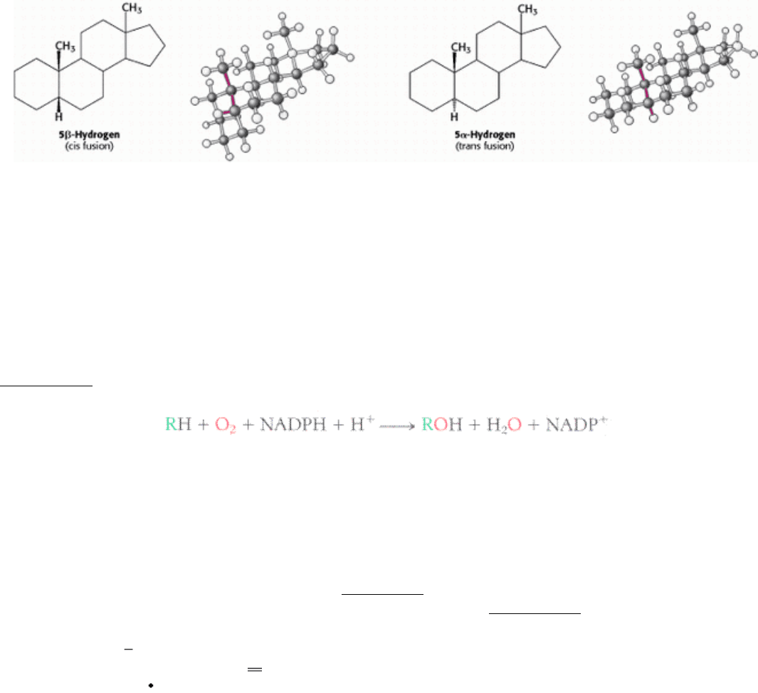
Carbon atoms in steroids are numbered as shown for cholesterol in (Figure 26.25). The rings in steroids are denoted by
the letters A, B, C, and D. Cholesterol contains two angular methyl groups: the C-19 methyl group is attached to C-10,
and the C-18 methyl group is attached to C-13. The C-18 and C-19 methyl groups of cholesterol lie above the plane
containing the four rings. A substituent that is above the plane is termed β oriented, whereas a substituent that is below
the plane is α oriented.
If a hydrogen atom is attached to C-5, it can be either α or β oriented. The A and B steroid rings are fused in a trans
conformation if the C-5 hydrogen is < oriented, and cis if it is < oriented. The absence of a Greek letter for the C-5
hydrogen atom on the steroid nucleus implies a trans fusion. The C-5 hydrogen atom is α oriented in all steroid
hormones that contain a hydrogen atom in that position. In contrast, bile salts have a β-oriented hydrogen atom at C-5.
Thus, a cis fusion is characteristic of the bile salts, whereas a trans fusion is characteristic of all steroid hormones that
possess a hydrogen atom at C-5. A trans fusion yields a nearly planar structure, whereas a cis fusion gives a buckled
structure.
26.4.2. Steroids Are Hydroxylated by Cytochrome P450 Monooxygenases That Utilize
NADPH and O
2
Hydroxylation reactions play a very important role in the synthesis of cholesterol from squalene and in the conversion of
cholesterol into steroid hormones and bile salts. All these hydroxylations require NADPH and O
2
. The oxygen atom of
the incorporated hydroxyl group comes from O
2
rather than from H
2
O. While one oxygen atom of the O
2
molecule goes
into the substrate, the other is reduced to water. The enzymes catalyzing these reactions are called monooxygenases (or
mixed-function oxygenases). Recall that a monooxygenase also participates in the hydroxylation of aromatic amino acids
(Section 23.5.7).
Hydroxylation requires the activation of oxygen. In the synthesis of steroid hormones and bile salts, activation is
accomplished by a cytochrome P450, a family of cytochromes that absorb light maximally at 450 nm when complexed in
vitro with exogenous carbon monoxide. These membraneanchored proteins (~50 kd) contain a heme prosthetic group.
Because the hydroxylation reactions promoted by P450 enzymes are oxidation reactions, it is at first glance surprising
that they also consume the reductant NADPH. NADPH transfers its high-potential electrons to a flavoprotein, which
transfers them, one at a time, to adrenodoxin, a nonheme iron protein. Adrenodoxin transfers one electron to reduce the
ferric (Fe
3+
) form of P450 to the ferrous (Fe
2+
) form (Figure 26.26). Without the addition of this electron, P450 will not
bind oxygen. Recall that only the ferrous form of hemoglobin binds oxygen (Section 10.2.1). The binding of O
2
to the
heme is followed by the acceptance of a second electron from adrenodoxin. The acceptance of this second electron leads
to cleavage of the O
O bond. One of the oxygen atoms is then protonated and released as water. The remaining oxygen
atom forms a highly reactive ferryl (Fe O) intermediate. This intermediate abstracts a hydrogen atom from the
substrate RH to form R
. This transient free radical captures the OH group from the iron atom to form ROH, the
hydroxylated product, returning the iron atom to the ferric state.
26.4.3. The Cytochrome P450 System Is Widespread and Performs a Protective
Function
The cytochrome P450 system, which in mammals is located primarily in the endoplasmic reticulum of the liver and
small intestine, is also important in the detoxification of foreign substances (xenobiotic compounds) by oxidative
metabolism. For example, the hydroxylation of phenobarbital, a barbiturate, increases its solubility and facilitates its
excretion. Likewise, polycyclic aromatic hydrocarbons are hydroxylated by P450, providing sites for conjugation with
highly polar units (e.g., glucuronate or sulfate), which markedly increase the solubility of the modified aromatic
molecule. One of the most relevant functions of the cytochrome P450 system to human beings is its role in drug
metabolism. Drugs such as caffeine and ibuprofen are oxidatively metabolized by these monooxygenases. Indeed, the
duration of action of many medications depends on their rate of inactivation by the P450 system. Despite its general
protective role in the removal of foreign chemicals, the action of the P450 system is not always beneficial. Some of the
most powerful carcinogens are generated from harmless compounds by the P450 system in vivo in the process of
metabolic activation. In plants, the cytochrome P450 system plays a role in the synthesis of toxic compounds as well as
the pigments of flowers.
The cytochrome P450 system is a ubiquitous superfamily of monooxygenases that is present in plants, animals,
and prokaryotes. The human genome encodes more than 50 members of the family, whereas the genome of the
plant Arabidopsis encodes more than 250 members. All members of this large family arose by gene duplication followed
by subsequent divergence that generated a range of substrate specificity. Indeed, the specificity of these enzymes is
encoded in delimited regions of the primary structure, and the substrate specificity of closely related members is often
defined by a few critical residues or even a single amino acid.
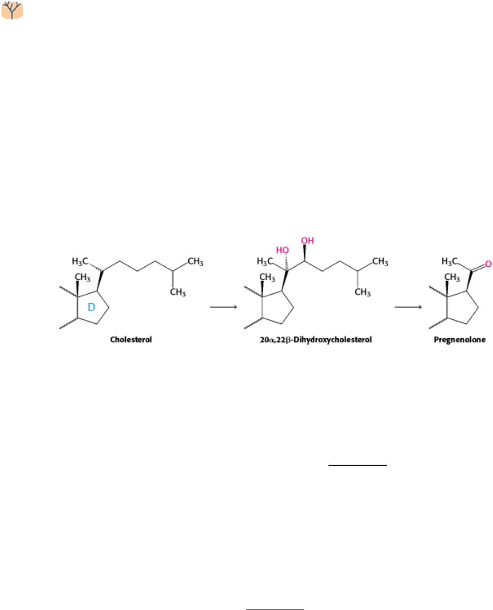
26.4.4. Pregnenolone, a Precursor for Many Other Steroids, Is Formed from
Cholesterol by Cleavage of Its Side Chain
Steroid hormones contain 21 or fewer carbon atoms, whereas cholesterol contains 27. Thus, the first stage in the
synthesis of steroid hormones is the removal of a six-carbon unit from the side chain of cholesterol to form
pregnenolone. The side chain of cholesterol is hydroxylated at C-20 and then at C-22, and the bond between these
carbon atoms is subsequently cleaved by desmolase. Three molecules of NADPH and three molecules of O
2
are
consumed in this remarkable six-electron oxidation.
Adrenocorticotropic hormone (ACTH, or corticotropin), a polypeptide synthesized by the anterior pituitary gland,
stimulates the conversion of cholesterol into pregnenolone, the precursor of all steroid hormones.
26.4.5. The Synthesis of Progesterone and Corticosteroids from Pregnenolone
Progesterone is synthesized from pregnenolone in two steps. The 3-hydroxyl group of pregnenolone is oxidized to a 3-
keto group, and the ∆
5
double bond is isomerized to a ∆
4
double bond (Figure 26.27). Cortisol, the major
glucocorticoid, is synthesized from progesterone by hydroxylations at C-17, C-21, and C-11; C-17 must be hydroxylated
before C-21 is, whereas C-11 can be hydroxylated at any stage. The enzymes catalyzing these hydroxylations are highly
specific, as shown by some inherited disorders. The initial step in the synthesis of aldosterone, the major
mineralocorticoid, is the hydroxylation of progesterone at C-21. The resulting deoxycorticosterone is hydroxylated at C-
11. The oxidation of the C-18 angular methyl group to an aldehyde then yields aldosterone.
26.4.6. The Synthesis of Androgens and Estrogens from Pregnenolone
Androgens and estrogens also are synthesized from pregnenolone through the intermediate progesterone. Androgens
contain 19 carbon atoms. The synthesis of androgens (Figure 26.28) starts with the hydroxylation of progesterone at C-
17. The side chain consisting of C-20 and C-21 is then cleaved to yield androstenedione, an androgen. Testosterone,
another androgen, is formed by the reduction of the 17-keto group of androstenedione. Testosterone, through its actions
in the brain, is paramount in the development of male sexual behavior. It is also important for maintenance of the testes
and development of muscle mass. Owing to the latter activity, testosterone is referred to as an anabolic steroid.
Testosterone is reduced by 5a-reductase to yield dihydrotestosterone (DHT), a powerful embryonic androgen that
instigates the development and differentiation of the male phenotype. Estrogens are synthesized from androgens by the
loss of the C-19 angular methyl group and the formation of an aromatic A ring. Estrone, an estrogen, is derived from

androstenedione, whereas estradiol, another estrogen, is formed from testosterone.
26.4.7. Vitamin D Is Derived from Cholesterol by the Ring-Splitting Activity of Light
Cholesterol is also the precursor of vitamin D, which plays an essential role in the control of calcium and phosphorus
metabolism. 7-Dehydrocholesterol (provitamin D
3
) is photolyzed by the ultraviolet light of sunlight to previtamin D
3
,
which spontaneously isomerizes to vitamin D
3
(Figure 26.29). Vitamin D
3
(cholecalciferol) is converted into calcitriol
(1,25-dihydroxycholecalciferol), the active hormone, by hydroxylation reactions in the liver and kidneys. Although not a
steroid, vitamin D acts in an analogous fashion. It binds to a receptor, structurally similar to the steroid receptors, to form
a complex that functions as a transcription factor, regulating gene expression.
Vitamin D deficiency in childhood produces rickets, a disease characterized by inadequate calcification of
cartilage and bone. Rickets was so common in seventeenth-century England that it was called the "children's
disease of the English." The 7-dehydrocholesterol in the skin of these children was not photolyzed to previtamin D
3
,
because there was little sunlight for many months of the year. Furthermore, their diets provided little vitamin D, because
most naturally occurring foods have a low content of this vitamin. Fish-liver oils are a notable exception. Cod-liver oil,
abhorred by generations of children because of its unpleasant taste, was used in the past as a rich source of vitamin D.
Today, the most reliable dietary sources of vitamin D are fortified foods. Milk, for example, is fortified to a level of 400
international units per quart (10 µg per quart). The recommended daily intake of vitamin D is 400 international units,
irrespective of age. In adults, vitamin D deficiency leads to softening and weakening of bones, a condition called
osteomalacia. The occurrence of osteomalacia in Bedouin Arab women who are clothed so that only their eyes are
exposed to sunlight is a striking reminder that vitamin D is needed by adults as well as by children.
26.4.8. Isopentenyl Pyrophosphate Is a Precursor for a Wide Variety of Biomolecules
Before this chapter ends, we will revisit isopentenyl pyrophosphate, the activated precursor of cholesterol. The
combination of isopentenyl pyrophosphate (C
5
) units to form squalene (C
30
) exemplifies a fundamental mechanism for
the assembly of carbon skeletons of biomolecules. A remarkable array of compounds is formed from isopentenyl
pyrophosphate, the basic five-carbon building block. The fragrances of many plants arise from volatile C
10
and C
15
compounds, which are called terpenes. For example, myrcene (C
10
H
16
) from bay leaves consists of two isoprene units,
as does limonene (C
10
H
15
) from lemon oil (Figure 26.30). Zingiberene (C
15
H
24
), from the oil of ginger, is made up of
three isoprene units. Some terpenes, such as gera-niol from geraniums and menthol from peppermint oil, are alcohols;
others, such as citronellal, are aldehydes. We shall see later (Chapter 32) how specialized sets of 7-TM receptors are
responsible for the diverse and delightful odor and taste sensations that these molecules induce.