Berg J.M., Tymoczko J.L., Stryer L. Biochemistry
Подождите немного. Документ загружается.

III. Synthesizing the Molecules of Life 27. DNA Replication, Recombination, and Repair 27.1. DNA Can Assume a Variety of Structural Forms
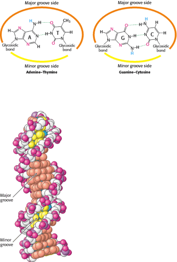
Figure 27.7. Major- and Minor-Groove Sides. Because the two glycosidic bonds are not diametrically opposite each
other, each base pair has a larger side that defines the major groove and a smaller side that defines the minor groove. The
grooves are lined by potential hydrogen-bond donors (blue) and acceptors (red).
III. Synthesizing the Molecules of Life 27. DNA Replication, Recombination, and Repair 27.1. DNA Can Assume a Variety of Structural Forms
Figure 27.8. Major and Minor Grooves in B-Form DNA. The major groove is depicted in orange, and the minor
groove is depicted in yellow. The carbon atoms of the backbone are shown in white.
III. Synthesizing the Molecules of Life 27. DNA Replication, Recombination, and Repair 27.1. DNA Can Assume a Variety of Structural Forms
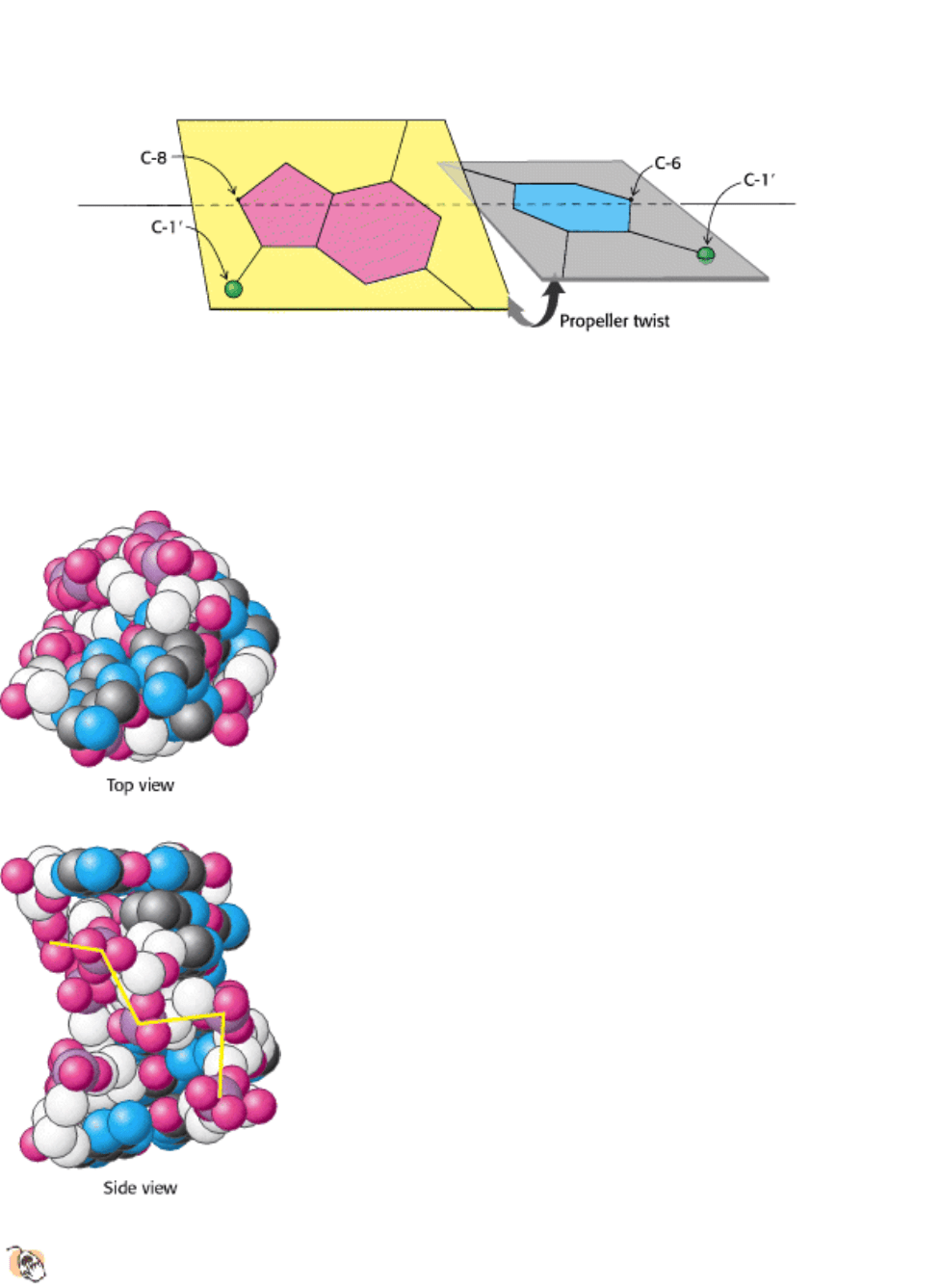
Figure 27.9. Propeller Twist. The bases of a DNA base pair are often not precisely coplanar. They are twisted with
respect to each other, like the blades of a propeller.
III. Synthesizing the Molecules of Life 27. DNA Replication, Recombination, and Repair 27.1. DNA Can Assume a Variety of Structural Forms
Figure 27.10. Z-DNA. DNA oligomers such as dCGCGCG adopt an alternative conformation under some conditions.
This conformation is called Z-DNA because the phosphate groups zigzag along the backbone.

III. Synthesizing the Molecules of Life 27. DNA Replication, Recombination, and Repair 27.1. DNA Can Assume a Variety of Structural Forms
Table 27.1. Comparison of A-, B-, and Z-DNA
Helix type
A B Z
Shape Broadest Intermediate Narrowest
Rise per base pair 2.3 Å 3.4 Å 3.8 Å
Helix diameter 25.5 Å 23.7 Å 18.4 Å
Screw sense Right-handed Right-handed Left-handed
Glycosidic bond anti anti alternating anti and
syn
Base pairs per turn of helix 11 10.4 12
Pitch per turn of helix 25.3 Å 35.4 Å 45.6 Å
Tilt of base pairs from normal to helix axis 19° 1° 9°
Major groove Narrow and very deep Wide and quite deep Flat
Minor groove Very broad and shallow Narrow and quite deep Very narrow and deep
III. Synthesizing the Molecules of Life 27. DNA Replication, Recombination, and Repair
27.2. DNA Polymerases Require a Template and a Primer
DNA polymerases catalyze the formation of polynucleotide chains through the addition of successive nucleotides
derived from deoxynucleoside triphosphates. The polymerase reaction takes place only in the presence of an appropriate
DNA template. Each incoming nucleoside triphosphate first forms an appropriate base pair with a base in this template.
Only then does the DNA polymerase link the incoming base with the predecessor in the chain. Thus, DNA polymerases
are template-directed enzymes.
DNA polymerases add nucleotides to the 3
end of a polynucleotide chain. The polymerase catalyzes the nucleophilic
attack of the 3
-hydroxyl group terminus of the polynucleotide chain on the α-phosphate group of the nucleoside
triphosphate to be added (see Figure 5.22). To initiate this reaction, DNA polymerases require a primer with a free 3 -
hydroxyl group already base-paired to the template. They cannot start from scratch by adding nucleotides to a free single-
stranded DNA template. RNA polymerase, in contrast, can initiate RNA synthesis without a primer (Section 28.1.4).
27.2.1. All DNA Polymerases Have Structural Features in Common
The three-dimensional structures of a number of DNA polymerase enzymes are known. The first such structure to be
determined was that of the so-called Klenow fragment of DNA polymerase I from E. coli (Figure 27.11). This fragment
comprises two main parts of the full enzyme, including the polymerase unit. This unit approximates the shape of a right
hand with domains that are referred to as the fingers, the thumb, and the palm. In addition to the polymerase, the Klenow
fragment includes a domain with 3
5 exonuclease activity that participates in proofreading and correcting the
polynucleotide product (Section 27.2.4).
DNA polymerases are remarkably similar in overall shape, although they differ substantially in detail. At least five
structural classes have been identified; some of them are clearly homologous, whereas others are probably the

products of convergent evolution. In all cases, the finger and thumb domains wrap around DNA and hold it across the
enzyme's active site, which comprises residues primarily from the palm domain. Furthermore, all the polymerases
catalyze the same polymerase reaction, which is dependent on two metal ions.
27.2.2. Two Bound Metal Ions Participate in the Polymerase Reaction
Like all enzymes with nucleoside triphosphate substrates, DNA polymerases require metal ions for activity. Examination
of the structures of DNA polymerases with bound substrates and substrate analogs reveals the presence of two metal ions
in the active site. One metal ion binds both the deoxynucleoside triphosphate (dNTP) and the 3
-hydroxyl group of the
primer, whereas the other interacts only with the 3
-hydroxyl group (Figure 27.12). The two metal ions are bridged by
the carboxylate groups of two aspartate residues in the palm domain of the polymerase. These side chains hold the metal
ions in the proper position and orientation. The metal ion bound to the primer activates the 3
-hydroxyl group of the
primer, facilitating its attack on the α-phosphate group of the dNTP substrate in the active site. The two metal ions
together help stabilize the negative charge that accumulates on the pentacoordinate transition state. The metal ion
initially bound to dNTP stabilizes the negative charge on the pyrophosphate product.
27.2.3. The Specificity of Replication Is Dictated by Hydrogen Bonding and the
Complementarity of Shape Between Bases
DNA must be replicated with high fidelity. Each base added to the growing chain should with high probability be the
Watson-Crick complement of the base in the corresponding position in the template strand. The binding of the NTP
containing the proper base is favored by the formation of a base pair, which is stabilized by specific hydrogen bonds.
The binding of a noncomplementary base is unlikely, because the interactions are unfavorable. The hydrogen bonds
linking two complementary bases make a significant contribution to the fidelity of DNA replication. However, DNA
polymerases replicate DNA more faithfully than these interactions alone can account for.
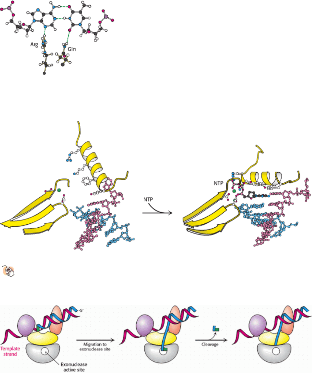
The examination of the crystal structures of various DNA polymerases indicated several additional mechanisms by
which replication fidelity is improved. First, residues of the enzyme form hydrogen bonds with the minor-groove side of
the base pair in the active site (Figure 27.13). In the minor groove, hydrogen-bond acceptors are present in the same
positions for all Watson-Crick base pairs. These interactions act as a "ruler" that measures whether a properly spaced
base pair has formed in the active site. Second, DNA polymerases close down around the incoming NTP (Figure 27.14).
The binding of a nucleoside triphosphate into the active site of a DNA polymerase triggers a conformational change: the
finger domain rotates to form a tight pocket into which only a properly shaped base pair will readily fit. The mutation of
a conserved tyrosine residue at the top of the pocket results in a polymerase that is approximately 40 times as error prone
as the parent polymerase.
27.2.4. Many Polymerases Proofread the Newly Added Bases and Excise Errors
Many polymerases further enhance the fidelity of replication by the use of proofreading mechanisms. As already noted,
the Klenow fragment of E. coli DNA polymerase I includes an exonuclease domain that does not participate in the
polymerization reaction itself. Instead, this domain removes mismatched nucleotides from the 3
end of DNA by
hydrolysis. The exonuclease active site is 35 Å from the polymerase active site, yet it can be reached by the newly
synthesized polynucleotide chain under appropriate conditions. The proofreading mechanism relies on the increased
probability that the end of a growing strand with an incorrectly incorporated nucleotide will leave the polymerase site
and transiently move to the exonuclease site (Figure 27.15).
How does the enzyme sense whether a newly added base is correct? First, an incorrect base will not pair correctly with
the template strand. Its greater structural fluctuation, permitted by the weaker hydrogen bonding, will frequently bring
the newly synthesized strand to the exonuclease site. Second, after the addition of a new nucleotide, the DNA
translocates by one base pair into the enzyme. The newly formed base pair must be of the proper dimensions to fit into a
tight binding site and participate in hydrogen-bonding interactions in the minor groove similar to those in the
polymerization site itself (see Figure 27.13). Indeed, the duplex DNA within the enzyme adopts an A-form structure,
allowing clear access to the minor groove. If an incorrect base is incorporated, the enzyme stalls, and the pause provides
additional time for the strand to migrate to the exonuclease site. There is a cost to this editing function, however: DNA
polymerase I removes approximately 1 correct nucleotide in 20 by hydrolysis. Although the removal of correct
nucleotides is slightly wasteful energetically, proofreading increases the accuracy of replication by a factor of
approximately 1000.
27.2.5. The Separation of DNA Strands Requires Specific Helicases and ATP
Hydrolysis
For a double-stranded DNA molecule to replicate, the two strands of the double helix must be separated from each other,
at least locally. This separation allows each strand to act as a template on which a new polynucleotide chain can be
assembled. For long double-stranded DNA molecules, the rate of spontaneous strand separation is negligibly low under
physiological conditions. Specific enzymes, termed helicases, utilize the energy of ATP hydrolysis to power strand
separation.
The detailed mechanisms of helicases are still under active investigation. However, the determination of the three-
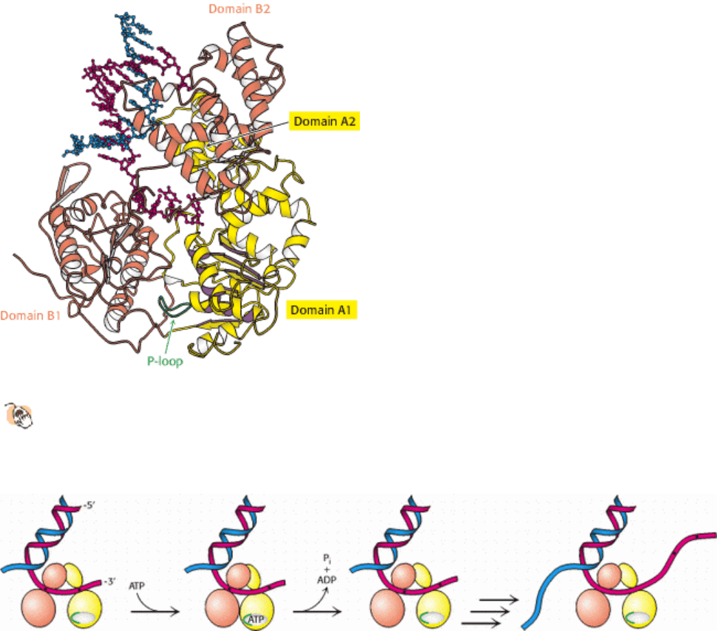
dimensional structures of several helicases has been a source of insight. For example, a bacterial helicase called PcrA
comprises four domains, hereafter referred to as domains A1, A2, B1, and B2 (Figure 27.16). Domain A1 contains a P-
loop NTPase fold, as was expected from amino acid sequence analysis. This domain participates in ATP binding and
hydrolysis. Domain B1 is homologous to domain A1 but lacks a P-loop. Domains A2 and B2 have unique structures.
From an analysis of a set of helicase crystal structures bound to nucleotide analogs and appropriate double- and single-
stranded DNA molecules, a mechanism for the action of these enzymes was proposed (Figure 27.17). Domains A1 and
B1 are capable of binding single-stranded DNA. In the absence of bound ATP, both domains are bound to DNA. The
binding of ATP triggers conformational changes in the P-loop and adjacent regions that lead to the closure of the cleft
between these two domains. To achieve this movement, domain A1 releases the DNA and slides along the DNA strand,
moving closer to domain B1. The enzyme then catalyzes the hydrolysis of ATP to form ADP and orthophosphate. On
product release, the cleft between domains A and B springs open. In this state, however, domain A1 has a tighter grip on
the DNA than does domain B1, so the DNA is pulled across domain B1 toward domain A1. The result is the
translocation of the enzyme along the DNA strand in a manner similar to the way in which an inchworm moves. In
regard to PcrA, the enzyme translocates in the 3
5 direction. When the helicase encounters a region of double-
stranded DNA, it continues to move along one strand and displaces the opposite DNA strand as it progresses.
Interactions with specific pockets on the helicase help destabilize the DNA duplex, aided by ATP-induced
conformational changes.
Helicases constitute a large and diverse class of enzymes. Some of these enzymes move in a 5 3 direction,
whereas others unwind RNA rather than DNA and participate in processes such as RNA splicing and the initiation
of mRNA translation. A comparison of the amino acid sequences of hundreds of these enzymes reveals seven regions of
striking conservation (Figure 27.18). Mapping these regions onto the PcrA structure shows that they line the ATP-
binding site and the cleft between the two domains, consistent with the notion that other helicases undergo
conformational changes analogous to those found in PcrA. However, whereas PcrA appears to function as a monomer,
other members of the helicase class function as oligomers. The hexameric structures of one important group are similar
to that of the F
1
component of ATP synthase (Section 18.4.1), suggesting potential mechanistic similarities.
III. Synthesizing the Molecules of Life 27. DNA Replication, Recombination, and Repair 27.2. DNA Polymerases Require a Template and a Primer
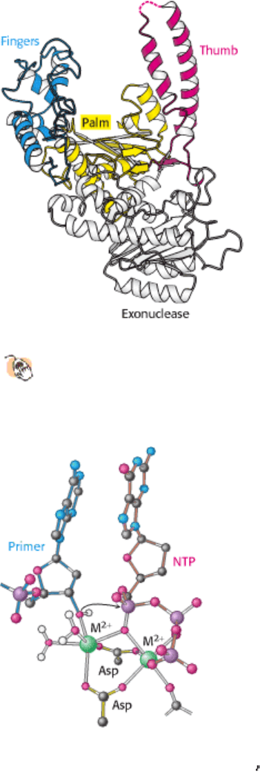
Figure 27.11. DNA Polymerase Structure.
The first DNA polymerase structure determined was that of a fragment of
E. coli DNA polymerase I called the Klenow fragment. Like other DNA polymerases, the polymerase unit
resembles a right hand with fingers (blue), palm (yellow), and thumb (red). The Klenow fragment also includes an
exonuclease domain.
III. Synthesizing the Molecules of Life 27. DNA Replication, Recombination, and Repair 27.2. DNA Polymerases Require a Template and a Primer
Figure 27.12. DNA Polymerase Mechanism. Two metal ions (typically, Mg
2+
) participate in the DNA polymerase
reaction. One metal ion coordinates the 3 -hydroxyl group of the primer, whereas the phosphate group of the nucleoside
triphosphate bridges between the two metal ions. The hydroxyl group of the primer attacks the phosphate group to form
a new O-P bond.
III. Synthesizing the Molecules of Life 27. DNA Replication, Recombination, and Repair 27.2. DNA Polymerases Require a Template and a Primer
Figure 27.13. Minor-Groove Interactions. DNA polymerases donate two hydrogen bonds to base pairs in the minor
groove. Hydrogen-bond acceptors are present in these two positions for all Watson-Crick base pairs including the A-T
base pair shown.
III. Synthesizing the Molecules of Life 27. DNA Replication, Recombination, and Repair 27.2. DNA Polymerases Require a Template and a Primer
Figure 27.14. Shape Selectivity.
The binding of a nucleoside triphosphate (NTP) to DNA polymerase induces a
conformational change, generating a tight pocket for the base pair consisting of the NTP and its partner on the
template strand. Such a conformational change is possible only when the NTP corresponds to the Watson-Crick
partner of the template base.
III. Synthesizing the Molecules of Life 27. DNA Replication, Recombination, and Repair 27.2. DNA Polymerases Require a Template and a Primer
Figure 27.15. Proofreading. The growing polynucleotide chain occasionally leaves the polymerase site of DNA
polymerase I and migrates to the exonuclease site. There, the last nucleotide added is removed by hydrolysis. Because
mismatched bases are more likely to leave the polymerase site, this process serves to proofread the sequence of the DNA
being synthesized.
III. Synthesizing the Molecules of Life 27. DNA Replication, Recombination, and Repair 27.2. DNA Polymerases Require a Template and a Primer
Figure 27.16. Helicase Structure.
The bacterial helicase PcrA comprises four domains: A1, A2, B1, and B2. The A1
domain includes a P-loop NTPase fold, whereas the B1 domain has a similar overall structure but lacks a P-loop
and does not bind nucleotides. Single-stranded DNA binds to the A1 and B1 domains near the interfaces with
domains A2 and B2.
III. Synthesizing the Molecules of Life 27. DNA Replication, Recombination, and Repair 27.2. DNA Polymerases Require a Template and a Primer
Figure 27.17. Helicase Mechanism. Initially, both domains A1 and B1 of PcrA bind single-stranded DNA. On binding
of ATP, the cleft between these domains closes and domain A1 slides along the DNA. On ATP hydrolysis, the cleft
opens up, pulling the DNA from domain B1 toward domain A1. As this process is repeated, double-stranded DNA is
unwound.
III. Synthesizing the Molecules of Life 27. DNA Replication, Recombination, and Repair 27.2. DNA Polymerases Require a Template and a Primer
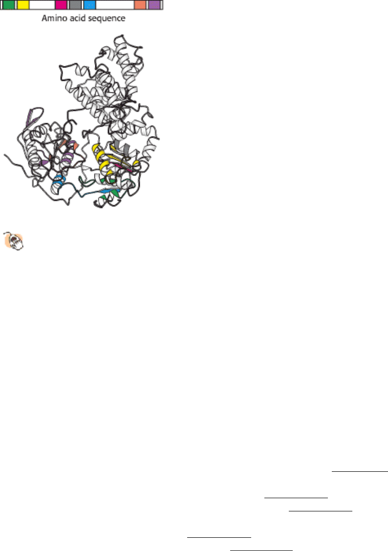
Figure 27.18. Conserved Residues among Helicases.
A comparison of the amino acid sequences of hundreds of
helicases revealed seven regions of strong sequence conservation (shown in color). When mapped onto the
structure of PcrA, these conserved regions lie along the interface between the A1 and B1 domains and along the
ATP binding surface.
III. Synthesizing the Molecules of Life 27. DNA Replication, Recombination, and Repair
27.3. Double-Stranded DNA Can Wrap Around Itself to Form Supercoiled Structures
The separation of the two strands of DNA in replication requires the local unwinding of the double helix. This local
unwinding must lead either to the overwinding of surrounding regions of DNA or to supercoiling. To prevent the strain
induced by overwinding, a specialized set of enzymes is present to introduce supercoils that favor strand separation.
27.3.1. The Linking Number of DNA, a Topological Property, Determines the Degree of
Supercoiling
In 1963, Jerome Vinograd found that circular DNA from polyoma virus separated into two distinct species when it was
centrifuged. In pursuing this puzzle, he discovered an important property of circular DNA not possessed by linear DNA
with free ends. Consider a linear 260-bp DNA duplex in the B-DNA form (Figure 27.19). Because the number of
residues per turn in an unstressed DNA molecule is 10.4, this linear DNA molecule has 25 (260/10.4) turns. The ends of
this helix can be joined to produce a relaxed circular DNA (Figure 27.19B). A different circular DNA can be formed by
unwinding the linear duplex by two turns before joining its ends (Figure 27.19C). What is the structural consequence of
unwinding before ligation? Two limiting conformations are possible: the DNA can either fold into a structure containing
23 turns of B helix and an unwound loop (Figure 27.19D) or adopt a supercoiled structure with 25 turns of B helix and 2
turns of right-handed (termed negative) superhelix (Figure 27.19E).
Supercoiling markedly alters the overall form of DNA. A supercoiled DNA molecule is more compact than a relaxed
DNA molecule of the same length. Hence, supercoiled DNA moves faster than relaxed DNA when analyzed by
centrifugation or electrophoresis. The rapidly sedimenting DNA in Vinograd's experiment was supercoiled, whereas the
slowly sedimenting DNA was relaxed because one of its strands was nicked. Unwinding will cause supercoiling in both
circular DNA molecules and in DNA molecules that are constrained in closed configurations by other means.
27.3.2. Helical Twist and Superhelical Writhe Are Correlated with Each Other
Through the Linking Number
Our understanding of the conformation of DNA is enriched by concepts drawn from topology, a branch of mathematics
dealing with structural properties that are unchanged by deformations such as stretching and bending. A key topological
property of a circular DNA molecule is its linking number (Lk), which is equal to the number of times that a strand of
DNA winds in the right-handed direction around the helix axis when the axis is constrained to lie in a plane. For the
relaxed DNA shown in Figure 27.19B, Lk = 25. For the partly unwound molecule shown in part D and the supercoiled
one shown in part E, Lk = 23 because the linear duplex was unwound two complete turns before closure. Molecules
differing only in linking number are topological isomers (topoisomers) of one another. Topoisomers of DNA can be
interconverted only by cutting one or both DNA strands and then rejoining them.
The unwound DNA and supercoiled DNA shown in Figure 27.19D and E are topologically identical but geometrically
different. They have the same value of Lk but differ in Tw (twist) and Wr (writhe). Although the rigorous definitions of
twist and writhe are complex, twist is a measure of the helical winding of the DNA strands around each other, whereas
writhe is a measure of the coiling of the axis of the double helix, which is called super-coiling. A right-handed coil is
assigned a negative number (negative supercoiling) and a left-handed coil is assigned a positive number (positive
supercoiling). Is there a relation between Tw and Wr? Indeed, there is. Topology tells us that the sum of Tw and Wr is
equal to Lk.
In Figure 27.19, the partly unwound circular DNA has Tw ~ 23 and Wr ~ 0, whereas the supercoiled DNA has Tw ~ 25
and Wr ~ -2. These forms can be interconverted without cleaving the DNA chain because they have the same value of
Lk; namely, 23. The partitioning of Lk (which must be an integer) between Tw and Wr (which need not be integers) is
determined by energetics. The free energy is minimized when about 70% of the change in Lk is expressed in Wr and
30% is expressed in Tw. Hence, the most stable form would be one with Tw = 24.4 and Wr = -1.4. Thus, a lowering of
Lk causes both right-handed (negative) supercoiling of the DNA axis and unwinding of the duplex. Topoisomers
differing by just 1 in Lk, and consequently by 0.7 in Wr, can be readily separated by agarose gel electrophoresis because
their hydrodynamic volumes are quite different supercoiling condenses DNA (Figure 27.20). Most naturally occurring
DNA molecules are negatively supercoiled. What is the basis for this prevalence? As already stated, negative
supercoiling arises from the unwinding or underwinding of the DNA. In essence, negative supercoiling prepares DNA
for processes requiring separation of the DNA strands, such as replication or transcription. Positive supercoiling
condenses DNA as effectively, but it makes strand separation more difficult.
27.3.3. Type I Topoisomerases Relax Supercoiled Structures
The interconversion of topoisomers of DNA is catalyzed by enzymes called topoisomerases which were discovered by
James Wang and Martin Gellert. These enzymes alter the linking number of DNA by catalyzing a three-step process: (1)
the cleavage of one or both strands of DNA, (2) the passage of a segment of DNA through this break, and (3) the
resealing of the DNA break. Type I topoisomerases cleave just one strand of DNA, whereas type II enzymes cleave both
strands. Both type I and type II topoisomerases play important roles in DNA replication and in transcription and
recombination.
Type I topoisomerases catalyze the relaxation of supercoiled DNA, a thermodynamically favorable process. Type II
topoisomerases utilize free energy from ATP hydrolysis to add negative supercoils to DNA. The two types of enzymes
have several common features, including the use of key tyrosine residues to form covalent links to the polynucleotide
backbone that is transiently broken.
The three-dimensional structures of several type I topoisomerases have been determined (Figure 27.21). These structures
reveal many features of the reaction mechanism. Human type I topoisomerase comprises four domains, which are
arranged around a central cavity having a diameter of 20 Å, just the correct size to accommodate a double-stranded DNA
molecule. This cavity also includes a tyrosine residue (Tyr 723), which acts as a nucleophile to cleave the DNA
backbone in the course of catalysis.
From analyses of these structures and the results of other studies, the relaxation of negatively supercoiled DNA