Berg J.M., Tymoczko J.L., Stryer L. Biochemistry
Подождите немного. Документ загружается.


(e) What can you conclude about the role of glutamate 3 in carnitine acyltransferase I function?
See answer
Media Problem
20.
E pluribus unum. The storage of chemical energy in the form of fatty acids requires carbon building blocks in
the form of acetyl-CoA and energy in the form of ATP and NADPH. Using the Conceptual Insights
overview of carbohydrate and fatty acid metabolism, estimate how many glucose molecules would be required to
form one molecule of palmitate (C
16
) if glucose were the sole carbon and energy source.
II. Transducing and Storing Energy 22. Fatty Acid Metabolism
Selected Readings
Where to start
S.J. Wakil. 1989. Fatty acid synthase, a proficient multifunctional enzyme Biochemistry 28: 4523-4530. (PubMed)
B.B. Rasmussen and R.R. Wolfe. 1999. Regulation of fatty acid oxidation in skeletal muscle Annu. Rev. Nutr. 19: 463-
484. (PubMed)
C.F. Semenkovich. 1997. Regulation of fatty acid synthase (FAS) Prog. Lipid Res. 36: 43-53. (PubMed)
H.S. Sul, C.M. Smas, D. Wang, and L. Chen. 1998. Regulation of fat synthesis and adipose differentiation Prog. Nucleic
Acid Res. Mol. Biol. 60: 317-345. (PubMed)
G. Wolf. 1996. Nutritional and hormonal regulation of fatty acid synthase Nutr. Rev. 54: 122-123. (PubMed)
M.R. Munday and C.J. Hemingway. 1999. The regulation of acetyl-CoA carboxylase: A potential target for the action of
hypolipidemic agents Adv. Enzyme Regul. 39: 205-234. (PubMed)
Books
Vance, D. E., and Vance, J. E. (Eds.), 1996. Biochemistry of Lipids, Lipoproteins, and Membranes . Elsevier.
Boyer, P. D. (Ed.), 1983. The Enzymes (3d ed.). Vol. 16, Lipid Enzymology . Academic Press.
Numa, S. (Ed.), 1984. Fatty Acid Metabolism and Its Regulation . Elsevier.
Fatty acid oxidation
J.J. Barycki, L.K. O'Brien, A.W. Strauss, and L.J. Banaszak. 2000. Sequestration of the active site by interdomain
shifting: Crystallographic and spectroscopic evidence for distinct conformations of
l-3-hydroxyacyl-CoA dehydrogenase
J. Biol. Chem. 275: 27186-27196. (PubMed)
R.R. Ramsay. 2000. The carnitine acyltransferases: Modulators of acyl-CoA-dependent reactions Biochem. Soc. Trans.
28: 182-186. (PubMed)
S. Eaton, K. Bartlett, and M. Pourfarzam. 1996. Mammalian mitochondrial beta-oxidation Biochem. J. 320: 345-357.
(PubMed)
C. Thorpe and J.J. Kim. 1995. Structure and mechanism of action of the acyl-CoA dehydrogenases FASEB J. 9: 718-725.
(PubMed)

D.W. Foster. 1984. From glycogen to ketones and back Diabetes 33: 1188-1199. (PubMed)
J.D. McGarry and D.W. Foster. 1980. Regulation of hepatic fatty acid oxidation and ketone body production Annu. Rev.
Biochem. 49: 395-420. (PubMed)
Fatty acid synthesis
Y.-M. Zhang, M.S. Rao, R.J. Heath, A.C. Price, A.J. Olson, C.O. Rock, and S.W. White. 2001. Identification and
analysis of the acyl carrier protein (ACP) docking site on beta-ketoacyl-ACP synthase III J. Biol. Chem. 276: 8231-8238.
(PubMed)
C. Davies, R.J. Heath, S.W. White, and C.O. Rock. 2000. The 1 8 Å crystal structure and active-site architecture of beta-
ketoacyl-acyl carrier protein synthase III (FabH) from Escherichia coli Structure Fold Des. 8: 185-195. (PubMed)
R.M. Denton, K.J. Heesom, S.K. Moule, N.J. Edgell, and P. Burnett. 1997. Signalling pathways involved in the
stimulation of fatty acid synthesis by insulin Biochem. Soc. Trans. 25: 1238-1242. (PubMed)
J.K. Stoops, S.J. Kolodziej, J.P. Schroeter, J.P. Bretaudiere, and S.J. Wakil. 1992. Structure-function relationships of the
yeast fatty acid synthase: Negative-stain, cryo-electron microscopy, and image analysis studies of the end views of the
structure Proc. Natl. Acad. Sci. USA 89: 6585-6589. (PubMed) (Full Text in PMC)
T.M. Loftus, D.E. Jaworsky, G.L. Frehywot, C.A. Townsend, G.V. Ronnett, M.D. Lane, and F.P. Kuhajda. 2000.
Reduced food intake and body weight in mice treated with fatty acid synthase inhibitors Science 288: 2379-2381.
(PubMed)
Acetyl CoA carboxylase
J.B. Thoden, C.Z. Blanchard, H.M. Holden, and G.L. Waldrop. 2000. Movement of the biotin carboxylase B-domain as
a result of ATP binding J. Biol. Chem. 275: 16183-16190. (PubMed)
K.H. Kim. 1997. Regulation of mammalian acetyl-coenzyme A carboxylase Annu. Rev. Nutr. 17: 77-99. (PubMed)
G.M. Mabrouk, I.M. Helmy, K.G. Thampy, and S.J. Wakil. 1990. Acute hormonal control of acetyl-CoA carboxylase:
The roles of insulin, glucagon, and epinephrine J. Biol. Chem. 265: 6330-6338. (PubMed)
K.H. Kim, C.F. Lopez, D.H. Bai, X. Luo, and M.E. Pape. 1989. Role of reversible phosphorylation of acetyl-CoA
carboxylase in long-chain fatty acid synthesis FASEB J. 3: 2250-2256. (PubMed)
L.A. Witters and B.E. Kemp. 1992. Insulin activation of acetyl-CoA carboxylase accompanied by inhibition of the 5 -
AMP-activated protein kinase J. Biol. Chem. 267: 2864-2867. (PubMed)
P. Cohen and D.G. Hardie. 1991. The actions of cyclic AMP on biosynthetic processes are mediated indirectly by cyclic
AMP-dependent protein kinases Biochim. Biophys. Acta 1094: 292-299. (PubMed)
F. Moore, J. Weekes, and D.G. Hardie. 1991. Evidence that AMP triggers phosphorylation as well as direct allosteric
activation of rat liver AMP-activated protein kinase: A sensitive mechanism to protect the cell against ATP depletion
Eur. J. Biochem. 199: 691-697. (PubMed)
Eicosanoids
M.G. Malkowski, S.L. Ginell, W.L. Smith, and R.M. Garavito. 2000. The productive conformation of arachidonic acid
bound to prostaglandin synthase Science 289: 1933-1937. (PubMed)
T. Smith, J. McCracken, Y.-K. Shin, and D. DeWitt. 2000. Arachidonic acid and nonsteroidal anti-inflammatory drugs
induce conformational changes in the human prostaglandin endoperoxide H2 synthase-2 (cyclooxygenase-2) J. Biol.
Chem. 275: 40407-40415. (PubMed)

A.S. Kalgutkar, B.C. Crews, S.W. Rowlinson, C. Garner, K. Seibert, and L.J. Marnett. 1998. Aspirin-like molecules that
covalently inactivate cyclooxygenase-2 Science 280: 1268-1270. (PubMed)
W.E. Lands. 1991. Biosynthesis of prostaglandins Annu. Rev. Nutr. 11: 41-60. (PubMed)
E. Sigal. 1991. The molecular biology of mammalian arachidonic acid metabolism Am. J. Physiol. 260: L13-L28.
(PubMed)
G. Weissmann. 1991. Aspirin Sci. Am. 264: (1) 84-90. (PubMed)
Vane, J. R., Flower, R. J., and Botting, R. M., 1990. History of aspirin and its mechanism of action. Stroke (12 suppl.):
IV12 IV23.
Genetic diseases
M. Brivet, A. Boutron, A. Slama, C. Costa, L. Thuillier, F. Demaugre, D. Rabier, J.M. Saudubray, and J.P. Bonnefont.
1999. Defects in activation and transport of fatty acids J. Inherited Metab. Dis. 22: 428-441. (PubMed)
R.J. Wanders, E.G. van Grunsven, and G.A. Jansen. 2000. Lipid metabolism in peroxisomes: Enzymology, functions and
dysfunctions of the fatty acid alpha- and beta-oxidation systems in humans Biochem. Soc. Trans. 28: 141-149. (PubMed)
R.J. Wanders, P. Vreken, M.E. den Boer, F.A. Wijburg, A.H. van Gennip, and L. IJlst. 1999. Disorders of mitochondrial
fatty acyl-CoA β-oxidation J. Inherited Metab. Dis. 22: 442-487. (PubMed)
J. Kerner and C. Hoppel. 1998. Genetic disorders of carnitine metabolism and their nutritional management Annu. Rev.
Nutr. 18: 179-206. (PubMed)
K. Bartlett and M. Pourfarzam. 1998. Recent developments in the detection of inherited disorders of mitochondrial β-
oxidation Biochem. Soc. Trans. 26: 145-152. (PubMed)
R.J. Pollitt. 1995. Disorders of mitochondrial long-chain fatty acid oxidation J. Inherited Metab. Dis. 18: 473-490.
(PubMed)
Roe, C. R., and Coates, P. M., 1995. Mitochondrial fatty acid oxidation disorders. In The Metabolic Basis of Inherited
Diseases (7th ed., pp. 1501 1534), edited by C. R. Striver, A. L. Beaudet, W. S. Sly, D. Valle, J. B. Stanbury, J. B.
Wyngaarden, and D. S. Fredrickson. McGraw-Hill.
II. Transducing and Storing Energy
23. Protein Turnover and Amino Acid Catabolism
The assembly of new proteins requires a source of amino acids. These building blocks are generated by the digestion of
proteins in the intestine and the degradation of proteins within the cell. Many cellular proteins are constantly degraded
and resynthesized. To facilitate this recycling, a complex system for the controlled turnover of proteins has evolved.
Damaged or unneeded proteins are marked for destruction by the covalent attachment of chains of a small protein,
ubiquitin. Polyubiquitinated proteins are subsequently degraded by a large, ATP-dependent complex called the
proteasome.
The primary uses of amino acids are as building blocks for protein and peptide synthesis and as a source of nitrogen for
the synthesis of other amino acids and other nitrogenous compounds such as nucleotide bases. Amino acids in excess of
those needed for biosynthesis cannot be stored, in contrast with fatty acids and glucose, nor are they excreted. Rather,
surplus amino acids are used as metabolic fuel. The α-amino group is removed, and the resulting carbon skeleton is
converted into a major metabolic intermediate. Most of the amino groups of surplus amino acids are converted into urea
through the urea cycle, whereas their carbon skeletons are transformed into acetyl CoA, acetoacetyl CoA, pyruvate, or
one of the intermediates of the citric acid cycle. Hence, fatty acids, ketone bodies, and glucose can be formed from
amino acids.
Several coenzymes play key roles in amino acid degradation, foremost among them is pyridoxal phosphate. This
coenzyme forms Schiff-base intermediates that allow α-amino groups to be shuttled between amino acids and ketoacids.
We will consider several genetic errors of amino acid degradation that lead to brain damage and mental retardation
unless remedial action is initiated soon after birth. Phenylketonuria, which is caused by a block in the conversion of
phenylalanine into tyrosine, is readily diagnosed and can be treated by removing phenylalanine from the diet. The study
of amino acid metabolism is especially rewarding because it is rich in connections between basic biochemistry and
clinical medicine.
II. Transducing and Storing Energy 23. Protein Turnover and Amino Acid Catabolism
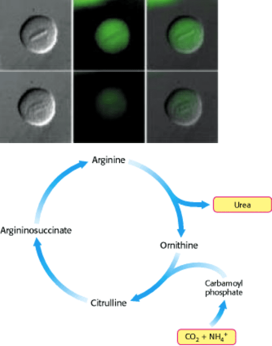
Degradation of cyclin B. This important protein in cell cycle regulation is visible as the green areas in the images above
(the protein was fused with green fluorescent protein). Cyclin B is prominent during metaphase, but is degraded in
anaphase to prevent premature initiation of another cell cycle. A large protease complex called the proteasome digests
the protein into amino acids. These are either reused, or further processed by the urea cycle, which removes the nitrogen
as urea. [(Above left) Courtesy of Jonathan Pines, University of Cambridge, Wellcome/CRC Institute of Cancer and
Developmental Biology.]
II. Transducing and Storing Energy 23. Protein Turnover and Amino Acid Catabolism
23.1. Proteins Are Degraded to Amino Acids
Dietary protein is a vital source of amino acids. Proteins ingested in the diet are digested into amino acids or small
peptides that can be absorbed by the intestine and transported in the blood. Another source of amino acids is the
degradation of defective or unneeded cellular proteins.
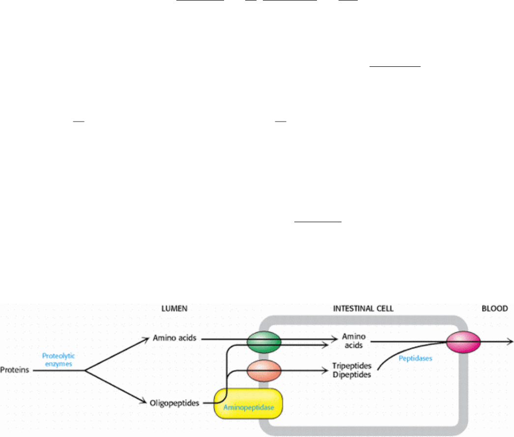
23.1.1. The Digestion and Absorption of Dietary Proteins
Protein digestion begins in the stomach, where the acidic environment favors protein denaturation. Denatured proteins
are more accessible as substrates for proteolysis than are native proteins. The primary proteolytic enzyme of the stomach
is pepsin, a nonspecific protease that, remarkably, is maximally active at pH 2. Thus, pepsin can be active in the highly
acidic environment of the stomach, even though other proteins undergo denaturation there.
Protein degradation continues in the lumen of the intestine owing to the activity of proteolytic enzymes secreted by the
pancreas. These proteins, introduced in Chapters 9 and 10 (Sections 9.1 and 10.5), are secreted as inactive zymogens and
then converted into active enzymes. The battery of enzymes displays a wide array of specificity, and so the substrates are
degraded into free amino acids as well as di- and tripeptides. Digestion is further enhanced by proteases, such as
aminopeptidase N, that are located in the plasma membrane of the intestinal cells. Aminopeptidases digest proteins from
the amino-terminal end. Single amino acids, as well as di- and tripeptides, are transported into the intestinal cells from
the lumen and subsequently released into the blood for absorption by other tissues (Figure 23.1).
23.1.2. Cellular Proteins Are Degraded at Different Rates
Protein turnover
the degradation and resynthesis of proteins takes place constantly in cells. Although some proteins
are very stable, many proteins are short lived, particularly those that are important in metabolic regulation. Altering the
amounts of these proteins can rapidly change metabolic patterns. In addition, cells have mechanisms for detecting and
removing damaged proteins. A significant proportion of newly synthesized protein molecules are defective because of
errors in translation. Even proteins that are normal when first synthesized may undergo oxidative damage or be altered in
other ways with the passage of time.
The half-lives of proteins range over several orders of magnitude (Table 23.1). Ornithine decarboxylase, at
approximately 11 minutes, has one of the shortest half-lives of any mammalian protein. This enzyme participates in the
synthesis of polyamines, which are cellular cations essential for growth and differentiation. The life of hemoglobin, on
the other hand, is limited only by the life of the red blood cell, and the lens protein, crystallin, by the life of the organism.
II. Transducing and Storing Energy 23. Protein Turnover and Amino Acid Catabolism 23.1. Proteins Are Degraded to Amino Acids
Figure 23.1. Digestion and Absorption of Proteins. Protein digestion is primarily a result of the activity of enzymes
secreted by the pancreas. Aminopeptidases associated with the intestinal epithelium further digest proteins. The amino
acids and di- and tripeptides are absorbed into the intestinal cells by specific transporters. Free amino acids are then
released into the blood for use by other tissues.
II. Transducing and Storing Energy 23. Protein Turnover and Amino Acid Catabolism 23.1. Proteins Are Degraded to Amino Acids
Table 23.1. Dependence of the half-lives of cytosolic yeast proteins on the nature of their amino-
terminal residues
Highly stabilizing residues
(t
1/2
> 20 hours)
Ala Cys Gly Met
Pro Ser Thr Val
Intrinsically destabilizing residues
(t
1/2
= 2 to 30 minutes)
Arg His Ile Leu
Lys Phe Trp Tyr
Destabilizing residues after chemical modification
(t
1/2
= 3 to 30 minutes)
Asn Asp Gln Glu
Source: J. W. Tobias, T. E. Schrader, G. Rocap, and A. Varshavsky. Science 254(1991):1374.
II. Transducing and Storing Energy 23. Protein Turnover and Amino Acid Catabolism
23.2. Protein Turnover Is Tightly Regulated
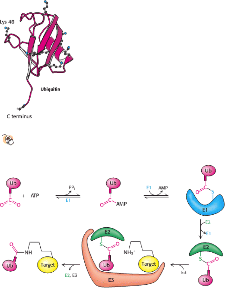
How can a cell distinguish proteins that are meant for degradation? Ubiq-uitin, a small (8.5-kd) protein present in all
eukaryotic cells, is the tag that marks proteins for destruction (Figure 23.2). Ubiquitin is the cellular equivalent of the
"black spot" of Robert Louis Stevenson's Treasure Island: the signal for death.
23.2.1. Ubiquitin Tags Proteins for Destruction
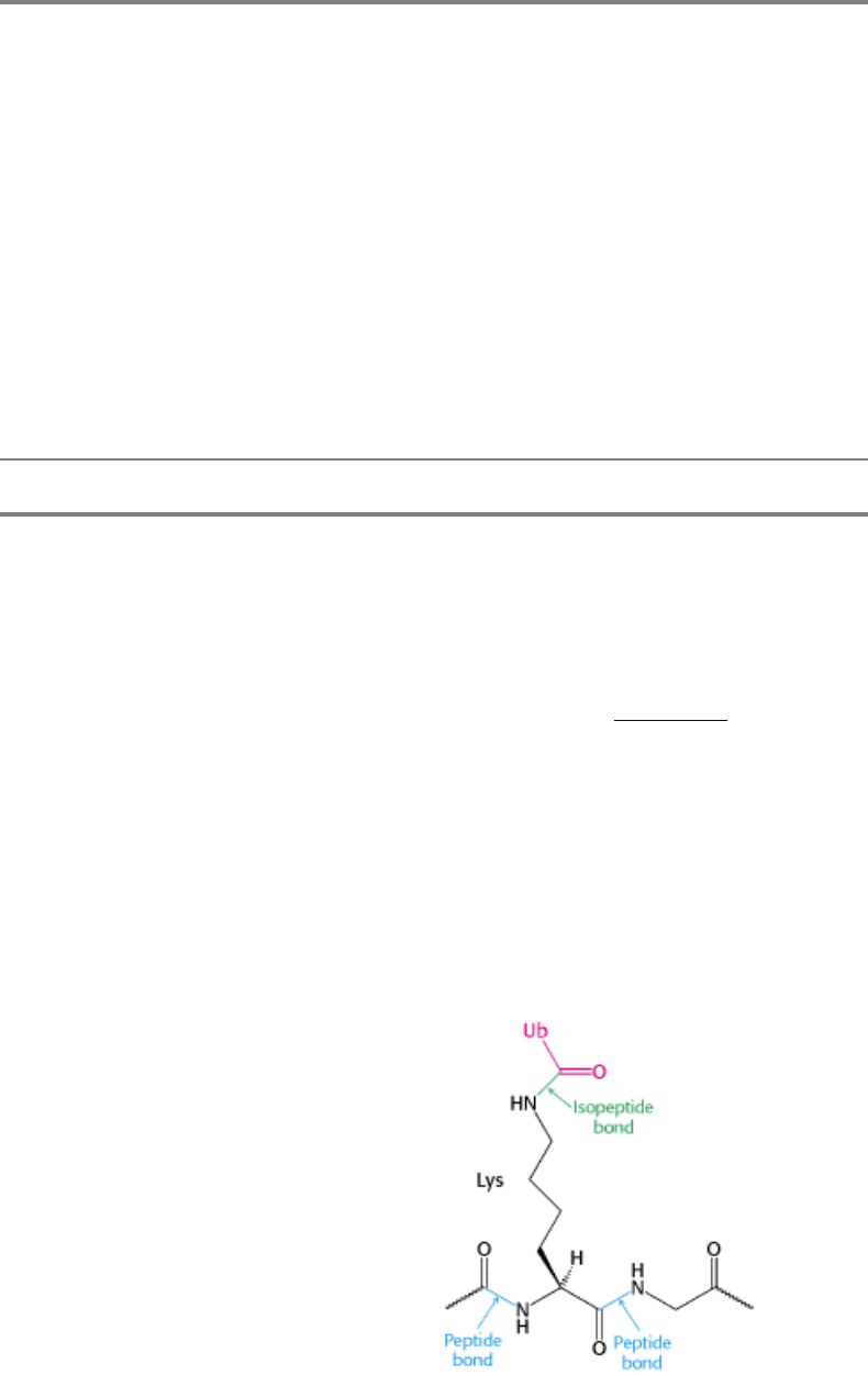
Ubiquitin is highly conserved in eukaryotes: yeast and human ubiquitin differ at only 3 of 76 residues. The carboxyl-
terminal glycine residue of ubiquitin (Ub) becomes covalently attached to the ε-amino groups of several lysine residues
on a protein destined to be degraded. The energy for the formation of these isopeptide bonds (iso because ε- rather than
α-amino groups are targeted) comes from ATP hydrolysis.

Three enzymes participate in the attachment of ubiquitin to each protein (Figure 23.3): ubiquitin-activating enzyme, or
E1, ubiquitin-conjugating enzyme, or E2, and ubiquitin-protein ligase, or E3. First, the terminal carboxylate group of
ubiquitin becomes linked to a sulfhydryl group of E1 by a thioester bond. This ATP-driven reaction is reminiscent of
fatty acid activation (Section 22.4.1). An adenylate is linked to the C-terminal carboxylate of ubiquitin with the release
of pyrophosphate, and the ubiquitin is transferred to a sulfhydryl group of a key cysteine residue in E1. Second, activated
ubiquitin is shuttled to a sulfhydryl group of E2. Finally, E3 catalyzes the transfer of ubiquitin from E2 to an ε-amino
group on the target protein.
The attachment of a single molecule of ubiquitin is only a weak signal for degradation. However, the ubiquitination
reaction is processive: chains of ubiquitin can be generated by the linkage of the ε-amino group of lysine residue 48 of
one ubiquitin molecule to the terminal carboxylate of another. Chains of four or more ubiquitin molecules are
particularly effective in signaling degradation (Figure 23.4). The use of chains of ubiquitin molecules may have at least
two advantages. First, the ubiquitin molecules interact with one another to form a binding surface distinct from that
created by a single ubiquitin molecule. Second, individual ubiquitin molecules can be cleaved off without loss of the
degradation signal.
Although most eukaryotes have only one or a small number of distinct E1 enzymes, all eukaryotes have many distinct E2
and E3 enzymes. Moreover, there appears to be only a single family of evolutionarily related E2 proteins but many
distinct families of E3 proteins. Although the E3 component provides most of the substrate specificity for ubiquitination,
the multiple combinations of the E2
E3 complex allow for more finely tuned substrate discrimination.
What determines whether a protein becomes ubiquitinated? One signal turns out to be unexpectedly simple. The half-life
of a cytosolic protein is determined to a large extent by its amino-terminal residue (see Table 23.1). This dependency is
referred to as the N-terminal rule. A yeast protein with methionine at its N terminus typically has a half-life of more than
20 hours, whereas one with arginine at this position has a half-life of about 2 minutes. A highly destabilizing N-terminal
residue such as arginine or leucine favors rapid ubiquitination, whereas a stabilizing residue such as methionine or
proline does not. E3 enzymes are the readers of N-terminal residues. Other signals thought to identify proteins for
degradation include cyclin destruction boxes, which are amino acid sequences that mark cell-cycle proteins for
destruction, and proteins rich in proline, glutamic acid, serine, and threonine (PEST sequences).
Some pathological conditions vividly illustrate the importance of the regulation of protein turnover. For example,
human papilloma virus (HPV) encodes a protein that activates a specific E3 enzyme. The enzyme ubiquitinates the
tumor suppressor p53 and other proteins that control DNA repair, which are then destroyed. The activation of this E3
enzyme is observed in more than 90% of cervical carcinomas. Thus, the inappropriate marking of key regulatory proteins
for destruction can trigger further events, leading to tumor formation.
23.2.2. The Proteasome Digests the Ubiquitin-Tagged Proteins
If ubiquitin is the mark of death, what is the executioner? A large protease complex called the proteasome or the 26S
proteasome digests the ubiquitinated proteins. This ATP-driven multisubunit protease spares ubiq-uitin, which is then
recycled. The 26S proteasome is a complex of two components: a 20S proteasome, which contains the catalytic activity,
and a 19S regulatory subunit.
The 20S complex is constructed from two copies each of 14 subunits and has a mass of 700 kd (Figure 23.5). All 14
subunits are homologous and adopt the same overall structure. The subunits are arranged in four rings of 7 subunits that
stack to form a structure resembling a barrel. The components of the two rings at the ends of the barrel are called the α
subunits and those of the two central rings the β subunits. The active sites of the protease are located at the N-termini of
certain β subunits on the interior of the barrel
specifically, those β chains having an N-terminal threonine or serine
residue. The hydroxyl groups of these amino acids are converted into nucleophiles with the assistance of their own
amino groups. These nucleophilic groups then attack the carbonyl groups of peptide bonds and form acyl-enzyme
intermediates (Section 9.1.2). The structure of the complex sequesters the proteolytic active sites from potential
substrates until they are directed into the barrel. Substrates are degraded in a processive manner without the release of

degradation intermediates, until the substrate is reduced to peptides ranging in length from seven to nine residues.
The 20S proteasome is a sealed barrel. Access to its interior is controlled by a 19S regulatory complex, itself a 700-kd
complex made up of 20 subunits. This complex binds to both ends of the 20S proteasome core to form the complete 26S
proteasome (Figure 23.6). The 19S subunit binds specifically to polyubiquitin chains. Key components of the 19S
complex are six distinct ATPases of the AAA class (ATPase associated with various cellular activities) characterized by
a conserved 230 amino acid ATP-binding domain of the P-loop NTPase family. This class of ATPase, found in all
kingdoms, is associated with a variety of cell functions including cell-cycle regulation and organelle biogenesis.
Although the exact role of the ATPase remains uncertain, ATP hydrolysis may assist the 19S complex to unfold the
substrate and induce conformational changes in the 20S proteasome so that the substrate can be passed into the center of
the complex. Finally, the 19S subunit also contains an isopeptidase that cleaves off intact ubiquitin molecules. Thus, the
ubiquitinization pathway and the proteasome cooperate to degrade unwanted proteins. The ubiquitin is recycled and the
peptide products are further degraded by other cellular proteases to yield individual amino acids.
23.2.3. Protein Degradation Can Be Used to Regulate Biological Function
Table 23.2 lists a number of physiological processes that are con trolled at least in part by protein degradation.
This control is exerted by dynamically altering the stability and abundance of regulatory proteins. Consider, for
example, control of the inflammatory response. A transcription factor called NF- κB (NF for nuclear factor) initiates the
expression of a number of the genes that take part in this response. This important factor is itself activated by the
degradation of an inhibitory protein, I- κB. NF- κB is maintained in the cytoplasm in its inactive state by association
with I-κB (I for inhibitor). In response to inflammatory signals, I- κ β is phosphorylated at two serine residues, creating
an E3 binding site. The binding of E3 leads to the ubiquitination and degradation of I- κB and thereby disrupts the
inhibitor's association with NF- κB. The liberated transcription factor migrates to the nucleus to stimulate transcription
of the target genes. The NF- κB-I- κB system illustrates the interplay of several key regulatory motifs: receptor-mediated
signal transduction, phosphorylation, compartmentalization, controlled and specific degradation, and selective gene
expression.
23.2.4. The Ubiquitin Pathway and the Proteasome Have Prokaryotic Counterparts
Both the ubiquitin pathway and the proteasome appear to be pres- ent in all eukaryotes. Homologs of the
proteasome are found in prokaryotes, although the physiological roles of these homologs have not been well
established. The proteasomes of some archaea are quite similar in overall structure to their eukaryotic counterparts and
similarly have 28 subunits (Figure 23.7). In the archaeal proteasome, however, all α subunits and all β subunits are
identical; in eukaryotes, each α or β subunit is one of seven different isoforms. This specialization provides distinct
substrate specificity.
Ubiquitin, however, has not been found in prokaryotes. Indeed, the high level of sequence similarity between the human
and yeast proteins suggests that ubiquitin, in its present form, diverged relatively recently in evolutionary terms.
Ubiquitin's molecular ancestors were recently identified in prokaryotes. Remarkably, these proteins take part not in
protein modification but in coenzyme biosynthesis (Figure 23.8). The biosynthesis of thiamine (Section 8.6.1) begins
with a sulfide ion derived from cysteine. This sulfide is added to the C-terminal carboxylate of the protein ThiS, which
had been activated as an acyl adenylate. The activation of ThiS and the addition of sulfide are catalyzed by the enzyme
ThiF. Human E1 includes two tandem regions of 160 amino acids that are 28% identical in amino acid sequence with a
region of ThiF from E. coli. The evolutionary relationships between these two pathways were cemented by the
determination of the three-dimensional structure of ThiS, which revealed a structure very similar to that of ubiquitin,
despite being only 14% identical in amino acid sequence (Figure 23.9). Thus, a eukaryotic system for protein
modification evolved from a preexisting prokaryotic pathway for coenzyme biosynthesis.
II. Transducing and Storing Energy 23. Protein Turnover and Amino Acid Catabolism 23.2. Protein Turnover Is Tightly Regulated
Figure 23.2. Ubiquitin.
The structure of ubiquitin reveals an extended carboxyl terminus that is activated and linked to
other proteins. Lysine residues also are shown, including lysine 48, the major site for linking additional ubiquitin
molecules.
II. Transducing and Storing Energy 23. Protein Turnover and Amino Acid Catabolism 23.2. Protein Turnover Is Tightly Regulated
Figure 23.3. Ubiquitin Conjugation. The ubiquitin-activating enzyme E1 adenylates ubiquitin (Ub) and transfers the
ubiquitin to one of its own cysteine residues. Ubiquitin is then transferred to a cysteine residue in the ubiquitin-
conjugating enzyme E2. Finally, the ubiquitin-protein ligase E3 transfers the ubiquitin to a lysine residue onthe target
protein.

II. Transducing and Storing Energy 23. Protein Turnover and Amino Acid Catabolism 23.2. Protein Turnover Is Tightly Regulated
Figure 23.4. Tetra Ubiquitin.
Four ubiquitin molecules are linked by isopeptide bonds. This unit is the primary signal
for degradation when linked to a target protein.
II. Transducing and Storing Energy 23. Protein Turnover and Amino Acid Catabolism 23.2. Protein Turnover Is Tightly Regulated
Figure 23.5. 20S Proteasome.
The 20S proteasome comprises 28 homologous subunits (α, red; β, blue), arranged in
four rings of 7 subunits each. Some of the β subunits (highlighted in yellow) include protease active sites at the
amino termini. The top view shows the approximate seven-fold symmetry of the structure.