Cook R.A., Stewart B. Colour Atlas of Anatomical Pathology
Подождите немного. Документ загружается.

RENAL
SYSTEM
Fig. 6.29
Fig. 6.25
Xanthogranulomatous
pyelonephritis.
F/60.
The
kidney
is
opened
to
show
the
features
of
acute pyelonephritis
already
displayed
in
Figure 6.23,
together
with large areas
of
haemorrhage
and
lipid accumulation. These features occur
in a
small
percentage
of
cases
of
pyelonephritis,
but do not
appear
to
have
any
special significance.
Fig. 6.26
Pyonephrosis.
M/26 weeks. This child
had
congenital abnormalities
of the
lower urinary tract which
predisposed
to
infection.
As
well
as
acute pyelonephritis there
is
a
large amount
of pus in the
calyceal system.
Fig. 6.27
Nephrolithiasis
and
hydronephrosis.
F/42.
Fragments
of a
staghorn calculus
are
impacted
in the
calyces
at
the
upper pole
of the
kidney.
Fig. 6.28 Renal
tuberculosis.
M/28.
In the
lower third
of the
kidney
there
is a
caseous inflammatory mass extending through
the
whole thickness
of the
renal cortex. Numerous acid-fast
bacilli were demonstrated
in the
microscopic sections. This
patient presented with
the
classic symptom
of
painless
haematuria. Investigations confirmed
the
diagnosis
of
tuberculosis,
and
nephrectomy
was
performed.
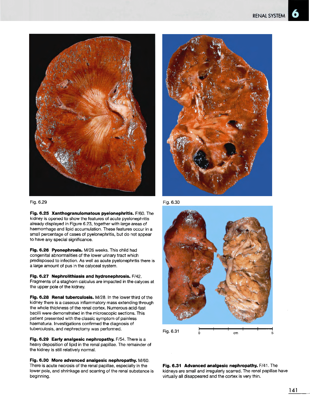
Fig. 6.29 Early
analgesic
nephropathy.
F/54. There
is a
heavy
deposition
of
lipid
in the
renal papillae.
The
remainder
of
the
kidney
is
still relatively normal.
Fig. 6.30
More
advanced
analgesic
nephropathy.
M/60.
There
is
acute necrosis
of the
renal papillae, especially
in the
lower
pole,
and
shrinkage
and
scarring
of the
renal substance
is
beginning.
Fig. 6.30
Fig. 6.31
Fig. 6.31 Advanced
analgesic
nephropathy.
F/41.
The
kidneys
are
small
and
irregularly scarred.
The
renal papillae have
virtually
all
disappeared
and the
cortex
is
very
thin.
141
RENAL
SYSTEM
Fig. 6.34
Fig. 6.33
Fig. 6.32 Oncocytoma (renal tubular adenoma). M/65.
Vertical
slice through
the
middle
of the
right kidney.
In the
anterior portion
of
the
right upper pole there
is a
well circumscribed spherical tumour
30 mm in
diameter.
It has a
dark reddish-brown, homogeneous
cut
surface.
These tumours
are
benign,
but
this kidney
was
removed
because
of a
radiological diagnosis
of
renal carcinoma.
Fig. 6.33
Intramedullary
fibroma.
F/58. This
is a
small, pale,
benign tumour which causes
no
clinical symptoms
and is
frequently found
in
examination
of
postmortem kidneys.
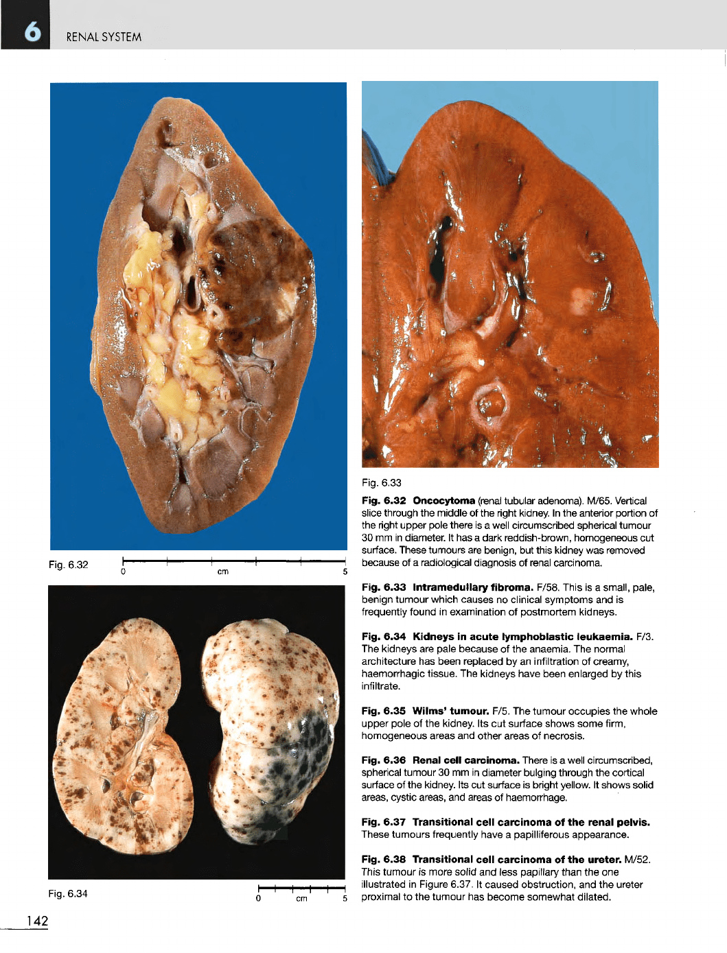
Fig. 6.34
Kidneys
in
acute
lymphoblastic
leukaemia.
F/3.
The
kidneys
are
pale because
of the
anaemia.
The
normal
architecture
has
been replaced
by an
infiltration
of
creamy,
haemorrhagic tissue.
The
kidneys have been enlarged
by
this
infiltrate.
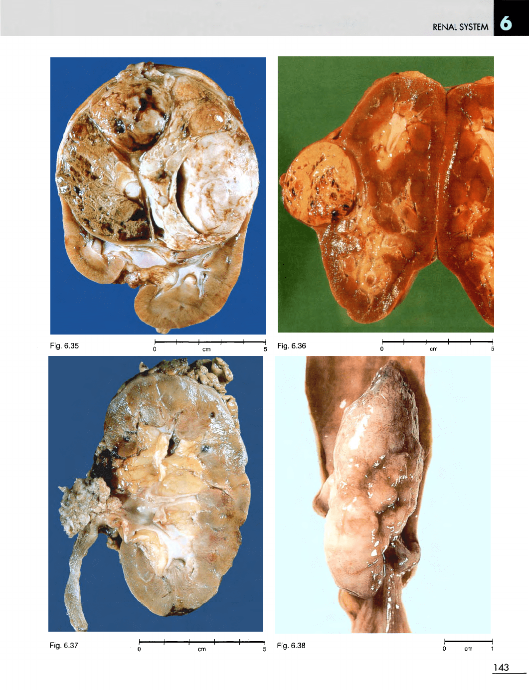
Fig. 6.35 Wilms'
tumour.
F/5.
The
tumour occupies
the
whole
upper pole
of the
kidney.
Its cut
surface shows some firm,
homogeneous areas
and
other areas
of
necrosis.
Fig. 6.36 Renal
cell
carcinoma.
There
is a
well circumscribed,
spherical tumour
30 mm in
diameter bulging through
the
cortical
surface
of the
kidney.
Its cut
surface
is
bright yellow.
It
shows solid
areas, cystic areas,
and
areas
of
haemorrhage.
Fig. 6.37
Transitional
cell
carcinoma
of the
renal
pelvis.
These tumours frequently
have
a
papilliferous appearance.
Fig. 6.38
Transitional
cell
carcinoma
of the
ureter.
M/52.
This
tumour
is
more
solid
and
less
papillary
than
the one
illustrated
in
Figure 6.37.
It
caused obstruction,
and the
ureter
5
proximal
to the
tumour
has
become somewhat dilated.
142
Fig. 6.32
RENAL
SYSTEM
Fig.
6.37
143
Fig.
6.36
Fig.
6.35
Fig.
6.38
RENAL SYSTEM
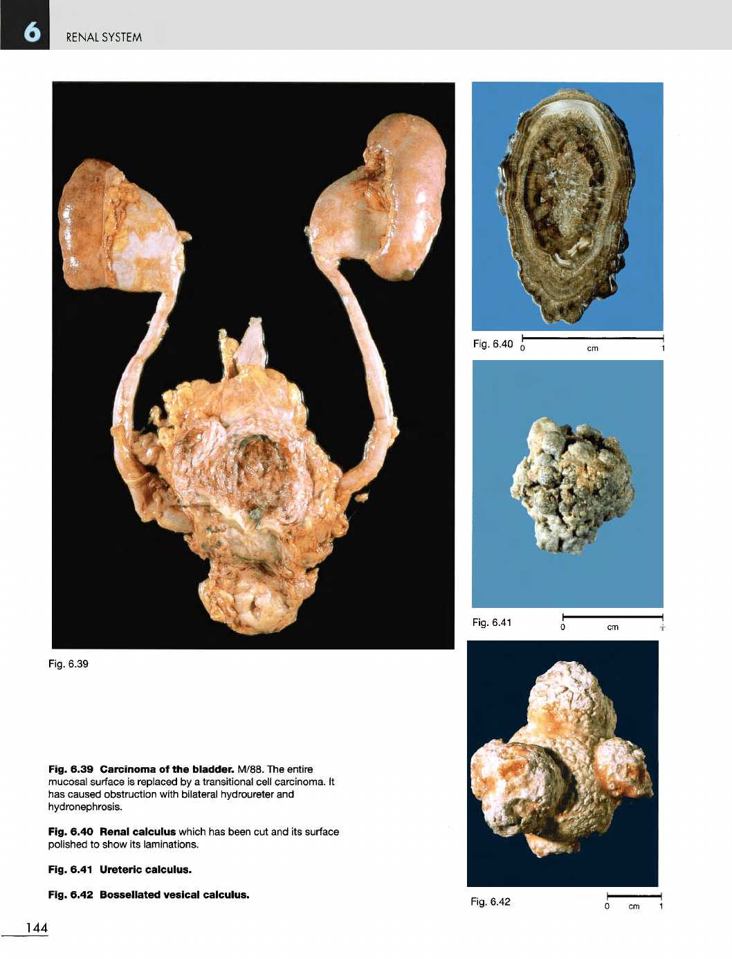
Fig. 6.39
Fig. 6.39
Carcinoma
of the
bladder.
M/88.
The
entire
mucosal surface
is
replaced
by a
transitional cell carcinoma.
It
has
caused obstruction with bilateral hydroureter
and
hydronephrosis.
Fig. 6.40 Renal
calculus
which
has
been
cut and its
surface
polished
to
show
its
laminations.
Fig. 6.41
Ureteric
calculus.
Fig. 6.42
Bossellated
vesical
calculus.
Fig.
6.42
cm
1
144
Fig.
6.40
Fig.
6.41

MALE
GENITAL
SYSTEM
7
MALE GENITAL
SYSTEM
Fig.
7.1
Fig.
7.2
Fig.
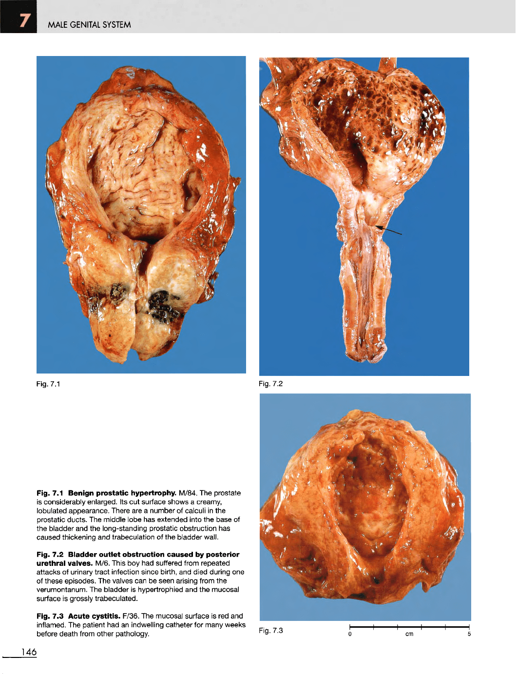
7.1
Benign
prostatic
hypertrophy. M/84.
The
prostate
is
considerably enlarged.
Its cut
surface shows
a
creamy,
lobulated appearance. There
are a
number
of
calculi
in the
prostatic ducts.
The
middle lobe
has
extended into
the
base
of
the
bladder
and the
long-standing prostatic obstruction
has
caused
thickening
and
trabeculation
of the
bladder
wall.
Fig.
7.2
Bladder
outlet
obstruction
caused
by
posterior
urethral
valves. M/6. This
boy had
suffered from repeated
attacks
of
urinary tract infection since birth,
and
died during
one
of
these episodes.
The
valves
can be
seen arising from
the
verumontanum.
The
bladder
is
hypertrophied
and the
mucosal
surface
is
grossly trabeculated.
Fig.
7.3
Acute
cystitis.
F/36.
The
mucosal surface
is red and
inflamed.
The
patient
had an
indwelling catheter
for
many weeks
before death from other pathology.
Fig.
7.3
146
MALE
GENITAL
SYSTEM
Fig.
7.4
Fig.
7.5
Fig.
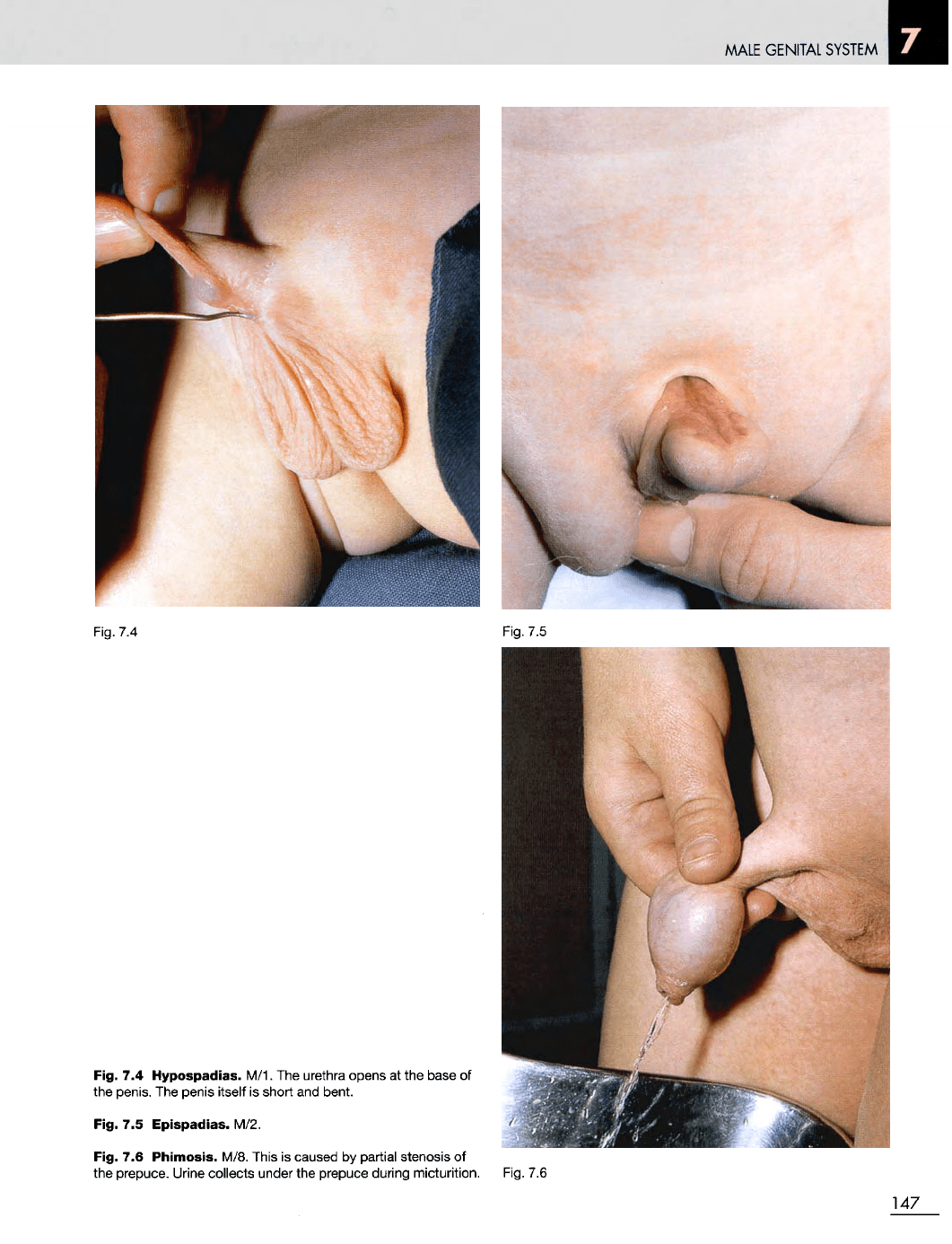
7.4
Hypospadias. M/1.
The
urethra opens
at the
base
of
the
penis.
The
penis itself
is
short
and
bent.
Fig.
7.5
Epispadias.
M/2.
Fig.
7.6
Phimosis.
M/8. This
is
caused
by
partial stenosis
of
the
prepuce. Urine collects under
the
prepuce during micturition. Fig.
7.6
147
MALE
GENITAL
SYSTEM
Fig.
7.7
Fig.
7.8
Fig.
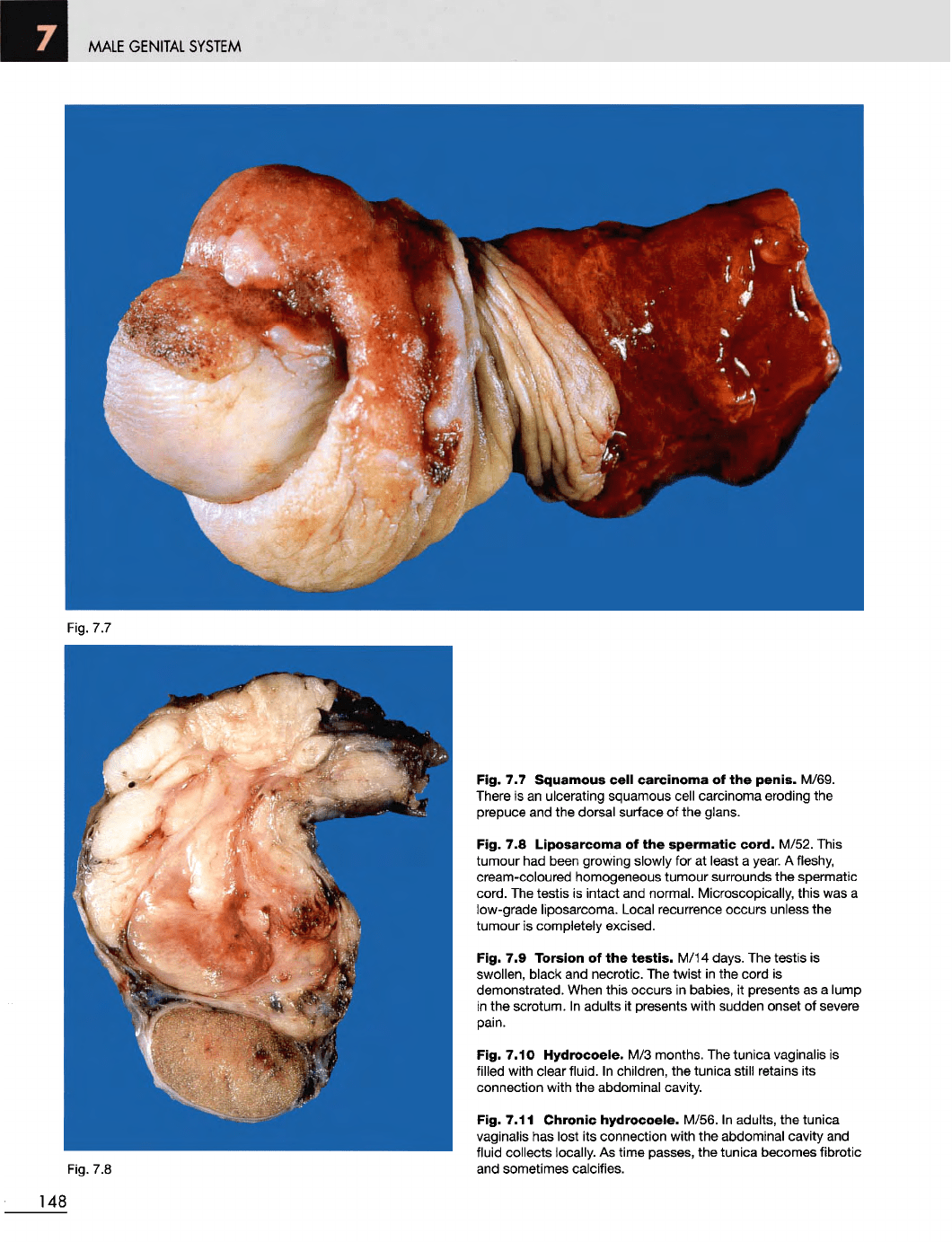
7.7
Squamous
cell
carcinoma
of the
penis.
M/69.
There
is an
ulcerating squamous cell carcinoma eroding
the
prepuce
and the
dorsal surface
of the
glans.
Fig.
7.8
Liposarcoma
of the
spermatic
cord.
M/52. This
tumour
had
been growing slowly
for at
least
a
year.
A
fleshy,
cream-coloured homogeneous tumour surrounds
the
spermatic
cord.
The
testis
is
intact
and
normal. Microscopically, this
was a
low-grade liposarcoma. Local recurrence occurs unless
the
tumour
is
completely excised.
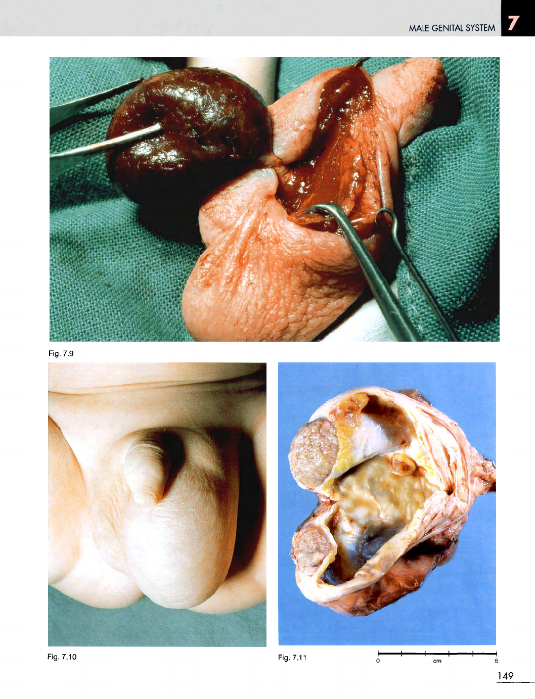
Fig.
7.9
Torsion
of the
testis.
M/14 days.
The
testis
is
swollen,
black
and
necrotic.
The
twist
in the
cord
is
demonstrated.
When
this occurs
in
babies,
it
presents
as a
lump
in
the
scrotum.
In
adults
it
presents with sudden onset
of
severe
pain.
Fig. 7.10
Hydrocoele.
M/3
months.
The
tunica vaginalis
is
filled with clear fluid.
In
children,
the
tunica
still
retains
its
connection with
the
abdominal cavity.
Fig. 7.11
Chronic
hydrocoele.
M/56.
In
adults,
the
tunica
vaginalis
has
lost
its
connection with
the
abdominal cavity
and
fluid collects locally.
As
time passes,
the
tunica becomes fibrotic
and
sometimes calcifies.
148
MALE
GENITAL SYSTEM
Fig. 7.10
Fig. 7.11
149
Fig.
7.9
MALE GENITAL SYSTEM
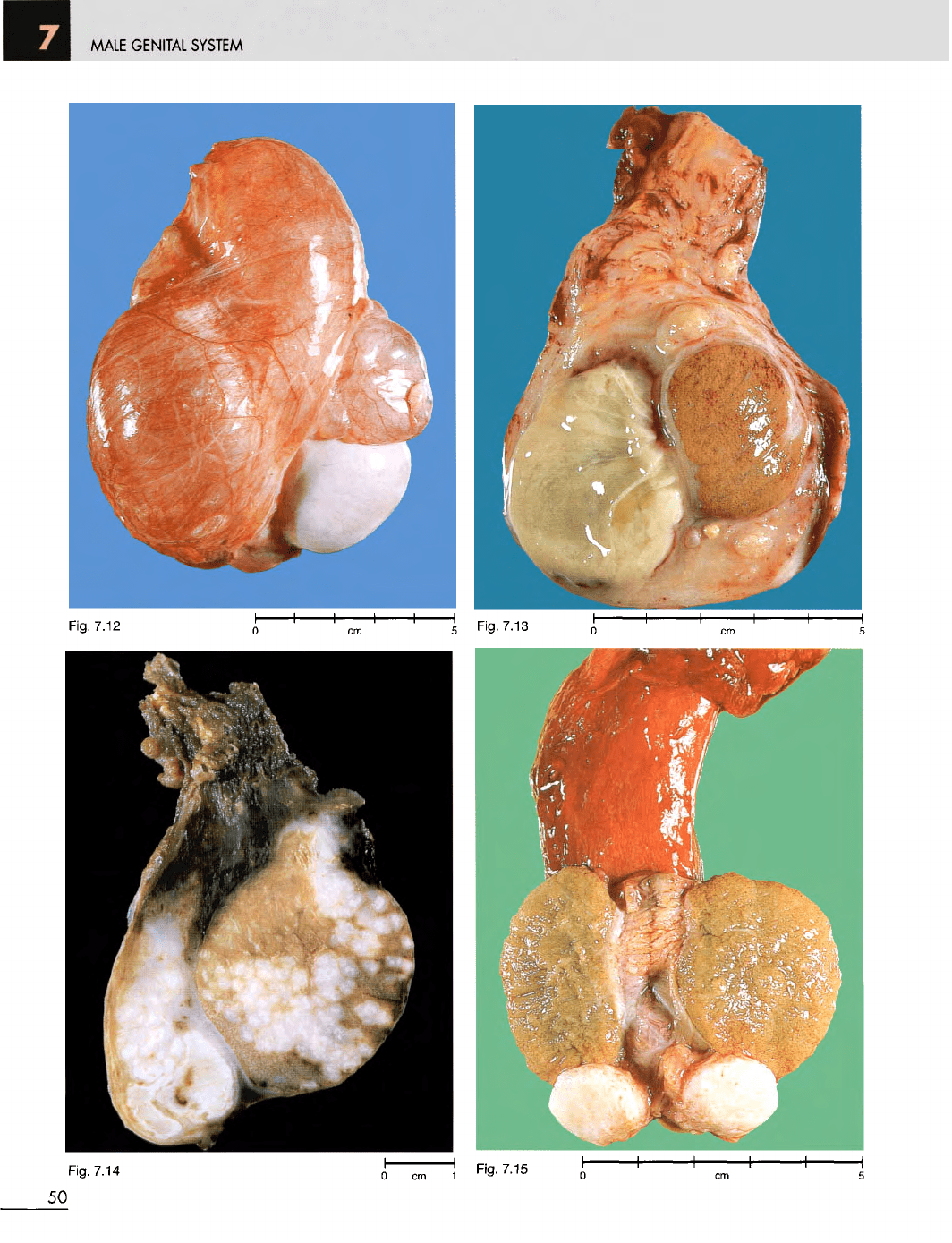
Fig. 7.14
150
Fig.
7.12
Fig.
7.13