Cook R.A., Stewart B. Colour Atlas of Anatomical Pathology
Подождите немного. Документ загружается.

MALE GENITAL
SYSTEM
Fig.
7.16
Fig. 7.12
Cyst
of the
epididymis.
M/34.
The
cyst
is
thin
walled,
multiloculated
and
filled with clear fluid.
It
developed
behind
the
testis.
Fig. 7.13 Abscess
in the
epididymis.
M/78. Thick purulent
material
is
replacing
the
epididymis.
The
patient presented with
a
mass
in the
scrotum.
Fig. 7.14
Tuberculosis
of the
testis
and
epididymis.
M/23.
The
testis contains
multiple
rounded granulomatous lesions.
The
epididymis
is
almost completely replaced
by
similar tissue.
The
patient
had
disseminated tuberculosis.
Fig. 7.15
Adenomatoid
tumour
of the
epididymis.
M/54.
There
is a
creamy, well circumscribed tumour
in the
epididymis.
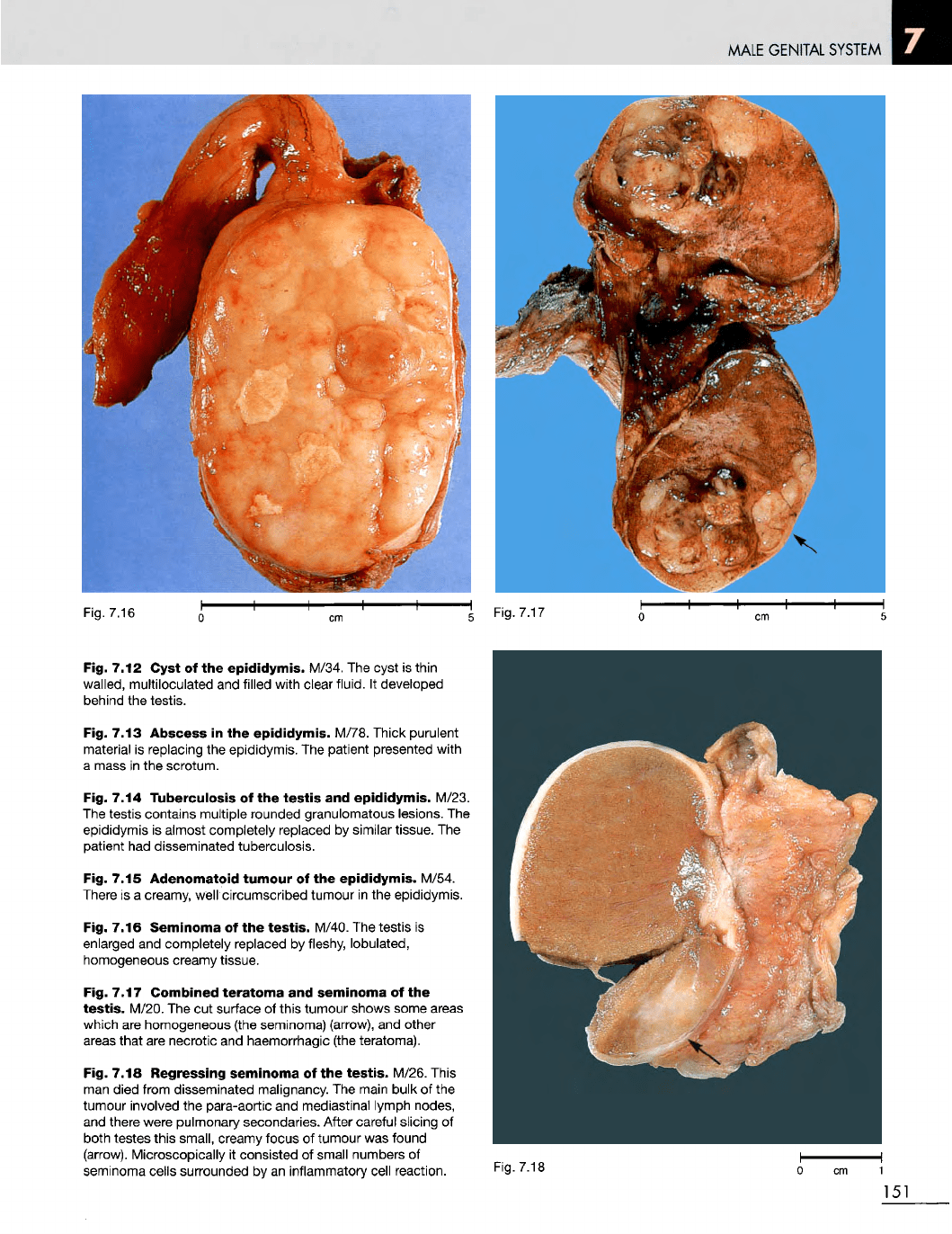
Fig. 7.16
Seminoma
of the
testis.
M/40.
The
testis
is
enlarged
and
completely replaced
by
fleshy, lobulated,
homogeneous creamy tissue.
Fig. 7.17
Combined
teratoma
and
seminoma
of the
testis.
M/20.
The cut
surface
of
this tumour shows some areas
which
are
homogeneous (the seminoma) (arrow),
and
other
areas
that
are
necrotic
and
haemorrhagic (the teratoma).
Fig. 7.18
Regressing
seminoma
of the
testis.
M/26. This
man
died from disseminated malignancy.
The
main bulk
of the
tumour
involved
the
para-aortic
and
mediastinal lymph nodes,
and
there were pulmonary secondaries. After careful slicing
of
both testes this small, creamy focus
of
tumour
was
found
(arrow).
Microscopically
it
consisted
of
small numbers
of
seminoma
cells surrounded
by an
inflammatory cell reaction.
Fig.
7.18
Fig. 7.17
MALE
GENITAL SYSTEM
Fig.
7.19
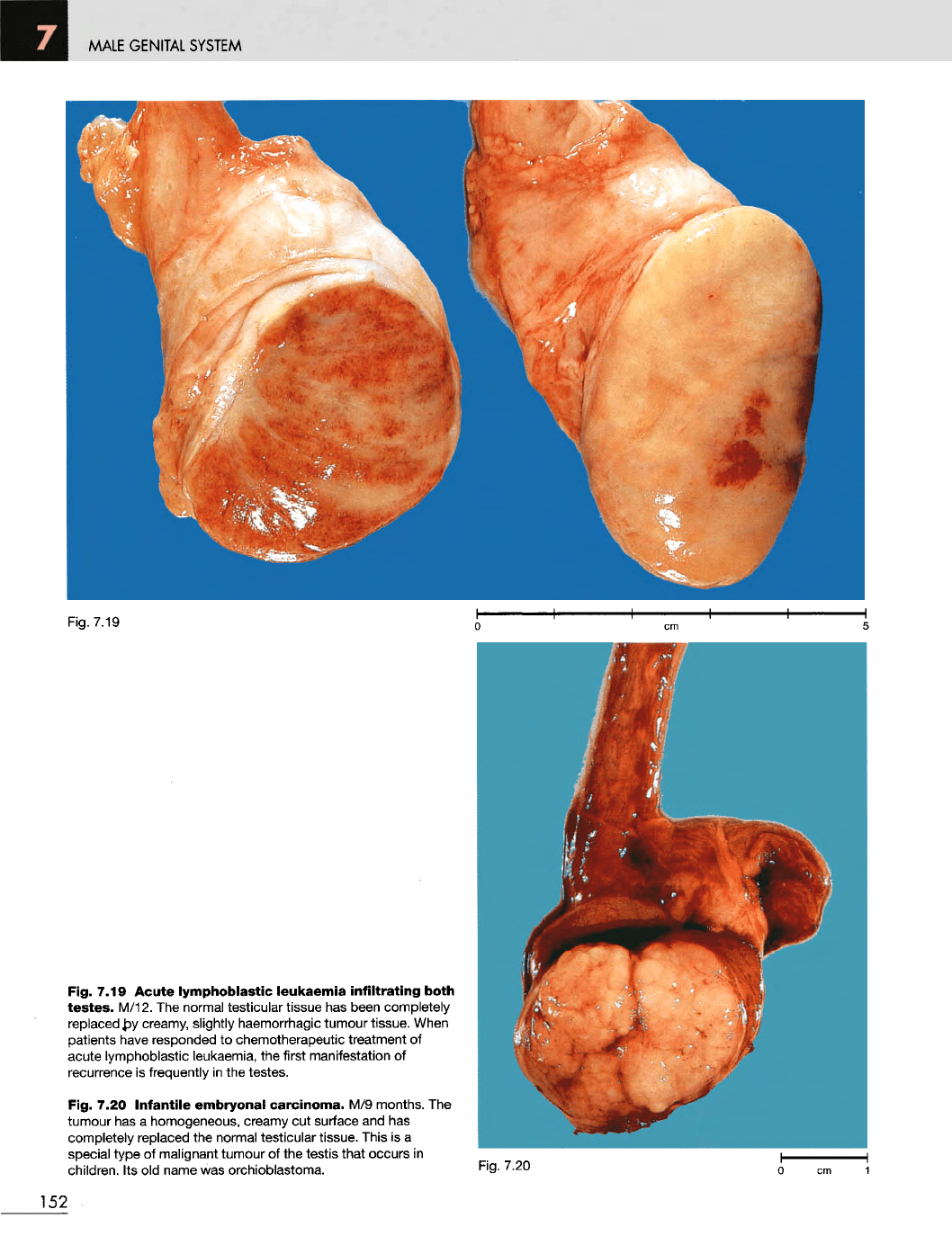
Fig. 7.19 Acute
lymphoblastic
leukaemia
infiltrating
both
testes.
M/12.
The
normal testicular tissue
has
been completely
replaced
by
creamy, slightly haemorrhagic tumour tissue.
When
patients
have
responded
to
chemotherapeutic treatment
of
acute lymphoblastic leukaemia,
the
first manifestation
of
recurrence
is
frequently
in the
testes.
Fig. 7.20
Infantile
embryonal
carcinoma.
M/9
months.
The
tumour
has a
homogeneous, creamy
cut
surface
and has
completely replaced
the
normal testicular tissue. This
is a
special
type
of
malignant tumour
of the
testis that occurs
in
children.
Its old
name
was
orchioblastoma.
Fig.
7.20
152

BREAST
AND
FEMALE
GENITAL
SYSTEM
8
BREAST
AND
FEMALE
GENITAL
SYSTEM
Fig.
8.2
Fig.
8.1
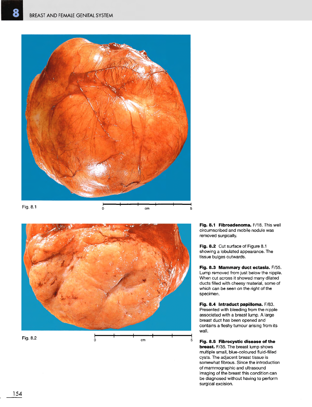
Fibroadenoma.
F/18. This
well
circumscribed
and
mobile nodule
was
removed
surgically.
Fig.
8.2 Cut
surface
of
Figure
8.1
showing
a
lobulated appearance.
The
tissue bulges outwards.
Fig.
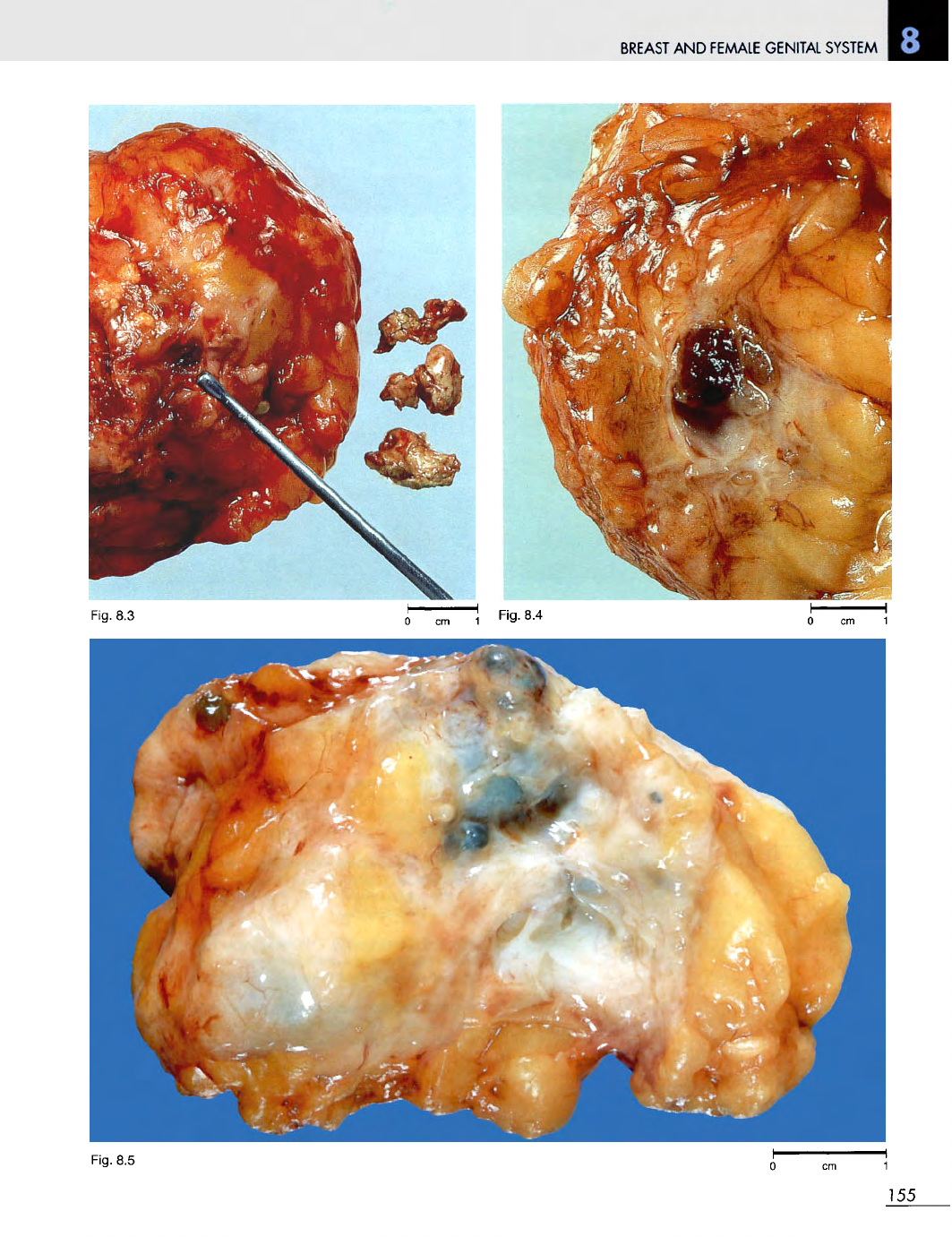
8.3
Mammary
duct
ectasia.
F/55.
Lump removed from just below
the
nipple.
When
cut
across
it
showed many dilated
ducts filled with cheesy material, some
of
which
can be
seen
on the
right
of the
specimen.
Fig.
8.4
Intraduct
papilloma.
F/83.
Presented
with bleeding from
the
nipple
associated with
a
breast lump.
A
large
breast
duct
has
been
opened
and
contains
a
fleshy tumour arising from
its
wall.
Fig.
8.5
Fibrocystic
disease
of the
breast.
F/35.
The
breast lump shows
multiple small, blue-coloured fluid-filled
cysts.
The
adjacent breast tissue
is
somewhat fibrous. Since
the
introduction
of
mammographic
and
ultrasound
imaging
of the
breast this condition
can
be
diagnosed without having
to
perform
surgical
excision.
154
Fig.
8.1
BREAST
AND
FEMALE GENITAL SYSTEM
155
Fig.
8.3
Fig.
8.4
Fig.
8.5
BREAST
AND
FEMALE
GENITAL SYSTEM
Fig.
8.6
Fig.
8.8
Fig.
8.7 o cm 1
Fig.
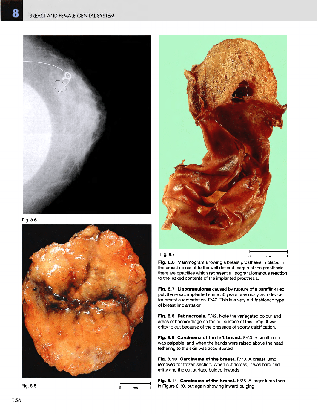
8.6
Mammogram showing
a
breast prosthesis
in
place.
In
the
breast adjacent
to the
well defined margin
of the
prosthesis
there
are
opacities which represent
a
lipogranulomatous reaction
to the
leaked contents
of the
implanted prosthesis.
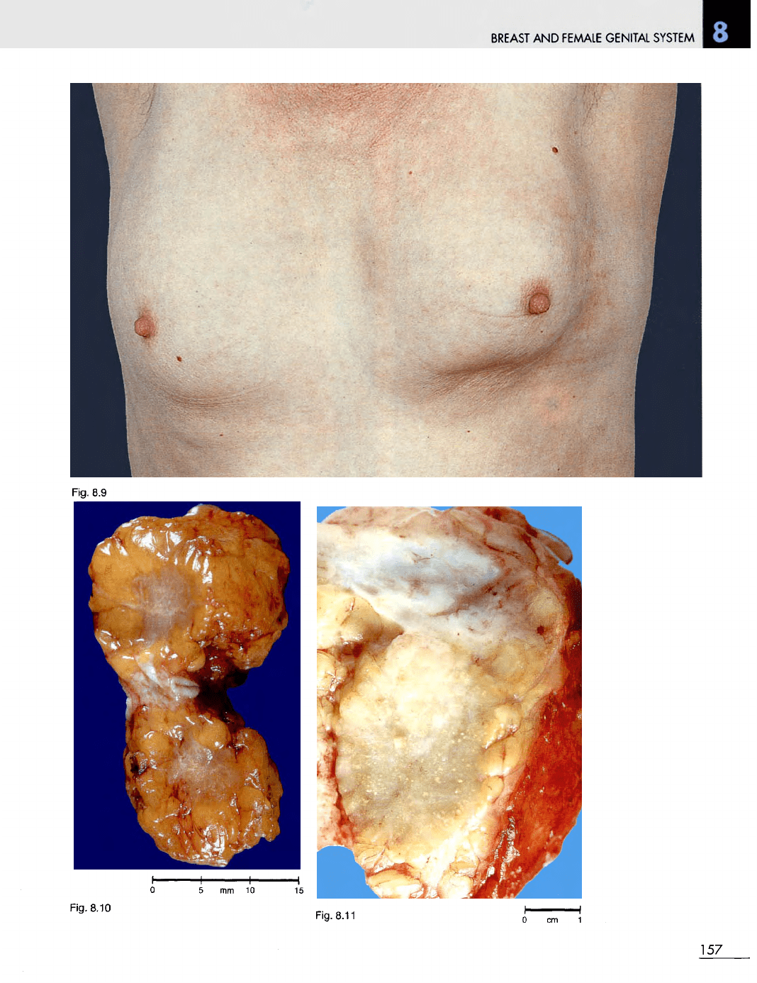
Fig.
8.7
Lipogranuloma
caused
by
rupture
of a
paraffin-filled
polythene
sac
implanted some
30
years previously
as a
device
for
breast augmentation. F/47. This
is a
very
old-fashioned type
of
breast
implantation.
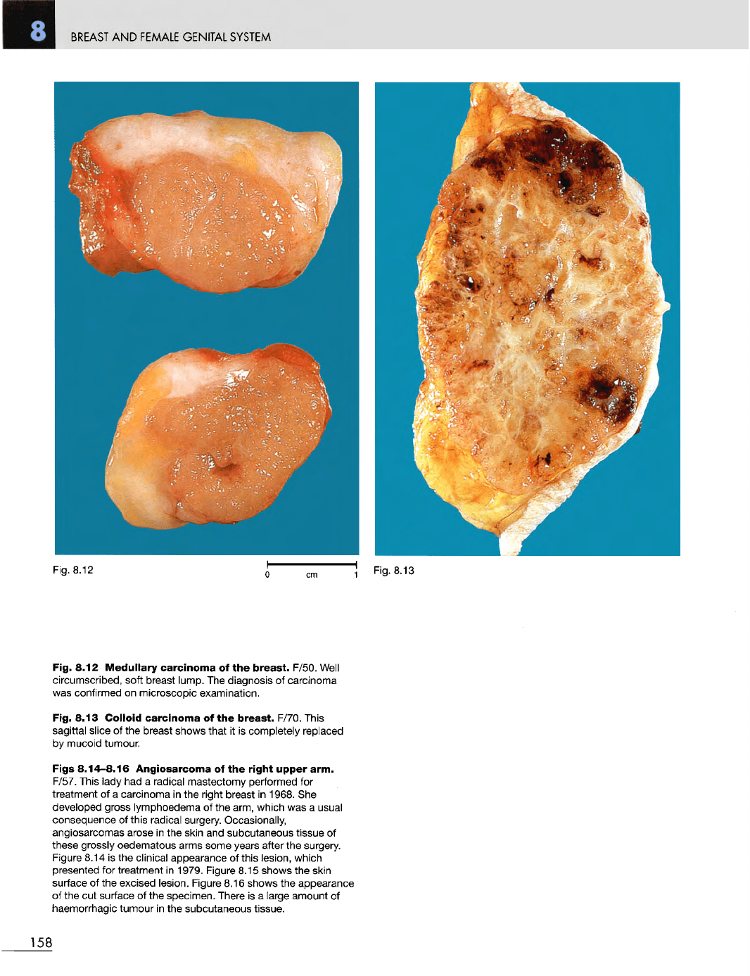
Fig.
8.8 Fat
necrosis.
F/42. Note
the
variegated colour
and
areas
of
haemorrhage
on the cut
surface
of
this lump.
It was
gritty
to cut
because
of the
presence
of
spotty
calcification.
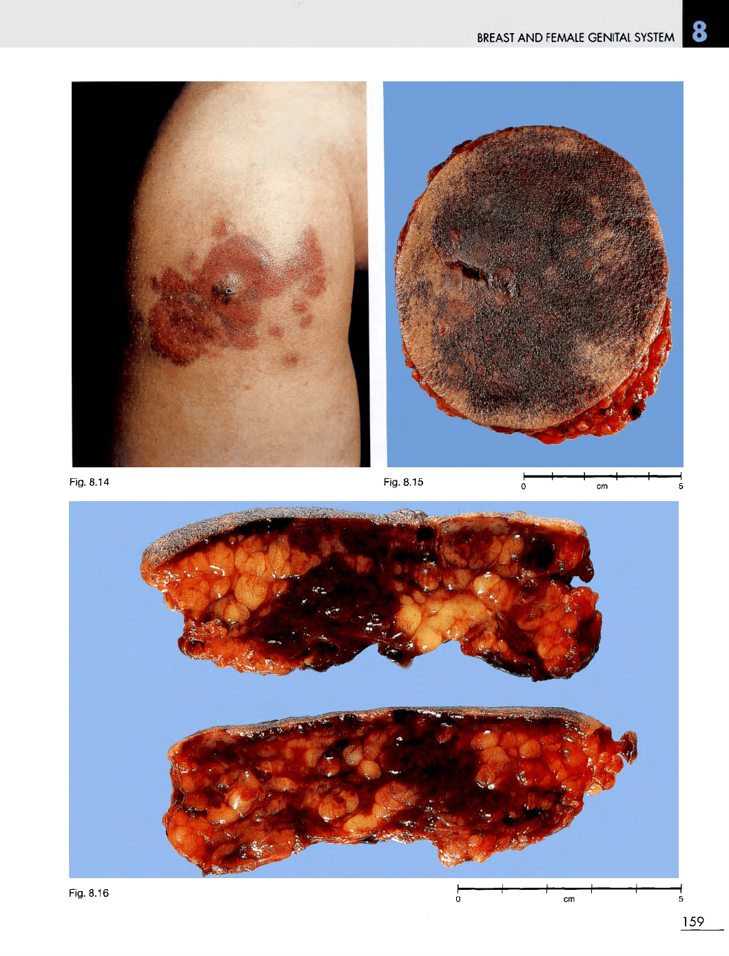
Fig.
8.9
Carcinoma
of the
left
breast.
F/60.
A
small lump
was
palpable,
and
when
the
hands were raised above
the
head
tethering
to the
skin
was
accentuated.
Fig. 8.10
Carcinoma
of the
breast.
F/70.
A
breast lump
removed
for
frozen section. When
cut
across,
it was
hard
and
gritty
and the cut
surface
bulged inwards.
Fig. 8.11
Carcinoma
of the
breast.
F/35.
A
larger lump than
in
Figure
8.10,
but
again showing inward bulging.
156
BREAST
AND
FEMALE
GENITAL
SYSTEM
157
Fig.
8.9
Fig. 8.10
Fig. 8.11
BREAST
AND
FEMALE
GENITAL
SYSTEM
Fig. 8.12
Medullary
carcinoma
of the
breast.
F/50.
Well
circumscribed, soft breast lump.
The
diagnosis
of
carcinoma
was
confirmed
on
microscopic examination.
Fig. 8.13
Colloid
carcinoma
of the
breast.
F/70. This
sagittal slice
of the
breast shows that
it is
completely replaced
by
mucoid tumour.
Figs
8.14-8.16
Angiosarcoma
of the
right
upper
arm.
F/57. This lady
had a
radical mastectomy performed
for
treatment
of a
carcinoma
in the
right breast
in
1968.
She
developed gross lymphoedema
of the
arm, which
was a
usual
consequence
of
this radical surgery. Occasionally,
angiosarcomas arose
in the
skin
and
subcutaneous tissue
of
these grossly oedematous arms some years after
the
surgery.
Figure
8.14
is the
clinical appearance
of
this
lesion, which
presented
for
treatment
in
1979. Figure 8.15 shows
the
skin
surface
of the
excised lesion. Figure 8.16 shows
the
appearance
of
the cut
surface
of the
specimen. There
is a
large amount
of
haemorrhagic tumour
in the
subcutaneous tissue.
158
Fig.
8.12
BREAST
AND
FEMALE
GENITAL
SYSTEM
Fig. 8.16
159
Fig. 8.14 Fig. 8.15
BREAST
AND
FEMALE
GENITAL
SYSTEM
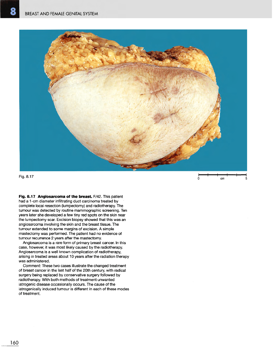
Fig. 8.17 Angiosarcoma
of the
breast.
F/42. This patient
had
a 1 -cm
diameter infiltrating duct carcinoma treated
by
complete local resection (lumpectomy)
and
radiotherapy.
The
tumour
was
detected
by
routine mammographic screening.
Ten
years
later
she
developed
a few
tiny
red
spots
on the
skin
near
the
lumpectomy scar. Excision biopsy showed that this
was an
angiosarcoma involving
the
skin
and the
breast tissue.
The
tumour
extended
to
some margins
of
excision.
A
simple
mastectomy
was
performed.
The
patient
had no
evidence
of
tumour recurrence
2
years
after
the
mastectomy.
Angiosarcoma
is a
rare form
of
primary breast cancer.
In
this
case,
however,
it was
most likely caused
by the
radiotherapy.
Angiosarcoma
is a
well known complication
of
radiotherapy,
arising
in
treated areas about
10
years
after
the
radiation therapy
was
administered.
Comment:
These
two
cases illustrate
the
changed treatment
of
breast cancer
in the
last half
of the
20th century, with radical
surgery
being replaced
by
conservative surgery followed
by
radiotherapy. With
both
methods
of
treatment unwanted
iatrogenic disease occasionally occurs.
The
cause
of the
iatrogenically induced tumour
is
different
in
each
of
these modes
of
treatment.
160
Fig.
8.17