Graef M. Introduction to conventional transmission electron microscopy
Подождите немного. Документ загружается.

658 Phase contrast microscopy
(a)
(b)
(c)
(d)
1 µm
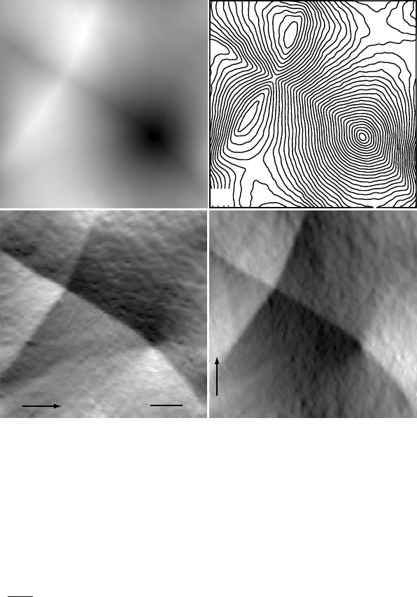
Fig. 10.38.
(a) Reconstructed phase for the square area outlined in Fig.
4.28(c). (b) Contour
plot of the phase φ(r); the vortex is clearly visible to the lower right. (c) B
x
and (d) B
y
maps, obtained by numerical differentiation of the phase. The maps are only qualitatively
correct, since the microscope defocus was not calibrated.
Exercises
10.1 Show that the paraxial image intensity in Lorentz mode is given by equation (10.78).
10.2 Derive equation (10.93) for the inverse Laplacian operator.
10.3 Show that the gradient of the aberration function χ(Q) reaches a minimum at Q =
√
D/3.
10.4 Consider the microscope I
2
from Table 10.1. If this machine
is equipped with a
1K×1K CCD camera with a pixel size of 25 × 25 µm
2
, then what microscope mag-
nification should be used to record images that contain spatial frequencies out to
the chromatic information limit (assuming the sample contains such spatial frequen-
cies)? How many pixels of the CCD camera cannot be used because of the detector
envelope function?
10.5 Adapt the
HREMExample.f90 program to use any pair of parameters as variables
for the output matrix. As an example, you may want to study the source code of the
ctf2.pro ION program on the website.
10.6 Final remarks 659
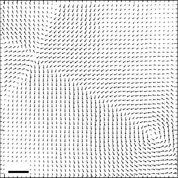
500 nm
Fig. 10.39. Vector map derived from the component images in Figs 10.38(c) and (d). Each
vector is an average over a 4 × 4 pixel square region (or 100 × 100 nm
2
).
10.6 Use an amorphous Ge film (or, if you do not have one,aCfilm)todetermine the
spherical aberration of the microscope you use for your observations. How accurately
can
you determine
C
s
?
10.7 Use your thin foil of GaAs to obtain a [110] high-resolution through-focus series
for two different diffraction aperture diameters. Simulate the images, and compare
simulations and experiments.
10.8 The transport-of-intensity equation can be used to reconstruct the phase of the elec-
tron wave for Lorentz-type observations. Can you think of a way to adapt the formal-
ism to high-resolution
images? What would happen if the formalism were applied
to images from a C
s
-corrected microscope?
10.6 Final remarks
Well, you have reached the end of this book. Hopefully, you found it to be a useful
book. If you were new to microscopy, it is hoped that this text has provided you
660 Phase contrast microscopy
1 µm
(a) (b) (c)
(d) (e) (f)
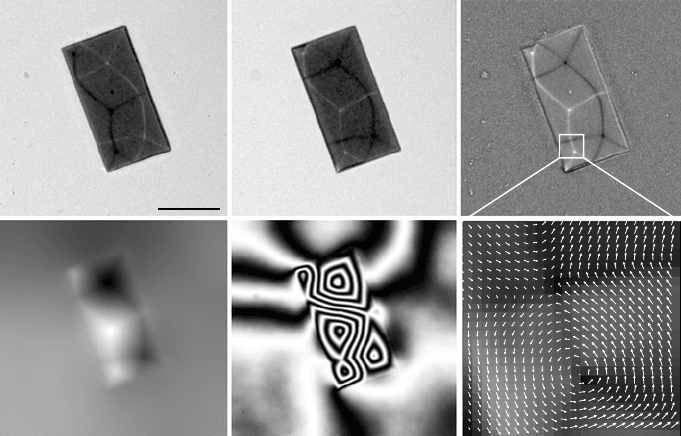
Fig. 10.40. (a) and (b) under- and over-focus Fresnel images (400 kV, zero-loss filtered)
ofa1× 2 µm Permalloy island on an Si
3
N
4
support membrane. The derivative ∂ I/∂ z is
shown in (c), along with the reconstructed phase φ in (d), and cos φ in (e). (f) is a vector
representation of the square area outlined in (c). (Sample courtesy of J. Chapman, Univ.
Glasgow.)
with a solid introduction to the physical principles underlying the interaction of
electrons with the specimen, and the subsequent propagation of those electrons
down the microscope column. If you like computers and simulations, then perhaps
the source code accompanying this book was just what you needed to get started. If
you are an experienced microscopist, it is hoped that you could find something new
in this text, perhaps a derivation that suddenly makes sense, or an illustration that
provoked what the German language so efficiently describes as an “Aha-Erlebnis”.
†
The field of electron optical methods is vast, and what you have learned in this
book is truly just an introduction to the operation and use of one the most important
instruments of materials science. The material covered in this text should give
you a basic knowledge of elastic scattering in crystalline matter. You can build on
this knowledge when you expand your studies to include inelastic scattering and
analytical observation methods.
†
An Aha-Erlebnis is “Ein eigenartiges im Denkverlauf auftretendes-lustbetontes Erlebnis, das sich bei pl¨otzlicher
Einsicht in einen zuerst undurchsichten Zusammenhang einstellt.” (K. B¨uhler, speech psychologist.)

Appendix A1
Explicit crystallographic equations
In Chapter 1, we have derived the general equations for angles and distances in both direct
and reciprocal space. Sometimes it is useful to have the explicit versions of those
equations for a particular crystal system. For instance, the d-spacing for the planes (hkl)
in a cubic crystal is given by
d
hkl
=
a
√
h
2
+ k
2
+l
2
,
and for manual calculations this is indeed easier to use than the more formal tensor
relations. For implementations in a computer program, it is much easier to use the tensor
formalism, because then one need not distinguish between the seven crystal systems (but,
beware of the hexagonal system!). The equations
for the hexagonal reference frame are
only valid for the
3-index system.
In this appendix, we list the explicit equations for the direct and reciprocal metric
tensors, the length of a vector t
uvw
(Table A1.1), the angle between two vectors t
u
1
v
1
w
1
and
t
u
2
v
2
w
2
(Table A1.2), the length of a reciprocal lattice vector g
hkl
(Table A1.3), and the
angle between two reciprocal lattice vectors g
h
1
k
1
l
1
and g
h
2
k
2
l
2
(Table A1.4). All equations
in the tables were directly derived from the expressions for the direct and reciprocal
metric tensors below.
(i) Direct metric tensors:
g
c
=
a
2
00
0 a
2
0
00a
2
, g
t
=
a
2
00
0 a
2
0
00c
2
,
g
o
=
a
2
00
0 b
2
0
00c
2
, g
h
=
a
2
−a
2
/20
−a
2
/2 a
2
0
00c
2
,
g
r
=
a
2
a
2
cos α a
2
cos α
a
2
cos α a
2
a
2
cos α
a
2
cos α a
2
cos α a
2
, g
m
=
a
2
0 ac cos β
0 b
2
0
ac cos β 0 c
2
,
661

662 Appendices
g
a
=
a
2
ab cos γ ac cos β
ba cos γ b
2
bc cos α
ca cos β cb cos α c
2
.
(ii) Reciprocal metric tensors :
g
∗
c
=
1/a
2
00
01/a
2
0
001/a
2
, g
∗
t
=
1/a
2
00
01/a
2
0
001/c
2
,
g
∗
o
=
1/a
2
00
01/b
2
0
001/c
2
, g
∗
h
=
4/3a
2
2/3a
2
0
2/3a
2
4/3a
2
0
001/c
2
,
g
∗
r
=
1
W
2
1 + cos α −
1−tan
2
α/2
2
−
1−tan
2
α/2
2
−
1−tan
2
α/2
2
1 + cos α −
1−tan
2
α/2
2
−
1−tan
2
α/2
2
−
1−tan
2
α/2
2
1 + cos α
,
with
W
2
= a
2
(1 + cos α − 2 cos
2
α),
g
∗
m
=
1
a
2
sin
2
β
0 −
cos β
ac sin
2
β
0
1
b
2
0
−
cos β
ac sin
2
β
0
1
c
2
sin
2
β
g
∗
a
=
1
2
b
2
c
2
sin
2
α abc
2
F(α, β, γ ) ab
2
cF(γ , α, β)
abc
2
F(α, β, γ ) a
2
c
2
sin
2
β a
2
bcF(β, γ, α)
ab
2
cF(γ , α, β) a
2
bcF(β, γ, α) a
2
b
2
sin
2
γ
where
F(α, β, γ ) = cos α cos β − cos γ
and
2
= a
2
b
2
c
2
(1 − cos
2
α − cos
2
β − cos
2
γ + 2 cos α cos β cos γ ).

A1 Explicit crystallographic equations 663
Table A1.1. Expressions for the length t of a vector t
uvw
in the seven crystal
systems. The primes on the hexagonal components indicate that the 3-index
notation must be used.
System l Expression
Cubic
c
ta{u
2
+ v
2
+ w
2
}
1/2
Tetragonal
t
t {a
2
(u
2
+ v
2
) + c
2
w
2
}
1/2
Orthorhombic
o
t {a
2
u
2
+ b
2
v
2
+ c
2
w
2
}
1/2
Hexagonal
h
t {a
2
(u
2
+ v
2
− u
v
) + c
2
w
2
}
1/2
Rhombohedral
r
ta{u
2
+ v
2
+ w
2
+ 2 cos α[uv + uw + vw]}
1/2
Monoclinic
m
t {a
2
u
2
+ b
2
v
2
+ c
2
w
2
+ 2acuw cos β}
1/2
Triclinic
a
t {a
2
u
2
+ b
2
v
2
+ c
2
w
2
+ 2bcvw cos α
+2acuw cos β + 2abuv cos γ }
1/2
Table A1.2. Expressions for the cosine of the angle α between two vectors t
u
1
v
1
w
1
and
t
u
2
v
2
w
2
in the seven crystal
systems. The primes on the he
xagonal components indicate that
the 3-index notation
must be used.
System cos α
Cubic
a
2
(u
1
u
2
+v
1
v
2
+w
1
w
2
)
c
t
1
×
c
t
2
Tetragonal
a
2
(u
1
u
2
+v
1
v
2
)+c
2
w
1
w
2
t
t
1
×
t
t
2
Orthorhombic
a
2
u
1
u
2
+b
2
v
1
v
2
+c
2
w
1
w
2
o
t
1
×
o
t
2
Hexagonal
a
2
(u
1
u
2
+v
1
v
2
−
1
2
(u
1
v
2
+v
1
u
2
))+c
2
w
1
w
2
h
t
1
×
h
t
2
Rhombohedral
a
2
(u
1
u
2
+v
1
v
2
+w
1
w
2
+cos α
[
u
1
(v
2
+w
2
)+v
1
(u
2
+w
2
)+w
1
(u
2
+v
2
)
]
)
r
t
1
×
r
t
2
Monoclinic
a
2
u
1
u
2
+b
2
v
1
v
2
+c
2
w
1
w
2
+ac(w
1
u
2
+u
1
w
2
) cos β
m
t
1
×
m
t
2
Triclinic
a
2
u
1
u
2
+b
2
v
1
v
2
+c
2
w
1
w
2
+bc(v
1
w
2
+v
2
w
1
) cos α+ac(u
1
w
2
+u
2
w
1
) cos β+ab(u
1
v
2
+u
2
v
1
) cos γ
a
t
1
×
a
t
2

664 Appendices
Table A1.3. Expressions for the length |g |=1/d
hkl
of a reciprocal lattice vector g
hkl
in
the seven crystal systems.
System g Expression
Cubic
c
g
1
a
h
2
+ k
2
+l
2
1/2
Tetragonal
t
g
1
a
2
(h
2
+ k
2
) +
1
c
2
l
2
1/2
Orthorhombic
o
g
1
a
2
h
2
+
1
b
2
k
2
+
1
c
2
l
2
1/2
Hexagonal
h
g
4
3a
2
(h
2
+ k
2
+ hk) +
1
c
2
l
2
1/2
Rhombohedral
r
g
1
a
7
(1+cos
2
α)(h
2
+k
2
+l
2
)−(1−tan
2
α/2)(hk+kl+lh)
1+cos α−2 cos
2
α
8
1/2
Monoclinic
m
g
7
1
a
2
h
2
sin
2
β
+
1
b
2
k
2
+
1
c
2
l
2
sin
2
β
−
2hl cos β
ac sin
2
β
8
1/2
Triclinic
a
g
1
{h
2
b
2
c
2
sin
2
α + k
2
a
2
c
2
sin
2
β + l
2
a
2
b
2
sin
2
γ
+ 2hkabc
2
F(α, β, γ ) + 2kla
2
bcF(β, γ, α)
+ 2lhab
2
cF(γ , α, β)}
1/2
with ={a
2
b
2
c
2
(1 − cos
2
α − cos
2
β − cos
2
γ
+ 2 cos α cos β cos γ )}
1/2
Table A1.4. Expressions for the cosine of the angle α between two vectors g
h
1
k
1
l
1
and
g
h
2
k
2
l
2
in the seven crystal systems.
System cos α
Cubic
1
a
2
h
1
h
2
+k
1
k
2
+l
1
l
2
c
g
1
×
c
g
2
Tetragonal
1
a
2
(h
1
h
2
+k
1
k
2
)+
1
c
2
l
1
l
2
t
g
1
×
t
g
2
Orthorhombic
1
a
2
h
1
h
2
+
1
b
2
k
1
k
2
+
1
c
2
l
1
l
2
o
g
1
×
o
g
2
Hexagonal
4
3a
2
(h
1
h
2
+k
1
k
2
+
1
2
(h
1
k
2
+k
1
h
2
))+
1
c
2
l
1
l
2
h
g
1
×
h
g
2
Rhombohedral
(1+cos α)(h
1
h
2
+k
1
k
2
+l
1
l
2
)−
1
2
(1−tan
2
α/2)(h
1
(k
2
+l
2
)+k
1
(h
2
+l
2
)+l
1
(h
2
+k
2
))
a
2
(1+cos α−2 cos
2
α)×
r
g
1
×
r
g
2
Monoclinic
1
a
2
sin
2
β
h
1
h
2
+
1
b
2
k
1
k
2
+
1
c
2
sin
2
β
l
1
l
2
+(l
1
h
2
+h
1
l
2
)
cos β
ac sin
2
β
m
g
1
×
m
g
2
Triclinic
1
2
×
a
g
1
×
a
g
2
h
1
h
2
b
2
c
2
sin
2
α + k
1
k
2
a
2
c
2
sin
2
β + l
1
l
2
a
2
b
2
sin
2
γ
+abc
2
(k
1
h
2
+ k
2
h
1
)F(α, β, γ )
+ab
2
c(h
1
l
2
+ h
2
l
1
)F(β, γ, α)
+a
2
bc(k
1
l
2
+ k
2
l
1
)F(γ, α, β)

Appendix A2
Physical constants
Table A2.1. Some physical constants and unit conversions often used
in electron microscopy.
Name Symbol V
alue Units
Speed of light in vacuum c 299 792 458 m s
−1
Planck’s constant h 6.626 075 × 10
−34
Js
h/e 4.135 669 × 10
−15
eV
s
Boltzmann’s constant k 1.380 658 × 10
−23
JK
−1
k/e 8.617 385 × 10
−5
eV K
−1
Permeability of vacuum µ
0
4π × 10
−7
–
Permittivity of vacuum
0
8.854 187 817 × 10
−12
Fm
−1
Magnetic flux quantum
0
= h/2e 2.067 834 × 10
−15
Wb
Elementary charge e 1.602 177 × 10
−19
C
e/ h 2.417 988 × 10
14
AJ
−1
Electron rest mass m
0
9.109 389 × 10
−31
kg
Electron rest energy m
0
c
2
×10 J
5.109 991 × 10
8
eV
Magnetic moment µ
e
9.284 770 × 10
−24
JT
−1
Electronvolt eV 1.602 177 × 10
−19
J
665

Appendix A3
Space group encoding and other software
This appendix contains the conversion table for the space group encoding scheme.
Table A3.1 lists the point symmetry parts of the 14 basic matrices, and Table A3.2 lists the
conversions for the components of translation vectors.
Due to space limitations, it is not possible to provide a detailed description of all the
Fortran-90 code provided with this book. The reader will find such a description in PDF
format on the
website.
Table A3.1. Explicit point symmetry matrices for the 14 matrices used to
encode the space group generators.
a =
100
010
001
b =
−100
0 −10
001
c =
−100
010
00−1
d =
001
100
010
e =
010
100
00−1
f =
0 −10
−100
00−1
g =
0 −10
100
001
h =
−100
0 −10
00−1
i =
100
010
00−1
j =
100
0 −10
001
k =
0 −10
−100
001
l =
010
100
001
m =
010
− 100
00−1
n =
0 −10
1 −10
001
Table A3.2. Conversions for the components of translation
vectors in the space group encoding scheme.
A =
1
6
B =
1
4
C =
1
3
D =
1
2
E =
2
3
F =
3
4
G =
5
6
O = 0 X =−
3
8
Y =−
1
4
Z =−
1
8
666
Appendix A4
Point groups and space groups
This appendix contains detailed information on the 32 crystallographic point groups. For
each point group we list: the group symbol (both International and Schœnflies notations),
the group order, the crystal class, the Laue class, the diffraction groups (see Chapter 5),
the number of space groups related to the point group, the stereographic projection, and a
rendered 3D view [DG98]. We also list the space group symbols, separated into the
symmorphic and non-symmorphic groups.
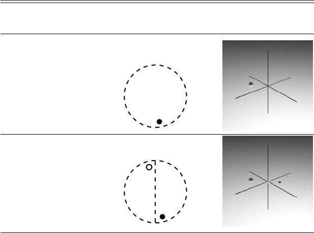
Table A4.1. The 32 point groups, with International–[Schœnflies] symbols,
stereographic projection (S.P.), rendered 3D view, and important properties.
Point group Stereographic
information projection Rendered 3D view
Order: 1
Crystal Class: a
Laue Class:
¯
1
Diffraction Groups:
1
# Space Groups: 1
1 –[C
1
]
Order: 2
Crystal Class: a
Laue Class:
¯
1
Diffraction Groups:
2
R
# Space Groups: 1
¯
1 –[C
i
]
667