Kenneth E. Gonsalves, Craig R. Halberstadt, Cato T. Laurencin, Lakshmi S. Nair. Biomedical Nanostructures
Подождите немного. Документ загружается.

62. Iozzo RV. Matrix proteoglycans: from molecular design to cellular function.
Annu Rev Biochem 1998;67:609652.
63. Iozzo RV, Cohen IR, Grassel S, Murdoch AD. The biology of perlecan: the
multifaceted heparan sulphate proteoglycan of basement membranes and
pericellular matrices. Biochem J 1994;302(Pt 3):625639.
64. Bhide VM, et al. Collagen phagocytosis by fibroblasts is regulated by decorin. J
Biol Chem 2005;280:2310323113.
65. Danielson KG, et al. Targeted disruption of decorin leads to abnormal collagen
fibril morphology and skin fragility. J Cell Biol 1997;136:729743.
66. Lin X. Functions of heparan sulfate proteoglycans in cell signaling during
development. Development 2004;131:60096021.
67. Hynes R. Molecular biology of fibronectin. Annu Rev Cell Biol 1985;1:6790.
68. Leahy DJ, Aukhil I, Erickson HP. 2.0 A crystal structure of a four-domain
segment of human fibronectin encompassing the RGD loop and synergy region.
Cell 1996;84:155164.
69. Groves JT. Learning the chemical language of cell-surface interactions. Sci STKE
2005;2005:E45.
70. Velling T, Risteli J, Wennerberg K, Mosher DF, Johansson S. Polymerization of
type I and III collagens is dependent on fibronectin and enhanced by integrins
alpha 11beta 1 and alpha 2beta 1. J Biol Chem 2002;277:3737737381.
71. Dallas SL, et al. Fibronectin regulates latent transforming growth factor-beta
(TGF beta) by controlling matrix assembly of latent TGF beta-binding protein-1.
J Biol Chem 2005;280:1887118880.
72. Nordahl J, Mengarelli-Widholm S, Hultenby K, Reinholt FP. Ultrastructural
immunolocalization of fibronectin in epiphyseal and metaphyseal bone of young
rats. Calcif Tissue Int 1995;57:442449.
73. Globus RK, et al. Fibronectin is a survival factor for differentiated osteoblasts. J
Cell Sci 1998;111(Pt 10):13851393.
74. Miner JH and Yurchenco PD. Laminin functions in tissue morphogenesis. Annu
Rev Cell Dev Biol 2004;20:255284.
75. Jones JC, Dehart GW, Gonzales M, Goldfinger LE. Laminins: an overview.
Microsc Res Tech 2000;51:211213.
76. Cheng YS, Champliaud MF, Burgeson RE, Marinkovich MP, Yurchenco PD.
Self-assembly of laminin isoforms. J Biol Chem 1997;272:3152531532.
77. Mayer U, Kohfeldt E, Timpl R. Structural and genetic analysis of laminin
nidogen interaction. Ann N Y Acad Sci 1998;857:130142.
78. Yurchenco PD, Amenta PS, Patton BL. Basement membrane assembly, Stability
and activities observed through a developmental lens. Matrix Biol 2004;22:
521538.
79. Timpl R, et al. Structure and function of laminin lg modules. Matrix Biol
2000;19:309317.
80. Timpl R and Brown JC. The laminins. Matrix Biol 1994;14:275281.
81. Quinn TM, Hunziker EB, Hauselmann HJ. Variation of cell and matrix
morphologies in articular cartilage among locations in the adult human knee.
Osteoarthritis Cartilage 2005;13:672678.
256
BIOMEDICAL NANOSTRUCTURES
82. Wong M and Carter DR. Articular cartilage functional histomorphology and
mechanobiology: a research perspective. Bone 2003;33:113.
83. Poole AR, et al. Composition and structure of articular cartilage: a template for
tissue repair. Clin Orthop Relat Res 2001;S26S33
84. Almarza AJ and Athanasiou KA. Design characteristics for the tissue engineering
of cartilaginous tissues. Ann Biomed Eng 2004;32:217
85. Meachim G and Stockwell RA. The Matrix. In: Freeman MAR, editor. Adult
Articular Cartilage. Pitman; London: 1973; p 150.
86. Stockwell RA and Meachim G. The chondrocytes. In: Freeman MAR, editor.
Adult Articular Cartilage. Pitman; London: 1973; p 5199.
87. Darling EM, Hu JC, Athanasiou KA. Zonal and topographical differences in
articular cartilage gene expression. J Orthop Res 2004;22:1182 1187.
88. Bottaro DP, Liebmann-Vinson A, Heidaran MA. Molecular signaling in
bioengineered tissue microenvironments. Ann N Y Acad Sci 2002;961:143153.
89. Ramirez F and Rifkin DB. Cell signaling events: a view from the matrix. Matrix
Biol 2003;22:101107.
90. Hyytiainen M, Penttinen C, Keski-Oja J. Latent TGF-beta binding proteins:
extracellular matrix association and roles in TGF-beta activation. Crit Rev Clin
Lab Sci 2004;41:233 264.
91. Maurer P and Hohenester E. Structural and functional aspects of calcium binding
in extracellular matrix proteins. Matrix Biol 1997;15:569580.
92. Cybulsky AV, McTavish AJ, Cyr MD. Extracellular matrix modulates epidermal
growth factor receptor activation in rat glomerular epithelial cells. J Clin Invest
1994;94:6878
93. Taipale J and Keski-Oja J. Growth factors in the extracellular matrix. FASEB J
1997;11:5159.
94. Bergers G, et al. Matrix metalloproteinase-9 triggers the angiogenic switch during
carcinogenesis. Nat Cell Biol 2000;2:737744.
95. Schenk S, et al. Binding to EGF receptor of a laminin-5 EGF-like fragment
liberated during MMP-dependent mammary gland involution. J Cell Biol
2003;161:197209.
96. Davis GE and Senger DR. Endothelial extracellular matrix: biosynthesis,
remodeling, and functions during vascular morphogenesis and neovessel stabi-
lization. Circ Res 2005;97:10931107.
97. Eliceiri BP and Cheresh DA. Adhesion events in angiogenesis. Curr Opin Cell Biol
2001;13:563568.
98. Serini G, Valdembri D, Bussolino F. Integrins and angiogenesis: a sticky business.
Exp Cell Res 2006;312:651658.
99. Stupack DG and Cheresh DA. ECM remodeling regulates angiogenesis:
endothelial integrins look for new ligands. Sci STKE 2002;2002:E7
100. Stupack DG and Cheresh DA. Apoptotic cues from the extracellular matrix:
regulators of angiogenesis. Oncogene 2003;22:90229029.
101. Koli K, Saharinen J, Hyytiainen M, Penttinen C, Keski-Oja J. Latency,
activation, and binding proteins of TGF-beta. Microsc Res Tech 2001;52:
354362.
ECM INTERACTIONS WITH CELLS FROM THE MACRO- TO NANOSCALE 257
102. Yu Q and Stamenkovic I. Cell surface-localized matrix metalloproteinase-9
proteolytically activates TGF-beta and promotes tumor invasion and angiogen-
esis. Genes Dev 2000;14:163176.
103. Takagi J. Structural basis for ligand recognition by rgd (Arg-Gly-Asp)-dependent
integrins. Biochem Soc Trans 2004;32:403406.
104. Lightner VA and Erickson HP. Binding of hexabrachion (tenascin) to the extra-
cellular matrix and substratum and its effect on cell adhesion. J Cell Sci
95:1990;(Pt 2):263277.
105. Maheshwari G, Brown G, Lauffenburger DA, Wells A, Griffith LG. Cell
adhesion and motility depend on nanoscale RGD clustering. J Cell Sci 2000;113(Pt
10):16771686.
106. Aota S, Nomizu M, Yamada KM. The short amino acid sequence Pro-His-Ser-
Arg-Asn in human fibronectin enhances cell-adhesive function. J Biol Chem
1994;269:2475624761.
107. Johansson S, Svineng G, Wennerberg K, Armulik A, Lohikangas L. Fibronectin-
integrin interactions. Front Biosci 1997;2:d126d146.
108. Schenk S and Quaranta V. Tales from the crypt [ic] sites of the extracellular
matrix. Trends Cell Biol 2003;13:366375.
109. Maquart FX, Pasco S, Ramont L, Hornebeck W, Monboisse JC. An introduction
to matrikines: extracellular matrix-derived peptides which regulate cell
activity, Implication in tumor invasion. Crit Rev Oncol Hematol 2004;49:
199202.
110. Ingham KC, Brew SA, Erickson HP. Localization of a cryptic binding site for
tenascin on fibronectin. J Biol Chem 2004;279:2813228135.
111. Marneros AG and Olsen BR. The role of collagen-derived proteolytic fragments
in angiogenesis. Matrix Biol 2001;20:337345.
112. Keselowsky BG, Collard DM, Garcia AJ. Integrin binding specificity regulates
biomaterial surface chemistry effects on cell differentiation. Proc Natl Acad Sci
USA 2005;102:59535957.
113. Curtis A and Wilkinson C. New depths in cell behaviour: reactions of cells to
nanotopography. Biochem Soc Symp 1999;65:1526.
114. Andersson AS, et al. Nanoscale features influence epithelial cell morphology and
cytokine production. Biomaterials 2003;24:34273436.
115. Orgel JP, et al. The in situ supermolecular structure of type I collagen. Structure
2001;9:10611069.
116. Curtis AS, et al. Cells react to nanoscale order and symmetry in their surroundings.
IEEE Trans Nanobioscience 2004;3:6165.
117. Cukierman E, Pankov R, Yamada KM. Cell interactions with three-dimensional
matrices. Curr Opin Cell Biol 2002;14:633639.
118. Cukierman E, Pankov R, Stevens DR, Yamada KM. Taking cellmatrix
adhesions to the third dimension. Science 2001;294:17081712.
119. Bershadsky AD, Balaban NQ, Geiger B. Adhesion-dependent cell mechan-
osensitivity. Annu Rev Cell Dev Biol 2003;19:677695.
120. Alenghat FJ and Ingber DE. Mechanotransduction: all signals point to
cytoskeleton, matrix, and integrins. Sci STKE 2002;2002:E6.
258
BIOMEDICAL NANOSTRUCTURES
121. Ingber DE. Cellular mechanotransduction: putting all the pieces together again.
FASEB J 2006;20:811827.
122. Giancotti FG. Integrin signaling: specificity and control of cell survival and cell
cycle progression. Curr Opin Cell Biol 1997;9:691700.
123. Hynes RO. Integrins: bidirectional, allosteric signaling machines. Cell 2002;110:
673687.
124. Zimmermann P and David G. The syndecans, tuners of transmembrane signaling.
FASEB J 1999;(13 Suppl):S91S100.
125. Hynes RO. Integrins: versatility, modulation, and signaling in cell adhesion. Cell
1992;69:1125.
126. Zamir E and Geiger B. Molecular complexity and dynamics of cellmatrix
adhesions. J Cell Sci 2001;114:35833590.
127. Giancotti FG and Ruoslahti E. Integrin signaling. Science 1999;285:10281032.
128. Emsley J, Knight CG, Farndale RW, Barnes MJ, Liddington RC. Structural basis
of collagen recognition by integrin alpha2beta1. Cell 2000;101:4756.
129. Geiger B, Bershadsky A, Pankov R, Yamada KM. Transmembrane crosstalk
between the extracellular matrixcytoskeleton crosstalk. Nat Rev Mol Cell Biol
2001;2793805.
130. Gilmore AP, Anoikis. Cell Death Differ 2005;12(2 Suppl):14731477.
131. Hynes RO. Cell adhesion: old and new questions. Trends Cell Biol 1999;9:
M33M37
132. Zamir E and Geiger B. Components of cellmatrix adhesions. J Cell Sci 2001;
114:35773579.
133. Lin CQ and Bissell MJ. Multi-faceted regulation of cell differentiation by extra-
cellular matrix. FASEB J 1993;7:737743.
134. Zhang Z, Vuori K, Reed JC, Ruoslahti E. The alpha 5 beta 1 integrin supports
survival of cells on fibronectin and up-regulates Bcl-2 expression. Proc Natl Acad
Sci USA 1995;92:61616165.
135. Moro L, et al. Integrins induce activation of EGF receptor: role in MAP kinase
induction and adhesion-dependent cell survival. EMBO J 1998;17:66226632.
136. Stamenkovic I. Extracellular matrix remodelling: the role of matrix metallopro-
teinases. J Pathol 2003;200:448464.
137. Vu TH and Werb Z. Matrix metalloproteinases: effectors of development and
normal physiology. Genes Dev 2000;14:21232133.
138. Sternlicht MD and Werb Z. How matrix metalloproteinases regulate cell
behavior. Annu Rev Cell Dev Biol 2001;17:463516.
139. Werb Z. ECM and cell surface proteolysis: regulating cellular ecology. Cell
1997;91:439442.
140. Fini ME, Cook JR, Mohan R, Brinckerhoff CE. Regulation of matrix
metalloproteinase gene expression. In: Mecham RP, Parks WC, editors.
Matrix Metalloproteinases. Academic Press; New York: 1998; p 299356.
141. Nakahara H, et al. Transmembrane/cytoplasmic domain-mediated membrane
type 1-matrix metalloprotease docking to invadopodia is required for cell invasion.
Proc Natl Acad Sci U S A 1997;94:79597964.
ECM INTERACTIONS WITH CELLS FROM THE MACRO- TO NANOSCALE 259
142. Giannelli G, Falk-Marzillier J, Schiraldi O, Stetler-Stevenson WG, Quaranta V.
Induction of cell migration by matrix metalloprotease-2 cleavage of laminin-5.
Science 1997;277:225228.
143. Mannello F, Luchetti F, Falcieri E, Papa S. Multiple roles of matrix
metalloproteinases during apoptosis. Apoptosis 2005;10:1924
144. Imai K, Hiramatsu A, Fukushima D, Pierschbacher MD, Okada Y. Degradation
of decorin by matrix metalloproteinases: identification of the cleavage sites. kinetic
analyses and transforming growth factor-beta1 release. Biochem J 1997;322(Pt 3):
809814.
260
BIOMEDICAL NANOSTRUCTURES

CHAPTER 10
Cell Behavior Toward Nanostructured
Surfaces
SANGAMESH G. KUMBAR, MICHELLE D. KOFRON, LAKSHMI S. NAIR, and
CATO T. LAURENCIN
10.1 INTRODUCTION
A basic understanding and knowledge of cellbiomaterial surface interaction
is of immense interest in tissue engineering and other related biomedical
applications. Studies have shown that cells respond to the surface topography
and their performances vary with topographic features [1]. Such studies on
cellbiomaterial surface interactions could better predict the cell behavior
under in vivo conditions. Based on these interactions, many biological
processes such as embryogenesis, angiogenesis, or tumor metastasis can also
be elucidated. This knowledge will be useful in designing various structures
with different surface topographies that can elicit the desired cellular behavior
when implanted in the body. Further, these cellular interactions with surface
topographies are also of prime importance in other related fields such as the
production of pharmaceuticals, toxicology screening, and design of various
prosthetic devices [2–4].
Basement membranes are unique extracellular matrices that support cell
adhesion and provide an environment for the cells to interact with the
surroundings. The extracellular matrix (ECM) is composed of specific proteins,
several functional groups, and growth factor reservoirs along with many tropic
agents that are responsible for cellular function. Several cellular activities such
as adhesion, proliferation, migration, differentiation, and cell shape are
influenced by the ECM in which they reside [5–9]. The ECM is principally
composed of hierarchical ly arranged collagen, laminin, other fibrils, and
proteoglycans in a complex topography in the nanometer range. Cells
BiomedicalNanostructures, Edited byKennethE.Gonsalves, CraigR. Halberstadt,CatoT.Laurencin,
and Lakshmi S. Nair
Copyright # 2008 John Wiley & Sons, Inc.
261
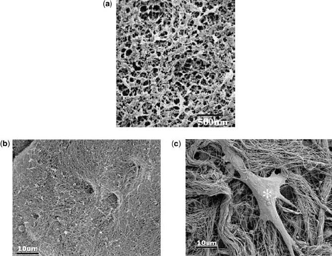
experience nanotopography and interact with nanofeatures in vivo as evident
from Fig. 10.1c. The basement membrane is an integrated component of the
ECM and measures approximately 200 nm in thickne ss. The basement
membrane separates various tissues such as epithelia, endothelia, muscle
fibers, and the nervous system from connective tissue compartments. The
basement membrane is characterized by a co mplex surface topography
consisting of intertwining fibers, ridges, and pores in the nanometer scale as
is evident from Fig. 10.1ac. The corneal membrane of rhesus macaque is
presented in Fig. 10.1a [10,11]. The topographical feature measured was
178 57 nm for the basement membrane height, the interpore dist ance was
127 54 nm, and fiber and pore diameters were 52 28 nm and 82 49 nm,
respectively [10,11]. These measured topographic feature values are similar to
FIGURE 10.1 Scanning electron micrographs (SEM) of the basement membrane
present (a) corneal of the rhesus macaque. The surface topography of the membrane is
a mixture of ridges, pores, and fibers in the nanometer scale. SEM micrographs of the
basement membrane from mice (b) and the dermis (c) present the dense collagen
meshwork consisting of fibers and pore structures in nanometer scale. SEM (c)
presents an attached fibroblast (asterisk) to the collagen meshwork (dermis). This
explains the actual nanotopographic features experienced by micrometer dimension
cells in vivo. (Reprinted with permission from (a) Reference [11], and (b and c), and
Reference [12].)
262
BIOMEDICAL NANOSTRUCTURES
the reported human basement membrane values [12, 13] SEM micrographs (b)
and (c) represent the dense collagen nanofiber bundles present in the basement
membrane and dermis of mouse skin. The collagen network appears less
compact in the dermis (Fig. 10.1c), and a fibroblast attached to collagen
nanofibrils can be seen. This explains the actual nanotopographic features
experienced by micrometer dimension cells in vivo. This complex three-
dimensional surface topography of the ECM provides not only the chemical
stimulus but also biophysical cues for the cells attached to it. The ECM
components and its topographic features are well characterized; however, the
mechanisms that can predict the ECMcell inter actions are not well
understood. The cellECM interaction based on integrin receptors that are
associated with the cell membrane to specific sequences such as (RGD)
arginineglycineaspartic acid part of the ECM is well characterized [14–17].
Cells produce ECM proteins to inter act with the surfaces or the substrate on
which they are growing. These ECM proteins act as transducers for
extracellular signals, namely physical and chemical, through the cytosol
membrane using focal contacts [18–20]. The cellECMsubstrate interaction
is shared among cell networks through intercellular communi cation. The
cellsubstrate interactions are critical for the integration and amplification of
extracellular signals [21, 22]. Change in substrate surface properties, namely
chemical composition, surface energy, surface roughness, and surface
topography, can significantly affect cellsubstrate interfacial characteristics
and potentially influence cellular behavior and function [23]. These factors are
more important when developing novel materials and surfaces for tissue
engineering applications as well as prosthetic devices . Cells produce complex
chemical and topographical cues in natural tissue and these cues will differ with
the synthetic surfaces normally used for in vitro culture. Cells may encounter
different surface topographie s and that might vary from macro- to nano-size
dimension, for example, macrotopographies experienced in bone or ligament
shape, microtopographies with the shapes of other cells, and nanotopographies
with protein folding and collagen banding [24].
Surface topography and biochemical cu es have been shown previously to
alter cell behaviors such as adhesion, orientation, cell activation, and migration
significantly [25–32]. Furthermore, these topographical cues are shown to
influence various modes of cell adhesion and consequently changes in cell
shape, growth, apoptosis and regulation of certain gene expressions [28, 29, 33–
35]. For instance, aligned fibroblasts on groove microtopography produced
higher quantities of the adhesive protein fibronectin than the nonaligned cells
on control plane surfaces [35, 36, 38].
Nanotopographical features of the ECM can significantly affect cellular
behavior and were the inspiration for the favored nanoscale dimensions used in
the design of new generation tissue engineering scaffolds and biomedical
implants. Several evidences documented in the literature clearly showed the
influence of nanotopography on cellular behavior that accounts for changes in
morphology [25, 30], adhesion [26], motility [34], proliferation [36], endo cytotic
activity [37], and gene regulation [35] of various cell types, namely fibroblasts
CELL BEHAVIOR TOWARD NANO STRUCTURED SURFACES 263
[36, 38], osteoblasts [39], osteoclasts [40], endothelial [41], smooth muscle [42],
epithelial [43, 44], and epitenon cells [45]. A detailed understanding of
nanotopographical surface interaction with precursor cells and their differ-
entiation is essential. In addition to fundamental understanding of
cellnanotopographic surface interactions, the nanotopographies may have
potential applications in various biomedical fields.
10.2 NANOTOPOGRAPHIC SURFACES: FABRICATION TECHNIQUES
Presently, the experiments perfor med on conventional tissue culture polystyr-
ene (TCPS) flat surfaces give an idea of cellsubstrate interaction; however,
they do not simulate the complex ECM topography and dimensions. Earlier
studies have shown the effect of micro- and nanoscaled surface topographies
on cellular functions including morphology, adhesion, motility, proliferation,
and gene regulation [25–35]. Thus, various topographic features namely pores,
ridges, grooves, fibers, nodes, and combinations of these features were created
using a wide range of fabrication techniques [1, 46–54].
Advances in nanotechnology have enabled the fabrication of various
structures in nanodimensions, and such structures vary from thin films to
genetic constructs that are used for building biological molecules. All the
nanofabrication techniques have been focused on two approaches, namely the
top-down and bottom-up. The top-down approach includes lithographic
techniques (e.g., soft, photo, colloidal, and electron beam lithography),
electrospinning, polymer demixing, phase separation, evaporation techniques,
and chemical etching. The bottom-up includes assembly process (supramole-
cular, assembly, monolayer, directed self-assembly), nanoparticle formation,
and probe lithography to name a few. The resulting nanostructures may result
in an ordered surface nanotopography or a ran dom topography that will affect
cellular functions, and thus cells behave according to the surface topography to
which they are exposed. Nanofabrication techniques such as photolithography
and electron beam lithography provide ordered nanotopographic surfaces.
Recently, electrospinning has emerged as a promising technique to create
nanofibers, and these nanofibers, can also be aligned to produce ordered
nanotopographies [54]. Other techniques such as polymer demixing, phase
separation, colloidal lithography, and self-assembly result in random surface
topographies. Random topographies are created spontaneously during the
process of fabrication or processing itself. These structures are randomly
organized, arranged without any control on the geometry and reproducibility.
However, creating these nanotopographic features is simple, inexpensive, and
spontaneous. On the contrary, fabrication of ordered nanotopographic
features requires complex and expensive equipments and sound technical
knowledge. Table 10.1 summarizes various popularly studied nanotopography
fabrication techniques, advantages, shortcomings, and observed changes in
cellular behavior. We will discuss some of the most commonly used
264 BIOMEDICAL NANOSTRUCTURES

TABLE 10.1 Summary of Nanotopographic Surfaces Created Using Different Fabrication Techniques and Observed Cellular Behavior
Technique
Topographic
features Merits Shortcomings Cell type used
Observed
change in
cellular
behaviors References
Ordered nanotopographic surfaces
1 Electron
beam
lithography
Square groove,
ridges, and
nanopillars
Dimensions
14 nm to
several microns
With the aid of
computer can
create precise
geometries and
patterns without
any mask
Expensive
equipment, more
time, lower
resolution, hard
to pattern large
surface area
Gingival
fibroblasts,
embryonic
Xenopus spinal
cord neurons,
rat hippocampal
neurons, corneal
epithelial cells,
keratocytes
Strong
alignment,
migration,
and
orientation
[52, 55–62]
2 (a) Photolithography
(near-UV)
coupled with
(b) etching
(a) Square- and
V-shaped
grooves,
ridges, round
nodes.
Dimensions
range between
30 nm to several
microns and
most frequently
dimensions with
few microns are
created
(b) Depends
on the nature and
time of etching
agent used
(a) Create precise
geometries and
patterns
(b) Fast,
simple, and
inexpensive
(a) Expensive and
complicated
instrumentation
and nanodimensions
at the higher side
(b) Difficult to
fabricate specific
geometric features
Rat dermal
fibroblasts,
P388D1
macrophages,
peritoneal
macrophages,
BHK, MDCK
cells, chick
embryo cerebral
neuron, Uromyces
appendiculatus
fungus, murine
macrophages, rat
astrocytes
Improved
orientation,
faster
spreading,
migration
into the
structures
and strong
alignment
[63–69]
265