Molina-Par?s C., Lythe G. (editors) Mathematical Models and Immune Cell Biology
Подождите немного. Документ загружается.

4 T Cell Activation and Function: Role of Signal Strength 83
the phenotype of Ctla4
=
mice which die from hyperproliferation of lymphocytes
by 4–6 weeks of age [20, 21]. Reducing Ctla4 expression in mice using small in-
terfering RNA causes onset of diabetes in mice [22]. This is important in light
of the fact that several autoimmune disorders map to mutations in CTLA4 in hu-
mans [23]. Several mechanisms have been proposed to explain the inhibition of
T cell activation by CTLA4. These can be broadly classified into two categories:
(1) by competing with CD28 for CD80/CD86 ligands or (2) generating its unique
set of signals. CTLA4 inhibits the TCR and CD28 induced raft formation, asso-
ciation with the tyrosine phosphatase SHP-2 and the serine/threonie phosphatase
PP2A which downregulates MAPK and ERK signalling. Increase in TGFˇ secre-
tion and increment in indoleamine dioxygenase (IDO) activity have been observed
with CTLA4 ligation. More recently, CTLA4 has been shown to decrease T cell–
APC dwell times by modulating intracellular Ca
2C
levels. Overall, the net effect of
CTLA4 ligation results in a decrease in IL-2 production and cell cycle progression
[24–27].
Some other positive costimulatory receptors are CD40, inducible-costimulator
(ICOS), OX40, 4-1BB, signalling lymphocyte activation molecule (SLAM). CD40
is expressed on B cells and other professional APCs and binds to its ligand CD40L
on T cells (Fig. 4.2)[28]. Productive T dependent B cell responses and isotype
switching requires CD40-CD40L interactions [29]. Cd40l
=
mice show reduced
antigen-induced inflammatory responses, e.g. lower IgE and IgG amounts, and are
unable to clear some microbial infections, e.g. Leishmania major [30, 31]. ICOS
is a member of the B7 family and binds to its ligand ICOS-L. It is induced on ac-
tivated T cells and Icos
=
mice have impaired germinal centre and Peyer’s patch
formation [32,33]. OX40 is a member of the tumor necrosis factor (TNF) family and
binding to its ligand, OX40L, sustains proliferation of T cell effectors. OX40 sig-
nalling enhances IL-2 and T
H
2 cytokine production. Ox40
=
mice have impaired
effector and primary response to keyhole limpet hemocyanin [34]. Also, OX40 sig-
nalling dampens Treg function, increases symptoms of autoimmunity but lowers
anti-tumor responses [35]. 4-1BB (CD137) is another tumor necrosis factor fam-
ily member which binds to its ligand 4-1BBL to enhance CD4
C
and CD8
C
T cell
responses and promote IFN production. Importantly, 4-1bb
=
mice have defec-
tive recall CD8
C
responses to some viruses like influenza [36]. Binding of CD27, a
member of the TNF family, to its ligand CD70 promotes proliferation and survival
of activated T cells. Constitutive activation of CD27 results in immunopathology
during autoimmunity and viral infections [37]. Signalling lymphocyte activation
molecule (SLAM) deficiency reduces IL-4 and marginally increases IFN produc-
tion in mice [38].
Other negative regulators of T cell activation, apart from CTLA4, are pro-
grammed death (PD)1 and B and T lymphocyte attenuator (BTLA). PD1 is a B7
family member and binds to its ligand PD-L1 and PD-L2. Pd1
=
mice develop
lupus-like disorders [39]. BTLA is also a B7 family member and a negative regula-
tor of B and T cell responses and Btla
=
mice develop exacerbated experimental
autoimmune encephalomyelitis (EAE) [40]. A regulatory role has recently been as-
signed to the T-cell Ig domain and mucin domain (TIM) family members TIM-1, 2, 3
84 A. Ahmed and D. Nandi
and 4. TIM1, 2 and 3 are expressed on T cells while TIM4 is primarily expressed on
APCs [41]. TIM-3 was identified as T
H
1 specific molecule responsible for regulat-
ing autoimmunity [42]. TIM-1, 3 and 4 can costimulate T cells, regulate apoptosis,
tolerance and clearance of apoptotic cells [41]. Links have been found between TIM
proteins and allergy, asthma and autoimmunity [43].
The proper functioning of positive and negative costimulatory molecules is cru-
cial for an optimal T cell response and is highlighted by the phenotypes of mice
deficient in any of these molecules. It is important to point out that costimulation
ensures that T cell activation occurs in a proper context e.g. during an ongoing im-
munological response and this safeguards against development of autoimmunity.
SOS and Its Intracellular Modulators
Two important factors during T cell activation are the affinity/avidity of the TCR
for the MHC–peptide complex and the time for which the two are engaged. Both
these factors along with the availability of co-stimulation determine the overall SOS
that is delivered to a T cell during activation. T cells express heterogeneous TCRs
which may bind to the same antigen with different affinities. Therefore, T cells get
activated with varying SOS, which has a profound impact during thymic education
and modulate T cell responses in the periphery with respect to proliferation, cytokine
secretion, survival and death. It is thought that optimal T cell activation requires
sustained signalling for several hours. To achieve this, TCRs need to be continuously
engaged or triggered at periodic intervals (serial triggering). The model of ‘temporal
summation’ proposes that each short-term TCR–MHC–peptide interaction leads to
accumulation of intracellular signalling intermediates until a threshold for optimal
activation is reached [44].
One of the signalling intermediates robustly modulated by signal strength is in-
tracellular Ca
2C
. The stronger the signal, the higher is the Ca
2C
flux [45]. Also,
intracellular amounts of Ca
2C
and ROS increase with the SOS in primary mouse
CD4
C
T cells [46]. The TCR interacting molecule (TRIM), a transmembrane adap-
tor protein expressed on T cells, stabilizes TCR surface levels after engagement
with MHC–peptide complexes and leads to higher intracellular Ca
2C
amounts, lead-
ing to enhanced activation [47]. The other intracellular integrator of signal strength
is ERK. There is evidence to suggest that strong signals lead to greater but tran-
sient ERK activation. On the other hand, weak signals result in lower but sustained
ERK phosphorylation [48]. Proteins such as Fos which have an ERK targeting DEF
motif are sensitive to transient versus sustained ERK activation and can trigger
gene transcription in accordance with the SOS [49]. Disruption of Mekk2 (an up-
stream MAPK kinase) in mice leads to enhanced IL-2 and IFN.Inthesemice
JNK activation, but not ERK activation, is affected and it is possible that MEKK
acts as a negative regulator of TCR signal strength by activating JNK [50]. An-
other negative regulator of TCR signal strength was identified as cyclophillin A, a
4 T Cell Activation and Function: Role of Signal Strength 85
peptidyl-prolyl isomerase (Ppia). Ppia interacts and inhibits Itk and Ppia
=
mice
have elevated levels IgE, develop allergic disorders and their T cells are hypersensi-
tive to activation [51].
Other cell surface molecules that are regulators of signal strength are CD5 and
the co-receptor CD8. CD5 is a negative regulator of TCR signalling and, hence,
protects against autoimmunity. T cells from Cd5
=
mice are hyperresponsive to
anti-CD3 mediated activation [52]. CD5 expression levels are also controlled by
the avidity of the TCR–MHC–peptide interaction and a strong interaction leads to
higher surface amounts of CD5. Massive deletion of thymocytes in transgenic mice
expressing high affinity TCR in a Cd5
=
background is observed due to a shift
from positive to negative selection [53]. Unlike CD5, CD8 expression is inversely
proportional to TCR signal strength. Experiments done to study this relationship
found that, as the affinity of TCR and self MHC–peptide complexes increased, CD8
surface expression is reduced. It is possible that reducing surface CD8 amounts
lowers the avidity of T cells for self ligands and prevents autoimmunity [54].
Although the molecular mechanisms and the intracellular modulators of signal
strength are not completely understood, progress has been made in identifying
several molecules that influence the SOS. However, the modulation of expression
of these molecules under varying SOS and their interaction with other molecules
affecting T cell responses need to be understood in greater detail. Nevertheless, the
impact of SOS on almost every aspect of T cell life is well established and discussed
below.
Role of Signal Strength in Cell Fate Decisions in the Thymus
The thymus is a bilobed structure located just above the heart comprised of support-
ing cells which constitute the stroma and T cells, in various stages of development,
known as thymocytes. Progenitor bone marrow cells migrate to the thymus under
the influence of chemotactic factors by day 11 of gestation in mice and the eighth
or ninth week in humans. Once in the thymus, progenitor T cells undergo a process
of maturation and education to yield mature single positive (SP) CD4
C
or CD8
C
T
cells that enter the peripheral circulation (Fig. 4.5). Approximately 99% of thymo-
cytes die by apoptosis during this process and only mature T cells that recognize
peptides presented in the context of either MHC class I or MHC class II are selected.
The importance of the thymus is highlighted in children suffering from DiGeorge
syndrome which results from deletions in chromosome 22q11.2. Similar deletions
in mice chromosome have highlighted the importance of the transcription factor
Tbx1 in this disorder. These children have an underdeveloped or completely absent
thymus and, along with several other defects, are highly susceptible to viral, fungal
and protozoan infections [55].
The development and maturation of thymocytes can be tracked by following the
changes in expression of cell surface molecules. During the early stages of differ-
entiation, thymocytes lack surface expression of CD4 and CD8 and are referred
to as CD4
CD8
or “double negative (DN)”. A small fraction of CD4
CD8
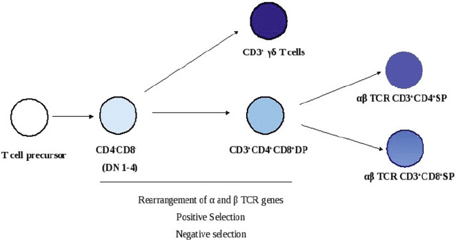
86 A. Ahmed and D. Nandi
Fig. 4.5 A simplified overview of thymic development and education. T cell precursors that enter
the thymus undergo a rigorous programme of development and education involving positive and
negative selection to emerge into the periphery as ı T cells or ˛ˇ TCR bearing CD4
C
or CD8
C
T cells
thymocytes make productive rearrangements of their and ı chain genes to ex-
press surface ı TCR together with the CD3 complex. ı T cells represent about
0.5–10% of peripheral T lymphocytes and are quite abundant in the skin, intestinal
epithelium and pulmonary epithelium in mice. These cells are capable of recogniz-
ing a broad range of peptide and non-peptide antigens but not necessarily in the
context of the MHC class I and class II molecules. They are believed to be members
of the earliest cell-mediated immune system and may have evolved to protect the
integrity of epithelial tissues [56].
A vast majority of thymocytes productively rearrange ˛ and ˇ TCR genes to ex-
press on their surface the ˛ˇ TCR–CD3 complex. TCR gene rearrangement occurs
in a manner similar to antibody gene rearrangement in B cells with the help of RAG-
1 and RAG-2 and other proteins required for DNA recombination. During these
stages, thymocytes also acquire surface CD4 and CD8 molecules and are referred to
as CD4
C
CD8
C
or “double positive (DP)” thymocytes. This stage is important for
thymic education as only those thymocytes which recognize self MHC molecules
are selected by a process known as “positive selection.” Thymocytes which bind to
self-MHC molecules with very high affinity are deleted by a process known as “neg-
ative selection.” This process is important as it reduces the risk of autoimmunity and
is well illustrated by the master transcription factor, AIRE, which is responsible for
expression of several self proteins in the thymus. The deficiency of AIRE results in
a multi-organ autoimmune disease known as autoimmune polyglandular syndrome,
type 1 [57]. Thymocytes that fail these selection processes die by apoptosis. The
MHC restricted self tolerant double-positive thymocytes lose either CD4 or CD8
co-receptor to become mature SP CD4
C
(T
H
)orCD8
C
(T
C
) T cells and enter the
peripheral circulation [3].
4 T Cell Activation and Function: Role of Signal Strength 87
Apart from positive and negative selection, two more cell fate decisions relevant
in terms of the SOS in the thymus are: (1) the development of T cells express-
ing either ı TCR or the ˛ˇ TCR and (2) differentiation of ˛ˇ TCR
C
cells
into CD4
C
or CD8
C
lineages. In both these cell fate decisions, the SOS deliv-
ered via the TCR plays crucial roles. Precursor cells in the thymus consisting of
c-kit
C
CD44
C
CD25
C
CD24
C
CD4
CD8
–
can differentiate into different lineages
and the DN stage is divided into 4 stages (1–4). As development progresses, thy-
mocytes express less c-kit, CD44, CD25 and CD24, acquire Thy1, CD4, CD8 and
begin to rearrange their TCR genes. Commitment to the T cell lineage takes place at
the DN stage 3 once precursors acquire Thy1 and express a fully functional preT˛
(an invariant ˛ chain of the TCR which is expressed early in the thymus) and rear-
range their and ı chains. This is also the stage when the choice between the two
development programs, i.e. ˛ˇ and ı, needs to be made. Those cells which fail to
productively rearrange their TCR genes or do not express surface TCR die by apop-
tosis. Two models have been proposed to explain how the choice between the ˛ˇ
TCR versus the ı TCR is made. The first model is the stochastic/selection model
according to which cell fate specification is made independent of signals induced
by the TCR, i.e. fate is decided before fully functional TCRs (ı and preT˛ˇ)are
expressed [58]. The second is the instructive model in which fate is decided based
on distinct signals generated through the preT˛ˇ and ı TCRs. One of the distin-
guishing factors in the generation of these two TCRs is SOS and the development
of one lineage should affect the development of the other, according to this model
[58–60]. The SOS model for lineage commitment was put forward by Hayes
et al. [61]. Using a model where fate specification was entirely mediated by the ı
TCR, this group showed that increasing signal strength through the ı TCR en-
hanced the development of ı T cells. Conversely, weakening of signal strength
resulted in the development of T cells belonging to the ˛ˇ lineage. Similar results
were reported by another group [62]. One way in which signal strength through
ı and preT˛ could be altered is through differences in their surface expression.
Reduction in ı TCR surface expression skewed development towards ˛ˇ T cells.
Concomitantly, increasing expression of the ı TCR increased the development
of ı T cells and reduced the development of ˛ˇ T cells [63]. Also, reducing ı
TCR signalling by reducing the number of ITAMs on the CD3 chain enhances ˛ˇ
commitment [64]. It has been shown that the magnitude of signalling downstream
of the ı TCR is greater compared to that generated by the pre-TCR [65].
A biochemical basis for the lineage commitment has also been explained in terms
of ERK activation. A strong signal as delivered by the ı
TCR leads to sustained
activation of ERK, while transient ERK activation by the weak pre-TCR signal
is more conducive for proliferation and transit of cells to the DP stage [61, 62].
Sustained ERK activation leads to enhanced induction of early growth response fac-
tor (Egr), a Zn
2C
finger transcription factor. Egr proteins are induced by both ı
and pre-TCR signals; however, the level of induction is regulated by the SOS. Over-
expression of Egr leads to more ı T cells and a decrease in ˛ˇ T cell numbers.
Downstream to Egr is Id3, a helix-loop-helix (HLH) protein. Induction of Id3 is
found to be concomitant with ı T cell lineage commitment [62]. One mechanism
88 A. Ahmed and D. Nandi
by which Id3 nudges cells towards the ı T cell lineage is by suppressing the activ-
ity of basic helix-loop-helix E proteins which are required for VDJ recombination
by the TCR ˇ chain [60,62].
In addition to differing SOS in determining ˛ˇ ‘vs’ ı cell fate, a role for Notch
signalling has also been proposed. According to this model, moderate pre-TCR sig-
nals synergize with Notch signals enabling generation and proliferation of ˛ˇ T
cells. While strong ı TCR signals are sufficient for the lineage commitment of ı
T cells, weak ı TCR signals can synergize with strong Notch signals to give rise
to ˛ˇ cells. In other words, it appears that development of ˛ˇ T cells depends more
on Notch signalling compared to ı T cells [66].
After commitment to the ˛ˇ lineage, DN stage 4 cells become CD4
C
CD8
C
or
DP in addition to the expression of the TCR ˇ chain. The ˛ TCR chain now un-
dergoes rearrangement. In fact, DP cells continue to express RAG proteins for an
extended period of time and multiple V/J recombinations occur at the same ˛ locus
so that several ˛ˇ TCRs can be produced per cell. DP thymocytes in the cortex
bearing the ˛ˇ TCR undergo selection processes that are dependent on signals
generated through the binding of TCR to the MHC–peptide complex. The affin-
ity/avidity model for positive and negative selection is the most widely accepted.
˛ˇ TCRs which have low to intermediate affinity for self MHC, undergo positive se-
lection and differentiate into CD4
C
or CD8
C
SP cells while those TCRs which have
high affinities are negatively selected and undergo clonal deletion. There are several
lines of evidence that indicate that the SOS through the TCR determines the fate of
DP thymocytes. Cells which are unable to engage MHC or do so with a very low
affinity die by neglect, a fate which befalls most DP thymocytes. Those which en-
gage MHC with an intermediate intensity are positively selected and those that bind
with too high an intensity are either clonally deleted, become anergic or differentiate
into regulatory T cells (Treg), which are peripheral immune regulators [67]. Those
thymocytes that survive the rigorous selection process and are MHC restricted are
present in the medulla and enter the peripheral circulation as mature T cells.
The binding affinities of TCR–MHC peptide complexes are reflected in the signal
strengths generated downstream of the TCR which trigger specific programs leading
to either positive or negative selection. Experiments have shown that by varying the
SOS, positive and negative selection decisions can be altered. In a study to exam-
ine the role of TCR signal intensity on positive and negative selection, TCR signal
potency was varied by substituting transgenic TCRs having 1, 2, 3 or no ITAMs
on the CD3 chain. Interestingly, decreasing the signalling ability in cells with low
affinity led to reduced selection. However, in cells which bound self-MHC with very
high affinity and would normally have undergone clonal deletion, decreasing TCR
signal potency by varying the number of ITAMs increased the chances of positive
selection [68]. Low affinity ligands generate the p21 form of CD3 which has only
2/3 ITAMs phosphorylated while high affinity ligands lead to phosphorylation of
all 3 ITAMs resulting in formation of the p23 form. The signalling molecules and
adaptor proteins recruited by p21 and p23 are different and may explain the dif-
ferences in the distinct signalling programs triggered during positive and negative
selection [69].
4 T Cell Activation and Function: Role of Signal Strength 89
The activation of ERK has been shown to integrate signal strength and cell fate
decisions in the thymus. Positive selecting ligands trigger low but sustained ERK
activation whereas negative selecting ligands lead to strong but transient ERK ac-
tivation. Also, in foetal thymic organ cultures, negative selection stimuli can be
converted to positive selection signals by use of pharmacological inhibitors of ERK.
One of the reasons behind differences in ERK activation during positive and nega-
tive selection is thought to be the time for which TCRs remain on the surface. High
affinity/negative selection ligands induce rapid internalization of TCR. However,
during low affinity interactions TCRs stay on the surface for longer times, lead-
ing to prolonged ERK activation which is essential for survival and proliferation
of positively selected DP thymocytes. Sustained ERK activation leads to phospho-
rylation and ubiquitin mediated proteosomal degradation of pro-apoptotic protein
Bim. On the other hand strong and transient ERK activation leads to upregulation of
genes such as Nur77 that lead to apoptosis [70–72]. Another line of evidence sug-
gesting the involvement of TCR-Ras-MEK-ERK pathway is the fact that targeted
deletion of RasGRP, a guanine nucleotide exchange factor for Ras, has a profound
effect on positive selection by virtue of reduced ERK activation [73]. A germline
knockout of ERK1 having a conditional deletion of ERK2 at the DN stage re-
sulted in developmental arrest at the DP stage, suggesting a role for ERK in positive
selection [74].
The process of commitment to either the helper (CD4
C
) or cytotoxic (CD8
C
) lin-
eages is crucial and DP thymocytes downregulate either CD4 or CD8 co-receptors
to become MHC class II or class I restricted. This process could be random and
independent of TCR signals such that cells which down regulate the mismatched
co-receptor are eliminated. However, the stochastic model could not explain why
constitutive co-receptor expression could not rescue cells which had mismatched
CD4 or CD8 expression. The instructive model, on the other hand, suggests that
commitment to the CD4 and CD8 lineage occurs in response to specific TCR sig-
nals [75]. Lck associates with CD4 more compared to CD8 and this may lead to
stronger signals being delivered by MHC class II compared to MHC class I re-
stricted cells [76]. Strong and weak signals lead to selective downmodulation of
either CD4 or CD8. Evidence suggests that weak signal stimulated cells develop into
CD8
C
T cells whereas strong signals stimulate differentiation into CD4
C
T cells.
Mice expressing an inactive Lck contain MHC class II restricted cells differenti-
atingintoCD8
C
T cells and constitutively active Lck can direct MHC-I restricted
cells to differentiate into the CD4 lineage [77]. Lineage commitment can also be de-
cided by signal duration where, longer and shorter duration signals lead to CD4 and
CD8 lineage commitment respectively [78]. Another model termed the ‘kinetic sig-
nalling model’ proposes that the duration of TCR signalling rather than the strength
plays a key role in determining the CD4
C
or CD8
C
T cell lineage. However, in this
case too, the quantity of signal delivered to a cell is the deciding factor. What this
model proposes is that all DP cells transiently terminate CD8 expression so that they
become CD4
C
CD8
lo
. If this transient population of cells receives sustained TCR
signalling, it develops to form CD4
C
cells. If the signalling is of a short duration,
transcription of CD8 is reinitiated, CD4 is silenced and CD8
C
SP cells develop [79].
90 A. Ahmed and D. Nandi
Consistent with this model, mice which expressed CD4 under the control of CD8
E8
III
enhancer, showed progressive downmodulation of CD4 after the DP stage.
Positive selection of MHC-II restricted cells was normal but the differentiation of
these cells was skewed towards the CD8 subtype by virtue of co-receptor downmod-
ulation [80].
Overall, the strength or duration of signal plays crucial roles during thymic de-
velopment and education: First, the conversion of DN cells to ı T cells or ˛ˇ T
cells; second, positive and negative selection of ˛ˇ TCR
C
DP cells and, finally, the
generation of mature CD4
C
or CD8
C
SP T cells.
Signal Strength Influences T
H
1/T
H
2Dichotomy
The encounter of an antigenic peptide presented on MHC class II triggers a series
of events leading to the differentiation of CD4
C
T cells into different T
H
subtypes,
which are characterized by distinct cytokine secretion patterns. Two primary sub-
types identified are T
H
1 which secrete IFN, IL-2, lymphotoxin ˇ and T
H
2which
secrete IL-4, IL-5, IL-6 and IL-13 [81,82]. Another recently identified effector sub-
set is T
H
17 which, primarily, produces IL-17. Their generation is regulated by IL-6
and TGFˇ [83] and their development is regulated by the master transcription fac-
tors Stat3 and ROR [84]. T
H
17 cells are crucial for combating several pathogens.
Mice lacking IL-17 are highly susceptible to infection with Klebsiella and Candida.
More importantly, T
H
17 cells have been associated with several autoimmune con-
ditions such as multiple sclerosis and rheumatoid arthritis. Indeed IL-17 deficient
mice develop less severe experimental autoimmune encephalomyelitis [85].
T
H
1 responses are crucial for clearing intracellular pathogens while T
H
2re-
sponses are effective during parasite infections and antibody production. Abnormal
or excessive T
H
activation leads to autoimmune disorders (T
H
1) or atopy (T
H
2).
The local cytokine milieu, co-stimulatory interactions, antigen concentration and
strength of TCR signals are the factors which govern T
H
differentiation. The
cytokine environment existing during priming has a far reaching effect on differ-
entiation. The presence of IL-12 skews differentiation towards T
H
1 whereas IL-4
results in greater T
H
2 responses [86]. The SOS, in terms of duration of engage-
ment and antigen dose, is a key determinant in T
H
fate decision [87, 88]. T cells
from allergic individuals produce IL-4 only when stimulated with a low dose of al-
lergen. When the allergen dose is increased, IL-4 production is reduced and may
form the basis for allergen immunotherapy [87]. On the other hand, IL-12 coupled
with a short duration TCR signal is sufficient for T
H
1 polarisation; however, pro-
longed TCR signalling is required for T
H
2 responses [88]. A weak intensity signal
provided by a low affinity altered peptide ligand favors T
H
2 whereas optimal con-
centrations of an agonist peptide which transduce a relatively strong signal favors
T
H
1 differentiation [89–91]. Agonist peptides trigger strong and more sustained
signalling events such as CD3 phosphorylation, Ca
2C
mobilization, JNK and
4 T Cell Activation and Function: Role of Signal Strength 91
MAPK activation as compared to altered peptide ligands [92–95]. The magnitude of
Ca
2C
fluxes downstream of weak and strong TCR signals is an important determi-
nant of T
H
subtypes. Weak TCR stimulation induce Ca
2C
signals that promote IL-4
production whereas strong signals induce MAPK activation that induce IFN [96].
Consistent with this observation, inhibition of MAPK and JNK in both human and
mouse T cells promotes T
H
2 and inhibits T
H
1 differentiation [97]. Constitutive ac-
tivation of p38 MAPK promotes T
H
1 but expression of a dominant negative form
of p38 MAPK inhibits T
H
1 differentiation [98]. Stimulation of JNK1 deficient cells
with a combination of anti-CD3 C anti-CD28 enhances T
H
2 differentiation [99].
JNK2 deficiency enhances T
H
2 differentiation but suppresses T
H
1 primarily due
to a lack of upregulation of the IL-12Rˇ2 subunit [100].
The key transcription factors required for T
H
1 differentiation are T-bet and Stat4
and those involved in T
H
2 differentiation are Stat6, c-Maf and GATA-3. T-bet and
GATA-3 are master regulators of T
H
1andT
H
2 differentiation respectively [86].
Over expression of T-bet induces T
H
2 cells to make IFN and T-bet
=
mice have
impaired T
H
1 differentiation, show increase in T
H
2 cytokines and develop asthma
like symptoms [101, 102]. On the other hand, deletion of GATA-3 leads to T
H
2
cytokine defects [103]. Stat4 is an amplifier of IL-12 signals and can induce T
H
1
differentiation even in absence of TCR signals, whereas Stat6, activated by IL-4, in
turn is essential for activation of GATA-3 [82, 86]. However, the transcription fac-
tors that are primarily modulated by the SOS belong to the NF-AT class. Activation
and nuclear localization of NF-AT is dependent on the Ca
2C
signal. Stronger Ca
2C
signals ensure longer nuclear dwell times for NF-AT. The SOS regulates amounts
of NF-ATp (NF-ATc2 or NF-AT1) and NF-ATc (NF-ATc1 or NF-AT2) in the nu-
cleus. Strong signals as opposed to weak ones lead to more NF-ATp accumulation
in the nucleus, which promotes GATA-3 suppression and T
H
1 differentiation. Al-
tered peptide ligands on the other hand have low NF-ATp but sufficient NF-ATc in
the nucleus to drive IL-4 transcription and T
H
2 differentiation. Both IL-4 and IL-2
have been shown to be essential for T
H
2 differentiation in response to low dose
antigenic peptide. Cells receiving low intensity signals upregulate GATA-3 which
can, in turn, enhance its own levels. GATA-3, along with IL-2 activated Stat5, drives
IL-4 transcription and T
H
2 differentiation [104].
Co-stimulation through CD28 has not been found to independently contribute
to T
H
differentiation. The addition of anti-CD28 enhances cytokine secretion but
does not change the kind of cytokines secreted. However, the ability of anti-CD28
to enhance IL-4 production in cells which receive a low intensity signal results in
greater differentiation of T
H
2 differentiated cells [91,95]. However what does have
a qualitative effect on cytokine pattern is interaction between LFA-1 and ICAM.
Blockade of this interaction promotes T
H
2 whereas increasing the LFA-1-ICAM
interaction promotes T
H
1 development due to a sustained Ca
2C
signal [105, 106].
Overall, it appears that SOS, along with other factors, is important during T
H
cell
differentiation. This aspect is relevant during T
H
responses as the dose and potency
of the antigen clearly modulate immune responses.
92 A. Ahmed and D. Nandi
Role of Signal Strength in Treg Function
Treg cells are important in the maintenance of peripheral tolerance. They have
been characterized as a subset (5–10%) of CD4
C
T cells expressing high levels
of CD25, the IL-2R˛ chain, and are capable of suppressing in vivo and in vitro T
cell responses [107]. The primary function of Treg cells is to regulate autoimmu-
nity and depletion of this population in mice has been shown to precipitate several
autoimmune disorders. Injection of CD4
C
CD25
C
cells reduces symptoms of sev-
eral autoimmune diseases such as colitis, gastritis, insulin dependent autoimmune
diabetes and thyroiditis in mouse models [107, 108].
Treg cells do not proliferate or produce IL-2 in response to TCR mediated stim-
ulation [109]. Most Treg cells are CD4
C
, express the transcription factor Foxp3 and
can be divided into two groups: natural Treg (nTreg) and induced Treg (iTreg). The
former arise in the thymus whereas the latter are derived from peripheral CD25
SP CD4
C
T cells under the influence of IL-2 and TGFˇ. Both express high levels
of CD25, glucocorticoid-induced tumor necrosis factor inhibitor (GITR), CTLA4,
CCR4, and CD62L [110]. Tregs are CD45RB
low
(mice) and CD45RO
low
(humans),
a sign that they are pre-activated [110]. Apart from the site of their generation, the
conditions that give rise to natural and induced Tregs are quite different. nTregs are
the result of strong TCR signals delivered by self peptides in the thymus [111]. Also
required for their generation are strong costimulatory signals because, in the absence
of CD28, the numbers of nTreg are reduced [112–114]. On the other hand, iTregs
arise in the periphery due to weak or suboptimal primary and secondary signals
[115,116]. However, absolutely essential in this case is CTLA4 signalling because
mice lacking CTLA4 do not have this category of Tregs [117]. IL-2 is the cytokine
that is crucial for generation and maintenance of Treg cells [118] as the expression
of Foxp3 is dependent on IL-2 signalling [119]. Indeed, Il-2
=
mice suffer from
fatal autoimmunity [120]. Treg cells are thought to mediate their suppressive effects
via direct cell–cell contact. Numerous mechanisms, including secretion of suppres-
sive cytokines IL-10 and TGFˇ, repression of IL-2 transcription and induction of
the tryptophan depleting enzyme indoleamine 2, 3 dioxygenase in target cells, have
been proposed to explain their mode of action [121].
Tregs, when isolated from peripheral blood, are anergic. As they have immense
therapeutic potential, the possibility of expanding them in vitro would be of great
benefit. What has been found is that TCR and costimulatory signals that drive pro-
liferation of na¨ıve CD4
C
T cells are insufficient for propagation of Tregs bearing
very high avidity TCRs for self antigens. Apart from strong TCR signals, Tregs re-
quire high intensity CD28 signals for expansion, but high amounts of IL-2 cannot
substitute for CD28 signalling [122]. These results are consistent with results ob-
tained with a superagonist anti-CD28 that selectively increased Treg numbers and
their suppressive activity in rats suffering from EAE. In fact, treatment with this an-
tibody was found to confer protection against EAE and was proposed as a therapy
for treating autoimmune disorders [123].
Signal strength, apart from controlling Treg expansion, also dictates resistance
or susceptibility to Treg mediated suppression. Using an in vitro model, it has been