Molina-Par?s C., Lythe G. (editors) Mathematical Models and Immune Cell Biology
Подождите немного. Документ загружается.

7 Modelling Lymphocyte Dynamics In Vivo 143
or the length of a cell’s telomeres. Others are administered for research purposes
and include the nucleoside analogue BrdU and different stable isotopes. Here we
will give an overview of these experimental methods, as well as some of the mathe-
matical models that have been developed to interpret the data.
Experiments of Nature
In the absence of specific labelling techniques, investigators have estimated the rate
of production and loss of different lymphocyte populations by following changes in
the numbers of cells under specific extreme “natural” circumstances. In patients with
extremely low lymphocyte numbers, e.g. because of severe regimens of chemother-
apy as treatment for cancer or prior to stem cell transplantation, the reconstitution of
the different lymphocyte compartments was followed over time and translated into
net rates of lymphocyte production [1, 2]. On the one hand, such rates may under-
estimate the true lymphocyte production rate, because cells may also die during the
reconstitution period, a process that may go unnoticed but which will decrease the
net rate of reconstitution. On the other hand, production rates in lymphopenic pa-
tients may exceed the actual rate at which cells are produced in a fully reconstituted
lymphocyte compartment in steady state, because cells undergo little competition
for survival factors.
Another extreme case that has been used to study the rate of lymphocyte turnover
is by measuring the loss of lymphocytes with chromosomal damage in patients after
treatment with radiotherapy for cancer. Assuming that no new lymphocytes with
chromosomal damage are produced after stopping radiotherapy, the average loss rate
of different lymphocyte populations has been estimated [3]. Again, extrapolation of
these estimates to the healthy situation has to be done cautiously, because these loss
rates may be influenced by the DNA damage of the cells under investigation and by
the low numbers of lymphocytes in these patients.
Despite the fact that the quantitative parameters resulting from these “experi-
ments of nature” may not translate exactly to the natural situation, these studies have
had a large impact on our insights into immunological memory. Both approaches
have clearly shown that the turnover rate of memory T lymphocytes exceeds that
of naive T cells, and have thereby suggested that immunological memory is not
maintained by a pool of long-lived memory cells, but by a highly dynamic pool of
memory T cells with a rapid rate of cellular turnover.
Static Markers of Cell Proliferation and Death
Instead of measuring net production rates or net loss rates of lymphocytes under
specific extreme circumstances, one can measure the natural expression of mark-
ers for cells undergoing cell proliferation or death. Ki67, for example, is a protein
144 B. Asquith and J.A.M. Borghans
whose expression is restricted to proliferating cells. It is expressed during all ac-
tive phases of the cell cycle, i.e. during the G
1
,S,G
2
and mitotic phase, but not
during the G
0
phase. Measuring the fraction of Ki67-expressing cells thus gives
insights into the fraction of cells that is undergoing proliferation. Similarly, cells
undergoing apoptosis can be measured by different methods. One of the plasma
membrane alterations characteristic of cells in the early phase of apoptosis is the
translocation of phosphatidylserines from the inner side of the membrane to the
outer side. This expression can be measured by Annexin V staining, a protein that
binds phosphatidylserine with high affinity. Alternatively, cells in apoptosis can be
distinguished by intracellular staining for certain caspases, which are proteases in-
volved in the cleavage of cellular proteins in cells undergoing apoptosis.
Measuring cell death and proliferation by such naturally expressed markers has
the advantage that the immune system does not need to be disturbed. These mea-
sures are therefore very useful in comparisons of lymphocyte dynamics between
individuals. Such static markers are, however, hard to translate into quantitative bi-
ological parameters such as the fraction of cells that proliferate or die per day. The
fraction of cells expressing markers of death or proliferation reflects not only the
fraction of cells undergoing death or proliferation per day, but is also influenced by
the period during which these markers are expressed. The problem becomes even
larger when the period of expression is not constant, e.g. because it is influenced
by the immunological situation under investigation. For example, if T cells in HIV
infected individuals were to die very rapidly, one might measure low fractions of
Annexin V positive T cells, while in fact many cells are killed. Likewise, it has been
suggested that T cells undergoing proliferation in HIV infected individuals may
get stuck in the cell cycle [4], and thereby cause higher levels of Ki67 expression
than one would expect from the fraction of cells that is actually producing progeny.
Avoiding such problems and obtaining more quantitative biological parameters re-
quires dynamic markers for cell proliferation and death.
Dynamic Markers of Cell Proliferation and Death
In order to study T lymphocyte production and death dynamically, a large number
of markers have been used, all with their own characteristics, advantages and dis-
advantages. Naturally occurring dynamic markers of T cell production and death
include the telomere length of a T cell population, a marker that is typically inter-
preted as reflecting the proliferative history of a cell population, and the expression
of T cell receptor excision circles (TRECs), a marker that has been proposed for the
measurement of T cell production in the thymus. Other dynamic markers are not
naturally expressed and include the administration of the nucleoside analogue BrdU
or stable isotopes such as deuterated water or deuterated glucose. By administration
of such labels and by following their incorporation in the DNA of newly-produced
cells during and after label administration, one can measure both the rate of pro-
liferation and loss of different lymphocyte populations. Below we will provide a
7 Modelling Lymphocyte Dynamics In Vivo 145
short summary of these different experimental techniques, as well as our favoured
mathematical interpretation of the resulting data. Note that for all these techniques
a number of different mathematical models have been developed. We focus on the
ones that we think most clearly illustrate the impact of mathematics on our insights
into lymphocyte dynamics.
T Cell Receptor Excision Circles
For decades, investigators have tried to quantify the contribution of T cells that are
newly formed by the thymus to the maintenance of the peripheral T cell pool. In
the late nineties a new assay was introduced to measure the number of cells that are
produced by the thymus. This assay is based on the occurrence of T cell receptor ex-
cision circles (TRECs) [5,6]. TRECs are by-products of V(D)J rearrangements that
occur in the thymus when the T cell receptor (TCR) is formed. During this genetic
rearrangement process, parts of the TCR genome are excised and form stable DNA
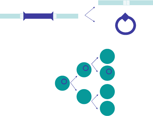
circles (Fig. 7.1a). These TRECs are not copied when a cell divides; they are simply
passed on to one of the daughter cells (Fig. 7.1b). TRECs are thus uniquely formed
in the thymus. Measurement of the average number of TRECs in a cell population
by quantitative PCR was therefore proposed as a direct measure for thymus output.
TREC DNA
V
J
V
J
TREC
a
b
Fig. 7.1 (a) TRECs are excised from the DNA, during T cell receptor V(D)J gene rearrangement
in the thymus. The excised pieces of DNA form stable circular DNA products, which fail to be
copied during DNA duplication. (b) As a consequence, the average number of TRECs (in blue)per
cell in a cell population decreases when cells proliferate

146 B. Asquith and J.A.M. Borghans
When the average number of TRECs per T cell was measured in individuals of
different ages, an exponential decline was observed [6]. These findings fitted exactly
to the common view that the thymus undergoesinvolution, and its output thereby de-
creases exponentially with age. Decreased TRECs per cell in HIV-infected patients
were similarly interpreted as evidence for impaired thymic output [6], and increased
TRECs per cell in individuals after haematopoietic stem cell transplantation as evi-
dence for thymic rebound to compensate for the lack of T lymphocytes [7].
A simple mathematical model pointed out, however, that TRECs do not directly
reflect thymic output [8–10]. The change in the average TREC content of a pop-
ulation of cells with age can be calculated from the change in the total number of
TRECs, T , and the change in the total number of cells N with age. The total number
of cells decreases by cell death at rate ı per day, and increases by T cell proliferation
at rate p per day and by thymic output, .t/, which itself decreases exponentially
with age at rate v per day:
dN
dt
D .t/ C pN ıN (7.1)
where
.t/ D .0/e
vt
:
The total number of TRECs also decreases by cell death and increases by thymic
output, but is not affected by T cell proliferation:
dT
dt
D c.t/ ıT; (7.2)
where c is the average TREC content of a cell that leaves the thymus. The average
TREC content A of a cell population can be calculated as A D T=N. It is easy to
demonstrate that when T and N are at equilibrium, the average TREC content A is:
A D c.ı p/=ı: (7.3)
In other words, the average TREC content of this cell population is totally inde-
pendent of the number of cells that are generated by the thymus per day. This
equation demonstrates that the rate of proliferation and death of this cell popula-
tion strongly influence its average TREC content. The more proliferation the cells
undergo, the lower their TREC content, and the larger the death rate of the cells,
the higher their TREC content. With hindsight, this can be understood intuitively,
because cell proliferation increases the number of cells but not TRECs, and because
cell death influences the average age of a cell population. The larger the death rate,
the younger the cell population, and hence the fewer rounds of proliferation have
occurred and diluted the average TREC content.
One thus has to be extremely careful when interpreting TREC data [8–10]. The
fact that T cell TREC contents decline with age probably reflects a homeostatic

7 Modelling Lymphocyte Dynamics In Vivo 147
response of the immune system to decreasing thymus output. While increased rates
of T cell proliferation or decreased rates of T cell death, both possibly contributing
to T cell homeostasis, strongly affect TREC contents, changes in thymic output per
se will not. Likewise, low TREC contents in HIV patients are more likely to reflect
increased rates of proliferation rather than decreased thymic output [8], an insight
with considerable implications for the HIV field. The above equations illustrate that
part of the problem can be solved by not only measuring average TREC contents
of cell populations (i.e. A), but also measuring total TREC numbers per ml blood
(i.e. T ), a measure that is independent of the proliferation rate of the population.
Nevertheless, even total TREC numbers do not directly reflect thymus output, be-
cause they are also influenced by cellular death rates. In summary, these analyses
have demonstrated that a marker that is so unique for T cell generation by the thy-
mus nevertheless also reflects all dynamical processes that T cells undergo in the
periphery.
Telomere Length
Another naturally occurring dynamic marker for cell proliferation and loss is the
length of a cell’s telomeres. Telomeres are unique structures at the end of chro-
mosomes, which consist of tandem DNA repeats. Because DNA polymerases fail
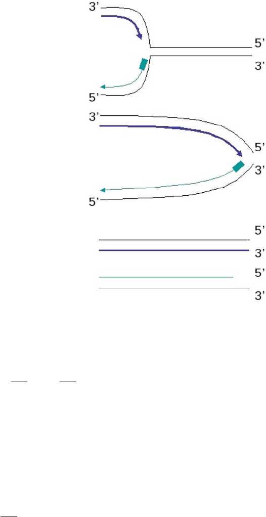
to copy the very ends of the chromosomes during cell division, telomeres shorten
with each cell division and thereby provide a record of a cell’s proliferation history
(Fig. 7.2)[11, 12]. Although it is very tempting to translate the decline in the aver-
age telomere length of a cell population directly into the number of cell divisions
the population has undergone, describing telomere dynamics with a mathematical
model has demonstrated that one has to be extremely cautious when interpreting
telomere data [13]. Furthermore, the activity of the enzyme telomerase which effec-
tively elongates telomeres can potentially destroy the relationship between telomere
length and the number of divisions a cell has undergone [14,15].
If one assumes that telomerase is not active then one can describe the shortening
of telomeres in a population of cells by giving each cell an index i, depending on
the number of cell divisions the cell has undergone, the so-called division index
[13]. When a cell with division index i divides, it produces two daughter cells with
division index i C 1. If the number of cells with division index i is denoted by
n
i
, and the total number of cells by N , the average division index of such a cell
population is:
D
1
N
1
X
iD0
in
i
: (7.4)
The average division index of a population increases with time. Its change over
time (d=dt) can be calculated by differentiating with respect to time, and by
substituting dN=dt, the change in the total number of cells N over time, and dn
i
=dt,
the change in the number of cells n
i
with division index i over time. If (7.4) applies
148 B. Asquith and J.A.M. Borghans
Fig. 7.2 Telomeres are
repetitive DNA patterns
found at the ends of the
chromosomes. During DNA
replication, the leading strand
(blue) can make a full copy of
the DNA, while the lagging
strand (green) fails to copy
the very end of the
chromosome, leading to
progressive telomere
shortening
to memory T cells, the change in the total number of memory T cells (N D M )is
described by [13]:
dM
dt
D
X
i
dm
i
dt
D M.p
M
ı
M
/ C CV; (7.5)
where p
M
denotes the proliferation rate of memory T cells, ı
M
denotes the rate at
which memory T cells die, the rate at which naive T cells are primed and enter the
memory compartment, C is the average number of cell divisions occurring during
clonal expansion (i.e. when naive T cells are triggered to become memory T cells)
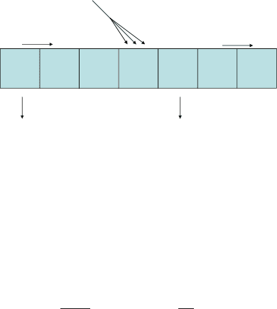
and V is the number of naive T cells. See Fig. 7.3. The change in the number of
memory cells m
i
with division index i is described by the following differential
equation [13]:
dm
i
dt
D 2p
M
m
i1
.p
M
C ı
M
/m
i
C V n
iK
; (7.6)
7 Modelling Lymphocyte Dynamics In Vivo 149
Naive cells V
γ
C
Memory cells M
p
M
p
M
d
M
d
M
m
0
m
1
m
2
m
3
m
4
m
5
m
...
Fig. 7.3 The total memory T cell population, M , consists of sub-populations of cells m
i
that have
undergone i cell divisions. Memory cells in any sub-population i divide at rate p
M
and produce
two daughter cells with division index i C 1, they die at rate ı
M
, and are produced when naive T
cells V are activated at rate and clonally expand into C memory cells
where K is the number of divisions made during clonal expansion. Substituting
these two equations into the derivative of (7.4), yields that the average division index
M
of the memory T cell population changes according to:
d
M
dt
D 2p
M
C
V
M
.
M
V
K/: (7.7)
The latter equation gives the important insight that the average telomere length of
the memory T cell population does not only reflect the proliferative history of the
memory T cell population, but also the transition of naive T cells into the memory
compartment [13]. Telomere loss in the memory T cell pool is in part compensated
by the influx of cells from the naive compartment, which – on average – have longer
telomeres than memory cells. Thus, the rate of telomere loss in the memory popu-
lation is to a large extent determined by the rate of telomere loss in the naive T cell
pool. It is therefore perhaps not surprising that the telomere loss of naive and mem-
ory T cells was found to occur at similar rates [16], even though naive and memory
T cells are thought to proliferate at different rates.
Similarly, one can argue that the rate at which naive T cells shorten their telom-
eres may not directly reflect their proliferation rate, but may also be influenced to a
large extent by the rate of telomere loss of progenitor cells that are exported from
the thymus into the periphery [13]. Since recent thymus emigrants form a permanent
source of cells with relatively long telomeres, they increase the average telomere
length of the naive T cell population. Ignoring this influx of cells from the thymus
into the periphery may therefore lead to an underestimation of the naive T cell divi-
sion rate.
In summary, although telomeric lengths may seem to be the best marker for the
proliferative history of a cell population, again, just like TREC contents, they are
not only influenced by T cell proliferation, but also by cellular death rates and input
from the thymus. In fact, TREC contents and telomere lengths thus reflect very
similar processes, even though they were originally proposed to be discriminative
for thymic output and cellular proliferation, respectively.
150 B. Asquith and J.A.M. Borghans
BrdU Labelling
T cell turnover rates have extensively been studied by the use of 5-bromo-2
0
-
deoxyuridine (BrdU), a nucleoside analogue that is incorporated into the DNA of
cells that are dividing, instead of the nucleoside thymidine [17–20]. Because of its
potential toxicity, BrdU has mainly been applied to rodents and non-human pri-
mates. It is typically administered via the drinking water and its incorporation in
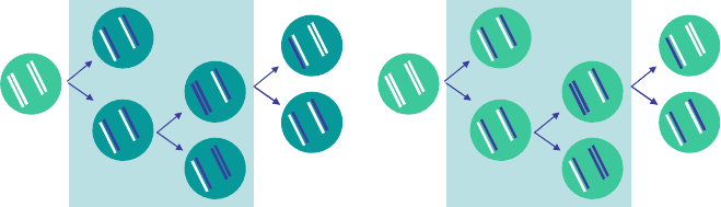
cells can be detected by flow cytometry. When cells divide in the presence of BrdU,
newly-formed DNA strands build in BrdU and are subsequently randomly dis-
tributed over the two daughter cells, resulting in two BrdU-labelled cells (Fig. 7.4).
When BrdU-labelled cells divide in the absence of BrdU, their label intensity de-
creases with every cell division. Nevertheless, BrdU labelling studies typically
report the percentage of BrdU-positive cells, i.e. the percentage of cells in which the
BrdU intensity exceeds a certain threshold, irrespective of their exact label intensity.
During BrdU intake, the percentage of cells that are BrdU-positive gradually
increases, and after BrdU is withdrawn from the drinking water the percentage of
BrdU-positive cells typically decreases. Simple, intuitive analyses interpret the rate
of increase of labelled cells during the labelling period as reflecting the rate of cell
proliferation and the rate of loss of labelled cells during the delabelling period as
the rate of cell death. Although this may sound very persuasive, in fact it is not so
straightforward. A simple mathematical model demonstrates where intuition goes
wrong [21–25]. One can describe the accrual of label during BrdU administration
either by the rate at which the fraction of labelled cells increases or by the rate at
which the fraction of unlabelled cells decreases. Since both rates should be equal,
one can simply use the easiest equation. During BrdU administration, it is easiest
to describe the number of unlabelled cells, U . Unlabelled cells are lost when they
ab
BrdU administration Deuterium administration
Fig. 7.4 BrdU labelling experiments follow the fraction of labelled cells, while stable isotope la-
belling experiments follow the fraction of labelled DNA strands. During BrdU administration (light
blue box), dividing cells build BrdU into their newly-formed DNA strands. Labelled DNA strands
(blue) are randomly distributed over the daughter cells. Upon BrdU-withdrawal, the label intensity
of the cells decreases with every cell division, but the fraction of BrdUC cells (dark green) does
not decrease. During stable isotope labelling (light blue box), dividing cells build deuterium into
their newly-formed DNA strands. Labelled DNA strands (blue) are randomly distributed over the
daughter cells. Upon deuterium withdrawal, the fraction of labelled DNA decreases with every cell
division

7 Modelling Lymphocyte Dynamics In Vivo 151
divide (because they become BrdU-labelled) at rate p per day, or when they die at
rate ı per day:
dU
dt
DpU ıU: (7.8)
This equation demonstrates that the fraction of unlabelled cells decreases, and hence
the fraction of labelled cells increases, during label administration at rate p Cı.Im-
portantly, and rather counter-intuitively, label accrual during label administration
thus does not only reflect proliferation but also loss of cells through death or matu-
ration.
When BrdU is no longer administered, it is easier to follow the loss of labelled
cells. Labelled cells are lost from the population by cell death at rate ı per day, but
they are also gained, by cell proliferation at rate p per day, even in the absence of
BrdU. Because previously labelled chromosomes are randomly distributed among
daughter cells, both daughter cells will be BrdU labelled (see Fig. 7.4), albeit with
lower BrdU intensity. Each BrdU labelled cell that divides thus adds another labelled
cell to the population, even in the absence of BrdU:
dL
dt
D pL ıL: (7.9)
The latter equation demonstrates that the fraction of labelled cells decreases at rate
p ı per day when label administration is stopped [21, 24]. Importantly, the rate
at which the fraction of labelled cells decreases after label cessation thus does not
directly reflect the rate of cell loss. For a population at steady state, one would
expect the rate of cell production p to be equal to the rate of cell loss ı,inother
words p ı D 0. Consequently, one would expect the fraction of BrdU labelled
cells to remain constant after label cessation. This is in contrast to what is routinely
observed: that labelled cells decrease after the end of the labelling period.
It is therefore rather surprising that most BrdU studies show a significant decline
in the proportion of labelled cells during de-labelling, suggesting that the average
proliferation rate of lymphocytes, p, is smaller than the average loss rate of BrdU
labelled cells, ı. Indeed, separate estimates of ı and p haveshownuptotenfold
larger rates of cell loss compared to the rate of T cell proliferation [21]. One possible
explanation for this discrepancy is that dividing cells are not at equilibrium although
the whole population is. Alternatively, if cell proliferation occurs in “bursts,” BrdU
labelling may have diluted to such an extent that cells are no longer recognized as
label-positive, after extensive T cell proliferation in the absence of label. Currently
ongoing research is aiming to distinguish between these options.
Stable Isotope Labelling
The most recently developed method to study lymphocyte kinetics in vivo is based
on stable isotope labelling. Stable isotopes are non-radioactive variants of a chemi-
cal element, with a different mass because of the presence of extra neutrons in the

152 B. Asquith and J.A.M. Borghans
nucleus. The stable isotope that is typically used in lymphocyte dynamic studies is
deuterium, the heavier variant of hydrogen, which contains one proton and one neu-
tron, and has twice the mass of hydrogen, which contains no neutrons. Deuterium
has been administered in the form of deuterated glucose (
2
H
2
-glucose) [26–33]or
deuterated water (
2
H
2
O) [34–36], and has proven to be safe and non-toxic when
given in low concentrations. The deuterium atoms from these stable isotope labelled
compounds are incorporated into the DNA of cells when they divide. Deuterium
incorporation can be measured by a combination of gas chromatography and mass
spectrometry (GC–MS) on the extracted DNA from sorted cells. In contrast to BrdU
labelling studies, in which the fraction of labelled cells is measured, stable isotope
labelling is measured in terms of fractions of labelled DNA fragments, not cells.
The mathematics required to analyse stable isotope labelling data therefore deviate
from those used in BrdU experiments [36–38].
The number of labelled DNA fragments in a population of cells increases by cell
proliferation at rate p per day, independent of whether the proliferating cells had
already incorporated deuterium or not. The extent to which label is incorporated
during cell proliferation depends on the availability of deuterium, D. In deuterated
glucose experiments, the availability is assumed to be either maximal, during label
intake, or zero, after label cessation, because the turnover rate of glucose is very
fast. In case of heavy water labelling, however, the supply of deuterium is more
variable, and hence should be explicitly taken into account (see below). The number
of labelled DNA fragments decreases by loss of labelled cells from the population
at rate ı per day, through differentiation, maturation or cell death. The change in the
total number of labelled DNA fragments per day can hence be described by [37]:
dL
dt
D pcND.t/ ıL (7.10)
where N denotes the total number of DNA fragments in the population, and c
reflects an amplification factor which needs to be introduced, because there are mul-
tiple hydrogen atoms in a single DNA fragment that can be replaced by deuterium.
Typically, deuterium is given in such low concentrations that the chance to double-
label a DNA fragment (and thereby miss it during mass spectrometry) is close to
zero. However, the presence of multiple hydrogen atoms does increase the chance
of labelling a DNA fragment at one position. Translating (7.10) into the fraction of
labelled DNA fragments (l D L=N ) yields:
dl
dt
D pcD.t/ ıl: (7.11)
If labelling is performed with deuterated glucose, then we can assume that D D 1
during the labelling period and D D 0 during the delabelling period and (7.11)has
the following solutions [37]:
l D
pc
ı
.1 e
ıt
/; during label administration .t < /