Watts J.F., Wolstenholme J. An Introduction to Surface Analysis by XPS and AES
Подождите немного. Документ загружается.

AUGER ELECTRON SPECTROSCOPY (AES)
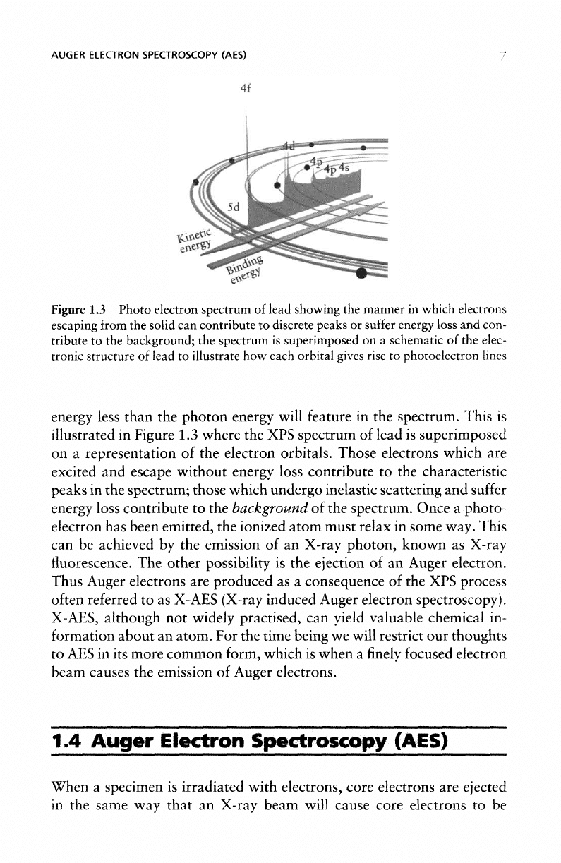
Figure
1.3
Photo electron spectrum
of
lead showing
the
manner
in
which electrons
escaping from
the
solid
can
contribute
to
discrete peaks
or
suffer
energy
loss
and
con-
tribute
to the
background;
the
spectrum
is
superimposed
on a
schematic
of the
elec-
tronic
structure
of
lead
to
illustrate
how
each orbital
gives
rise
to
photoelectron
lines
energy less
than
the
photon
energy will feature
in the
spectrum.
This
is
illustrated
in
Figure
1.3
where
the XPS
spectrum
of
lead
is
superimposed
on a
representation
of the
electron orbitals.
Those
electrons which
are
excited
and
escape without energy loss contribute
to the
characteristic
peaks
in the
spectrum;
those
which undergo inelastic
scattering
and
suffer
energy loss contribute
to the
background
of the
spectrum. Once
a
photo-
electron
has
been emitted,
the
ionized atom must relax
in
some way. This
can
be
achieved
by the
emission
of an
X-ray
photon,
known
as
X-ray
fluorescence.
The
other
possibility
is the
ejection
of an
Auger
electron.
Thus Auger electrons
are
produced
as a
consequence
of the XPS
process
often
referred
to as
X-AES
(X-ray induced Auger electron spectroscopy).
X-AES,
although
not
widely practised,
can
yield valuable chemical
in-
formation about
an
atom.
For the
time being
we
will
restrict
our
thoughts
to AES in its
more common form, which
is
when
a
finely
focused electron
beam
causes
the
emission
of
Auger
electrons.
1.4
Auger
Electron
Spectroscopy
(AES)
When
a
specimen
is
irradiated with electrons, core electrons
are
ejected
in
the
same
way
that
an
X-ray beam will cause
core
electrons
to be
ELECTRON
SPECTROSCOPY:
SOME
BASIC
CONCEPTS
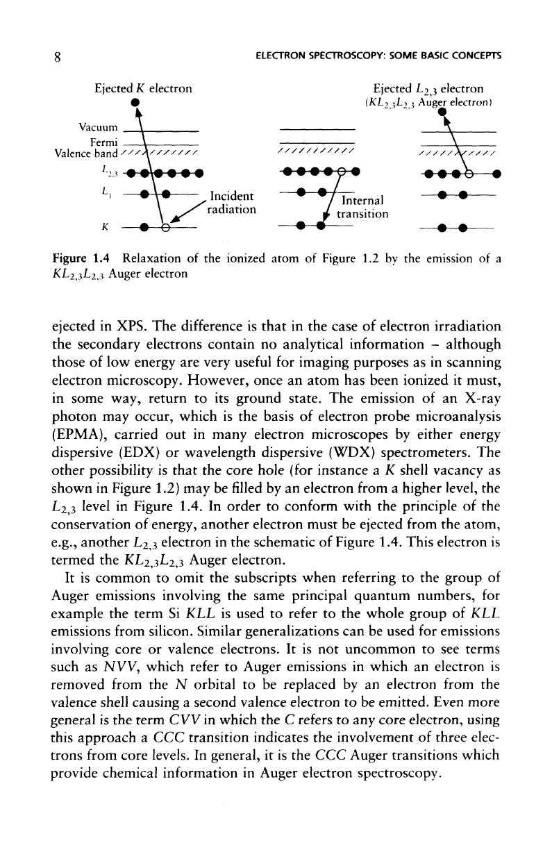
Ejected
K
electron Ejected L
2,3
electron
•
(KL
2
,
3
L
2,
3
Auger electron)
Vacuum
Fermi
Valence band
L,,
Incident
• • /
Internal
• •
radiation
1
transition
Figure
1.4
Relaxation
of the
ionized atom
of
Figure
1.2 by the
emission
of a
KL
2,3
L
2,3
Auger electron
ejected
in
XPS.
The
difference
is
that
in the
case
of
electron irradiation
the
secondary electrons contain
no
analytical information
-
although
those
of low
energy
are
very
useful
for
imaging purposes
as in
scanning
electron microscopy. However, once
an
atom
has
been ionized
it
must,
in
some way, return
to its
ground state.
The
emission
of an
X-ray
photon
may
occur,
which
is the
basis
of
electron probe microanalysis
(EPMA),
carried
out in
many electron microscopes
by
either energy
dispersive (EDX)
or
wavelength dispersive (WDX)
spectrometers.
The
other
possibility
is
that
the
core
hole (for instance
a K
shell vacancy
as
shown
in
Figure 1.2)
may be
filled
by an
electron from
a
higher level,
the
L
2,3
level
in
Figure 1.4.
In
order
to
conform with
the
principle
of the
conservation
of
energy, another electron must
be
ejected
from
the
atom,
e.g., another L
2
,
3
electron
in the
schematic
of
Figure 1.4. This electron
is
termed
the
KL
2
,
3
L
2
,
3
Auger electron.
It is
common
to
omit
the
subscripts when
referring
to the
group
of
Auger
emissions
involving
the
same principal quantum numbers,
for
example
the
term
Si KLL is
used
to
refer
to the
whole group
of KLL
emissions
from
silicon.
Similar
generalizations
can be
used
for
emissions
involving
core
or
valence electrons.
It is not
uncommon
to see
terms
such
as NW,
which
refer
to
Auger emissions
in
which
an
electron
is
removed from
the N
orbital
to be
replaced
by an
electron
from
the
valence shell causing
a
second valence electron
to be
emitted. Even more
general
is the
term
CVV in
which
the C
refers
to any
core
electron, using
this approach
a CCC
transition indicates
the
involvement
of
three elec-
trons
from
core
levels.
In
general,
it is the CCC
Auger transitions which
provide chemical information
in
Auger electron spectroscopy.
AUGER ELECTRON SPECTROSCOPY (AES)
9
The
kinetic energy
of a
KL
2,3
L
2,3
Auger electron
is
approximately
equal
to the
difference
between
the
energy
of the
core hole
and the
energy
levels
of the two
outer electrons,
EL
2,3
(the term L
2
,
3
is
used
in
this
case because,
for
light elements,
L
2
and L
3
cannot
be
resolved):
« E
K
—
E
L2,3
—
EL ,
This equation does
not
take into account
the
interaction energies
be-
tween
the
core holes (L
2,3
and
L
2,3
)
in the final
atomic state
nor the
inter-
and
extra-relaxation energies which come
about
as a
result
of the
additional
core screening needed. Clearly,
the
calculation
of the
energy
of
Auger
electron transitions
is
much more complex than
the
simple
model outlined above,
but
there
is a
satisfactory empirical approach
which
considers
the
energies
of the
atomic
levels
involved
and
those
of
the
next element
in the
periodic table.
Following this empirical approach,
the
Auger electron energy
of
tran-
sition
KL
1
L
2,3
for an
atom
of
atomic number
Z is
written:
E
KL1L2,3
(Z)
=
E
K
(Z)
-
1
/2[E
Ll
(Z) + E
Ll
(Z
+ 1)]
(Z)+E
L2,3
(Z
+ 1)]
Clearly
for the
KL
2,3
L
2,3
transition
the
second
and
third
terms
of the
above equation
are
identical
and the
expression
is
simplified
to:
E
K
L
2
,L
2
,(Z)
=
E
K
(Z)
-
[E
L2,3
(Z)+E
L2,3
(Z
+
1)]
It is the
kinetic energy
of
this Auger electron
(EKL
2,3
L
2,3
)
that
is the
characteristic material quantity irrespective
of the
primary beam com-
position (i.e., electrons, X-rays, ions)
or its
energy.
For
this reason Auger
spectra
are
always plotted
on a
kinetic energy scale.
The use of a finely
focused
electron
beam
for AES
enables
us to
achieve
surface
analysis
at a
high spatial resolution,
in a
manner analo-
gous
to
EPMA
in the
scanning electron microscope.
By
combining
an
electron spectrometer with
an
ultra-high vacuum (UHV)
SEM it be-
comes possible
to
carry
out
scanning Auger
microscopy.
In
this mode
of
operation various imaging
and
chemical mapping procedures become
possible.
10
ELECTRON SPECTROSCOPY:
SOME
BASIC CONCEPTS
1.5
Scanning
Auger
Microscopy
(SAM)
In
the
scanning Auger microscope various modes
of
operation
are
avail-
able,
the
variable quantities being
the
position
of the
electron probe
on
the
specimen
(x and y) and the
setting
of the
electron energy analyser
(£)
corresponding
to the
energy
of
emitted electrons
to be
analysed.
The
various possibilities
are
summarized
in
Table 1.3.
As
the
Auger electron yield
is
very sensitive
to the
electron
take-off
angle,
an
image
of
Auger electron intensities
will
invariably
reflect
the
surface
topography
of the
specimen, possibly more strongly than
the
chemical
variations,
as
illustrated
(Figure
1.5)
in the
Auger
map of
carbon
fibres
(Figure
1.5(b)) which
is
very
similar
to the SEM
image
Figure
1.5
Scanning Auger microscopy
of
carbon
fibres: (a) SEM
image,
(b)
peak
map
(P) of
carbon
Auger
electrons,
(c)
peak-background
map (P - B), B
recorded
40 eV
from Auger
peak,
(d)
correction
for
topographic
effects using
(P -
B)/B
algorithm;
the
diameter
of the fibres is 7 urn
THE
DEPTH
OF
ANALYSIS
IN
ELECTRON SPECTROSCOPY
Table
1.3
Modes
of
analysis available with
SAM
Mode
of
analysis
Point analysis
Line
scan
Chemical
map
Scanned
E
X
x, y
Fixed
x,y
E, y
E
(Figure
1.5(a)).
The
problem
is
overcome
by
recording
a
background
(B)
as
well
as the
Auger peak
(P)
map. However,
a
simple subtraction
of the
background
counts
from
the
peak intensity
(P - B) is not
sufficient
as
shown
by the (P
—
B) map of
Figure 1.5(c).
The use of a
simple algo-
rithm such
as (P -
B)/B, allows
correction
for the
effects
of
surface
topography
(Figure
1.5(d))
where variation
in
intensity
due to the cy-
lindrical
shape
of the
fibres
has
been completely suppressed
and
only
chemical
information remains.
1.6
The
Depth
of
Analysis
in
Electron
Spectroscopy
_
The
depth
of
analysis
in
both
XPS and AES
varies with
the
kinetic
energy
of the
electrons under consideration.
It is
determined
by a
quan-
tity
known
as the
attenuation length
(A)
of the
electrons, which
is
related
to the
inelastic mean
free
path
(IMFP).
It
varies
as
E
0.5
in the
energy
range
of
interest
in
electron spectroscopy
and
various relationships have
been
suggested which relate
A to
electron energy
and
material proper-
ties.
One
such equation
proposed
by
Seah
and
Dench (1979)
of the
National Physical Laboratory,
UK, is
given below:
where
E
A
is the
energy
of the
electron
in eV, a
3
A
is the
volume
of the
atom
in nm
3
and A is in nm.
Values
for
IMFP
may be
derived using optical spectroscopy,
or
reflec-
tion
electron
energy loss
spectroscopy
(REELS),
those
for A are
generally
deduced
from
XPS and
Auger spectroscopy measurements.
In
general,
12
ELECTRON SPECTROSCOPY: SOME BASIC CONCEPTS
the
attenuation length
is
about
10 per
cent less than
the
IMFP. Various
databases exist
from
which values
of
IMFP
and
attenuation length
can
be
obtained.
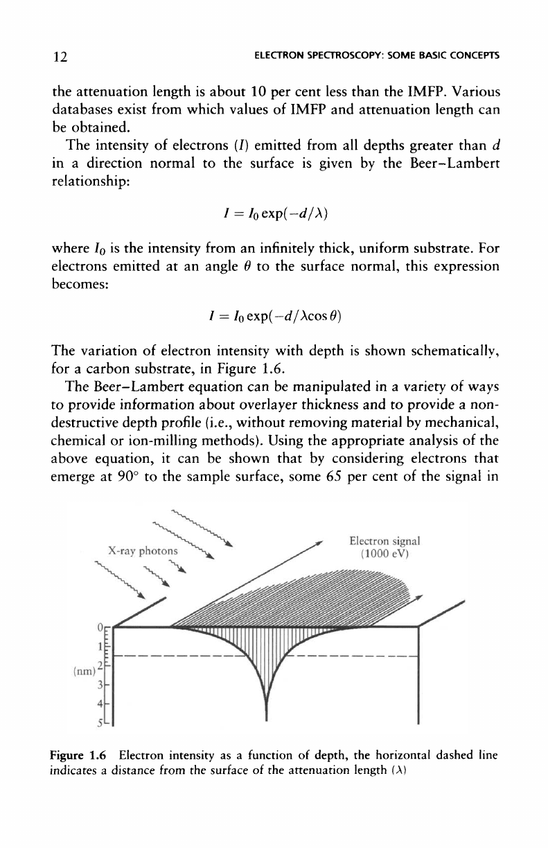
The
intensity
of
electrons
(/)
emitted
from
all
depths greater than
d
in
a
direction normal
to the
surface
is
given
by the
Beer-
Lambert
relationship:
where
I
0
is the
intensity from
an
infinitely
thick, uniform
substrate.
For
electrons emitted
at an
angle
9 to the
surface
normal, this expression
becomes:
I =
I
0
exp(—
d/XcosO)
The
variation
of
electron intensity with depth
is
shown schematically,
for
a
carbon substrate,
in
Figure 1.6.
The
Beer-Lambert
equation
can be
manipulated
in a
variety
of
ways
to
provide information
about
overlayer thickness
and to
provide
a
non-
destructive depth profile (i.e., without removing material
by
mechanical,
chemical
or
ion-milling methods). Using
the
appropriate
analysis
of the
above equation,
it can be
shown that
by
considering electrons that
emerge
at 90° to the
sample
surface,
some
65 per
cent
of the
signal
in
Figure
1.6
Electron intensity
as a
function
of
depth,
the
horizontal dashed line
indicates
a
distance
from
the
surface
of the
attenuation length
(A)
COMPARISON
OF
XPS
AND
AES/SAM
j 3
electron spectroscopy will emanate
from
a
depth
of
less than
A,
85 per
cent from
a
depth
of < 2A, and 95 per
cent from
a
depth
of
< 3A, as
illustrated
in
Figure 1.6.
The
values
of the
inelastic mean
free
paths
are of the
order
of a few
nanometers
and
this
is the
mechanism
by
which
the
signal
from
the
vast
majority
of the
atoms present
in the
sample
is
filtered
out
(stated
as a
requirement
for a
surface
analysis
technique
in
Section 1.1).
However,
the
depth
from
which information
can be
derived
is a few
nanometers,
a
fact
which
can be
exploited
in
angle resolved measure-
ments
to
obtain compositional depth
profiles.
This will
be
discussed
in
more
detail later.
1.7
Comparison
of XPS and
AES/SAM
Although
it is
difficult
to
make
a
comparison
of
techniques before they
are
described
and
discussed
in
detail,
it is
pertinent
at
this point
to
outline
the
strengths
and
weaknesses
of
each
to
provide
background
information.
XPS
is
also known
by the
acronym ESCA (electron spectroscopy
for
chemical analysis).
It is
this chemical specificity which
is the
major
strength
of XPS as an
analytical technique,
one for
which
it has
become deservedly popular.
By
this
we
mean
the
ability
to
define
not
only
the
elements present
in the
analysis
but
also
the
chemical state.
In
the
case
of
iron,
for
instance,
the
spectra
of
Fe°,
Fe
2+
,
and
Fe
3+
are
all
slightly
different
and to the
expert
eye are
easily distinguishable.
However,
such information
is
attainable
in XPS
only
at the
expense
of
spatial resolution,
and XPS is
usually regarded
as an
area averaging
technique.
Small area
XPS
(SAXPS
or
even SAX)
is
available
on
most modern instruments and, when operating
in
this mode,
a
spectro-
scopic spatial resolution
of
about
10 um is
possible. Many modern
in-
struments
offer
imaging
XPS and
such imaging
may
have
a
spatial
resolution
of
<3fim.
Set
beside
a
spatial resolution
of 10 to
15nm
which
can be
achieved
on the
latest
commercial Auger
microprobes,
it
becomes
clear that
the XPS is not the way to
proceed
for
surface analysis
at
very
high spatial resolution,
but the
advantages
of the
levels
of
information
available
from
an XPS
analysis (the ease with which
a
14
ELECTRON
SPECTROSCOPY:
SOME
BASIC
CONCEPTS
quantitative
analysis
can be
achieved,
its
applicability
to
insulators,
and
the
ready availability
of
chemical state information)
will
often
offset
this.
In
addition
to the
chemical state information referred
to
above,
XPS
spectra
can be
quantified
in a
very straightforward manner
and
meaningful
comparisons
can be
made between specimens
of a
similar
type. Quantification
of
Auger data
is
rather more complex
and the
accuracy obtained
is
generally
not so
good.
Because
of the
complemen-
tary nature
of the two
methods
and the
ease with which Auger
and
photoelectron analyses
can be
made
on the
same instrument,
the two
methods have come
to be
regarded
as the
most important methods
of
surface analysis
in the
context
of
materials science.
All
manufacturers
of
electron
spectrometers
offer
both
XPS and AES
options
for
their
systems.
1.8 The
Availability
of
Surface
Analytical
Equipment
The
capital cost
of an
XPS/AES/SAM spectrometer
is
high when com-
pared with
most
electron
microscopes,
and at the
time
of
writing
is of
the
order
of
£0.3 million
to
£0.5 million
for a
comprehensive system.
This, allied
to the
fairly
steep learning curve that
the
newcomer must
ascend before confidence
in the
technique
is
obtained,
has
lead
to the
development
of
laboratories
offering
surface analysis
as a
service
facil-
ity; such laboratories
may be
found throughout
the
world
and
they
are
often
associated
with universities
but the
balance between academic
and
industrial
work
varies greatly.
The use of
service facilities
presents
a
very
attractive proposition
to
inexperienced users
in
that expert advice
is
always
on
hand
to
ensure
the
efficient
use of
instrument time
- a
factor
that
is of
paramount importance since
the
daily
charge
for the
use
of
such
a
facility
can
exceed
£1500.
It is not
unusual
for
analysts
to
'cut their
teeth'
on the field of
surface analysis
in
such
a
way; once
the
need
within their
own
company (and their
own
personal expertise)
has
been
established,
a
surface analysis system
can
then
be
specified
for
their
own
particular needs.
Although originally
the
exclusive preserve
of
research laboratories
and
academic institutions, surface analysis
facilities
are now
frequently
THE
AVAILABILITY
OF
SURFACE
ANALYTICAL
EQUIPMENT
1 5
to be
found
in
trouble-shooting
and
quality assurance roles.
As the
techniques
find
wider applications,
so the
market
grows
and
manufac-
turers
are
very
willing
to
continue developing their spectrometers. Thus,
the
future
for XPS and AES
seems assured well into
the
future.

This page intentionally left blank