Berg J.M., Tymoczko J.L., Stryer L. Biochemistry
Подождите немного. Документ загружается.


10.5.3. The Generation of Trypsin from Trypsinogen Leads to the Activation of Other
Zymogens
The structural changes accompanying the activation of trypsinogen, the precursor of the proteolytic enzyme trypsin, are
somewhat different from those in the activation of chymotrypsinogen. X-ray analyses have shown that the conformation
of four stretches of polypeptide, constituting about 15% of the molecule, changes markedly on activation. These regions,
called the activation domain, are very flexible in the zymogen, whereas they have a well-defined conformation in trypsin.
Furthermore, the oxyanion hole (Section 9.1.3) in trypsinogen is too far from histidine 57 to promote the formation of
the tetrahedral transition state.
The digestion of proteins in the duodenum requires the concurrent action of several proteolytic enzymes, because each is
specific for a limited number of side chains. Thus, the zymogens must be switched on at the same time. Coordinated
control is achieved by the action of trypsin as the common activator of all the pancreatic zymogens
trypsinogen,
chymotrypsinogen, proelastase, procarboxypeptidase, and prolipase, a lipid degrading enzyme. To produce active
trypsin, the cells that line the duodenum secrete an enzyme, enteropeptidase, that hydrolyzes a unique lysine-isoleucine
peptide bond in trypsinogen as the zymogen enters the duodenum from the pancreas. The small amount of trypsin
produced in this way activates more trypsinogen and the other zymogens (Figure 10.34). Thus, the formation of trypsin
by enteropeptidase is the master activation step.
10.5.4. Some Proteolytic Enzymes Have Specific Inhibitors
The conversion of a zymogen into a protease by cleavage of a single peptide bond is a precise means of switching on
enzymatic activity. However, this activation step is irreversible, and so a different mechanism is needed to stop
proteolysis. Specific protease inhibitors accomplish this task. For example, pancreatic trypsin inhibitor, a 6-kd protein,
inhibits trypsin by binding very tightly to its active site. The dissociation constant of the complex is 0.1 pM, which
corresponds to a standard free energy of binding of about -18 kcal mol
-1
(-75 kJ mol
-1
). In contrast with nearly all known
protein assemblies, this complex is not dissociated into its constituent chains by treatment with denaturing agents such as
8 M urea or 6 M guanidine hydrochloride.
The reason for the exceptional stability of the complex is that pancreatic trypsin inhibitor is a very effective substrate
analog. X-ray analyses showed that the inhibitor lies in the active site of the enzyme, positioned such that the side chain
of lysine 15 of this inhibitor interacts with the aspartate side chain in the specificity pocket of trypsin. In addition, there
are many hydrogen bonds between the main chain of trypsin and that of its inhibitor. Furthermore, the carbonyl group of
lysine 15 and the surrounding atoms of the inhibitor fit snugly in the active site of the enzyme. Comparison of the
structure of the inhibitor bound to the enzyme with that of the free inhibitor reveals that the structure is essentially
unchanged on binding to the enzyme (Figure 10.35). Thus, the inhibitor is preorganized into a structure that is highly
complementary to the enzyme's active site. Indeed, the peptide bond between lysine 15 and alanine 16 in pancreatic
trypsin inhibitor is cleaved but at a very slow rate: the half-life of the trypsin-inhibitor complex is several months. In
essence, the inhibitor is a substrate, but its intrinsic structure is so nicely complementary to the enzyme's active site that
it binds very tightly and is turned over slowly.
Why does trypsin inhibitor exist? Indeed, the amount of trypsin is much greater that that of the inhibitor. Under
what circumstances is it beneficial to inhibit trypsin? Recall that trypsin activates other zymogens. Consequently,
it is vital that even small amounts of trypsin be prevented from initiating the cascade prematurely. Trypsin molecules
activated in the pancreas or pancreatic ducts could severely damage those tissues, leading to acute pancreatitis. Tissue
necrosis may result from the activation of proteolytic enzymes (as well as prolipases) by trypsin, and hemorrhaging may
result from its activation of elastase. We see here the physiological need for the tight binding of the inhibitor to trypsin.
Pancreatic trypsin inhibitor is not the only important protease inhibitor. α
1
-Antitrypsin (also called α
1
-antiproteinase),
a 53-kd plasma protein, protects tissues from digestion by elastase, a secretory product of neutrophils (white blood cells
that engulf bacteria). Antielastase would be a more accurate name for this inhibitor, because it blocks elastase much

more effectively than it blocks trypsin. Like pancreatic trypsin inhibitor, α
1
-antitrypsin blocks the action of target
enzymes by binding nearly irreversibly to their active sites. Genetic disorders leading to a deficiency of α
1
-antitrypsin
show that this inhibitor is physiologically important. For example, the substitution of lysine for glutamate at residue 53
in the type Z mutant slows the secretion of this inhibitor from liver cells. Serum levels of the inhibitor are about 15% of
normal in people homozygous for this defect. The consequence is that excess elastase destroys alveolar walls in the lungs
by digesting elastic fibers and other connective-tissue proteins.
The resulting clinical condition is called emphysema (also known as destructive lung disease). People with emphysema
must breathe much harder than normal people to exchange the same volume of air, because their alveoli are much less
resilient than normal. Cigarette smoking markedly increases the likelihood that even a type Z heterozygote will develop
emphysema. The reason is that smoke oxidizes methionine 358 of the inhibitor (Figure 10.36), a residue essential for
binding elastase. Indeed, this methionine side chain is the bait that selectively traps elastase. The methionine sulfoxide
oxidation product, in contrast, does not lure elastase, a striking consequence of the insertion of just one oxygen atom into
a protein. We will consider another protease inhibitor, antithrombin III, when we examine the control of blood clotting.
10.5.5. Blood Clotting Is Accomplished by a Cascade of Zymogen Activations
Enzymatic cascades are often employed in biochemical systems to achieve a rapid response. In a cascade, an initial
signal institutes a series of steps, each of which is catalyzed by an enzyme. At each step, the signal is amplified. For
instance, if a signal molecule activates an enzyme that in turn activates 10 enzymes and each of the 10 enzymes in turn
activates 10 additional enzymes, after four steps the original signal will have been amplified 10,000-fold. Blood clots are
formed by a cascade of zymogen activations: the activated form of one clotting factor catalyzes the activation of the next
(Figure 10.37). Thus, very small amounts of the initial factors suffice to trigger the cascade, ensuring a rapid response to
trauma.
There are two means of initiating clotting: the intrinsic and extrinsic pathways. The intrinsic clotting pathway is
activated by exposure of anionic surfaces on rupture of the endothelial lining of the blood vessels. These surfaces serve
as binding sites for factors in the clotting cascade. Substances that are released from tissues as a consequence of trauma
to them trigger the extrinsic clotting pathway. The extrinsic and intrinsic pathways converge on a common sequence of
final steps to form a clot composed of the protein fibrin. The two pathways interact with each other in vivo. Indeed, both
are needed for proper clotting, as evidenced by clotting disorders caused by a deficiency of a single protein in one of the
pathways. Note that the active forms of the clotting factors are designated with a subscript "a."
10.5.6. Fibrinogen Is Converted by Thrombin into a Fibrin Clot
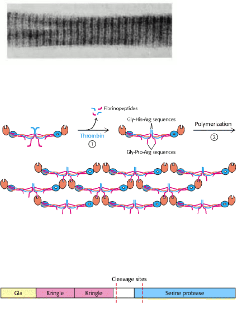
The best-characterized part of the clotting process is the final step in the cascade: the conversion of fibrinogen into fibrin
by thrombin, a proteolytic enzyme. Fibrinogen is made up of three globular units connected by two rods (Figure 10.38).
This 340-kd protein consists of six chains: two each of Aα, Bβ, and γ. The rod regions are triple-stranded α-helical
coiled coils, a recurring motif in proteins. Thrombin cleaves four arginine-glycine peptide bonds in the central globular
region of fibrinogen. On cleavage, an A peptide of 18 residues from each of the two Aα chains and a B peptide of 20
residues from each of the two Bβ chains are released from the globular region. These A and B peptides are called
fibrinopeptides. A fibrinogen molecule devoid of these fibrinopeptides is called a fibrin monomer and has the subunit
structure (α β γ)
2
.
Fibrin monomers spontaneously assemble into ordered fibrous arrays called fibrin. Electron micrographs and low-angle
x-ray patterns show that fibrin has a periodic structure that repeats every 23 nm (Figure 10.39). Higher-resolution images
reveal the detailed structure of the fibrin monomer, how the fibrin monomers come together, and how this assembly is
facilitated by removal of the fibrinopeptides. The homologous β and γ chains have globular domains at the carboxyl-
terminal ends (Figure 10.40). These domains have binding "holes" that interact with peptides. The β domain is specific
for sequences of the form H
3
N
+
-Gly-His-Arg-, whereas the γ domain binds H
3
N
+
-Gly-Pro-Arg-. Exactly these
sequences (sometimes called "knobs") are exposed at the amino-terminal ends of the β and α chains, respectively, on
thrombin cleavage. The knobs of the α subunits fit into the holes on the γ subunits of another monomer to form a
protofibril. This protofibril is extended when the knobs of the β subunits fit into holes of β subunits of other protofibrils.
Thus, analogous to the activation of chymotrypsinogen, peptide-bond cleavage exposes new amino termini that can
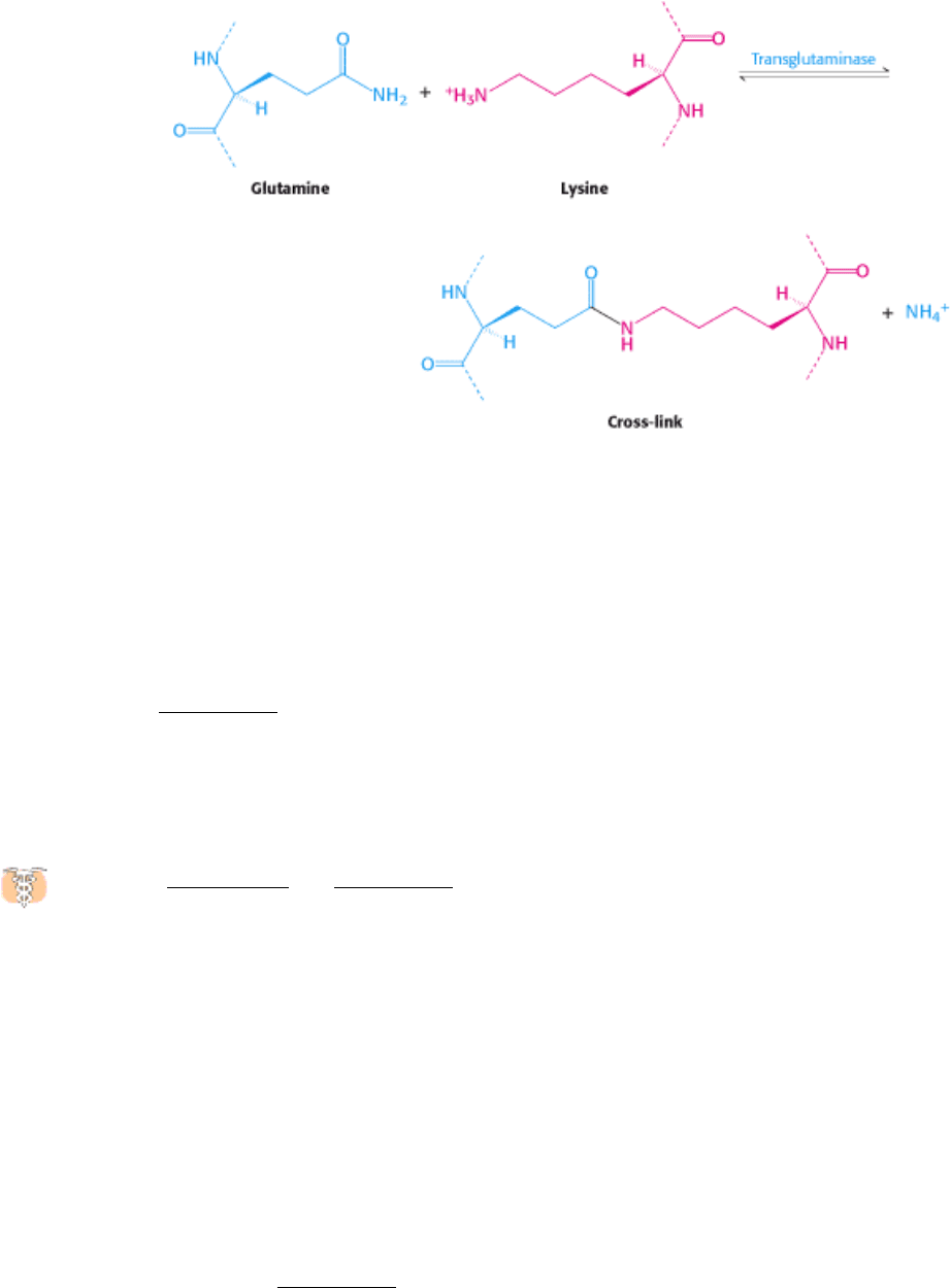
participate in specific interactions. The newly formed clot is stabilized by the formation of amide bonds between the side
chains of lysine and glutamine residues in different monomers.
This cross-linking reaction is catalyzed by transglutaminase (factor XIII
a
), which itself is activated from the
protransglutaminase form by thrombin.
10.5.7. Prothrombin Is Readied for Activation by a Vitamin K-Dependent Modification
Thrombin is synthesized as a zymogen called prothrombin, which comprises four major domains, with the serine
protease domain at its carboxyl terminus. The first domain is called a gla domain, whereas domains 2 and 3 are called
kringle domains (Figure 10.41). These domains work in concert to keep prothrombin in an inactive form and to target it
to appropriate sites for its activation by factor X
a
(a serine protease) and factor V
a
(a stimulatory protein). Activation is
accomplished by proteolytic cleavage of the bond between arginine 274 and threonine 275 to release a fragment
containing the first three domains and by cleavage of the bond between arginine 323 and isoleucine 324 (analogous to
the key bond in chymotrypsinogen) to yield active thrombin.
Vitamin K (Section 8.6.2 and Figure 10.42) has been known for many years to be essential for the synthesis of
prothrombin and several other clotting factors. The results of studies of the abnormal prothrombin synthesized in
the absence of vitamin K or in the presence of vitamin K antagonists, such as dicoumarol, revealed the mode of action of
this vitamin.Dicoumarolis found in spoiled sweet clover and causes a fatal hemorrhagic disease in cattle fed on this hay.
This coumarin derivative is used clinically as an anticoagulantto prevent thromboses in patients prone to clot formation.
Dicoumarol and such related vitamin K antagonists as warfarinalso serve as effective rat poisons. Cows fed dicoumarol
synthesize an abnormal prothrombin that does not bind Ca
2+
, in contrast with normal prothrombin. This difference was
puzzling for some time because abnormal prothrombin has the same number of amino acid residues and gives the same
amino acid analysis after acid hydrolysis as does normal prothrombin.
Nuclear magnetic resonance studies revealed that normal prothrombin contains γ-carboxyglutamate, a previously
unknown residue that evaded detection because its second carboxyl group is lost on acid hydrolysis. The abnormal
prothrombin formed subsequent to the administration of anticoagulants lacks this modified amino acid. In fact, the first
10 glutamate residues in the amino-terminal region of prothrombin are carboxylated to γ-carboxyglutamate by a vitamin
K-dependent enzyme system (Figure 10.43). The vitamin K-dependent carboxylation reaction converts glutamate, a
weak chelator of Ca
2
+
, into γ-carboxyglutamate, a much stronger chelator. Prothrombin is thus able to bind Ca
2+
, but

what is the effect of this binding? The binding of Ca
2+
by prothrombin anchors the zymogen to phospholipid membranes
derived from blood platelets after injury. The binding of prothrombin to phospholipid surfaces is crucial because it
brings prothrombin into close proximity to two clotting proteins that catalyze its conversion into thrombin. The
proteolytic activation of prothrombin removes the calcium-binding domain and frees thrombin from the membrane so
that it can cleave fibrinogen and other targets.
10.5.8. Hemophilia Revealed an Early Step in Clotting
Some important breakthroughs in the elucidation of clotting path ways have come from studies of patients with
bleeding disorders. Classic hemophilia, or hemophilia A, the best-known clotting defect, is genetically transmitted
as a sex-linked recessive characteristic. In classic hemophilia, factor VIII (antihemophilic factor) of the intrinsic pathway
is missing or has markedly reduced activity. Although factor VIII is not itself a protease, it markedly stimulates the
activation of factor X, the final protease of the intrinsic pathway, by factor IX
a
, a serine protease (Figure 10.44). Thus,
activation of the intrinsic pathway is severely impaired in hemophilia.
In the past, hemophiliacs were treated with transfusions of a concentrated plasma fraction containing factor VIII. This
therapy carried the risk of infection. Indeed, many hemophiliacs contracted hepatitis and AIDS. A safer preparation of
factor VIII was urgently needed. With the use of biochemical purification and recombinant DNA techniques, the gene
for factor VIII was isolated and expressed in cells grown in culture. Recombinant factor VIII purified from these cells
has largely replaced plasma concentrates in treating hemophilia.
An account of a hemorrhagic disposition existing in certain
families-
"About seventy or eighty years ago, a woman by the name of Smith
settled in the vicinity of Plymouth, New Hampshire, and transmitted
the following idiosyncrasy to her descendants. It is one, she
observed, to which her family is unfortunately subject and has been
the source not only of great solicitude, but frequently the cause of
death. If the least scratch is made on the skin of some of them, as
mortal a hemorrhage will eventually ensue as if the largest wound is
inflicted. . .. It is a surprising circumstance that the males only are
subject to this strange affection, and that all of them are not liable to
it. . .. Although the females are exempt, they are still capable of
transmitting it to their male children."
J
ohn Otto (1803)
10.5.9. The Clotting Process Must Be Precisely Regulated

There is a fine line between hemorrhage and thrombosis. Clots must form rapidly yet remain confined to the area
of injury. What are the mechanisms that normally limit clot formation to the site of injury? The lability of clotting
factors contributes significantly to the control of clotting. Activated factors are short-lived because they are diluted by
blood flow, removed by the liver, and degraded by proteases. For example, the stimulatory proteins factors V
a
and VIII
a
are digested by protein C, a protease that is switched on by the action of thrombin. Thus, thrombin has a dual function: it
catalyzes the formation of fibrin and it initiates the deactivation of the clotting cascade.
Specific inhibitors of clotting factors are also critical in the termination of clotting. The most important one is
antithrombin III, a plasma protein that inactivates thrombin by forming an irreversible complex with it. Antithrombin III
resembles α
1
-antitrypsin except that it inhibits thrombin much more strongly than it inhibits elastase. Antithrombin III
also blocks other serine proteases in the clotting cascade
namely, factors XII
a
, XI
a
, IX
a
, and X
a
. The inhibitory action
of antithrombin III is enhanced by heparin, a negatively charged polysaccharide found in mast cells near the walls of
blood vessels and on the surfaces of endothelial cells (Figure 10.45). Heparin acts as an anticoagulant by increasing the
rate of formation of irreversible complexes between antithrombin III and the serine protease clotting factors. Antitrypsin
and antithrombin are serpins, a family of serine protease inhibitors.
The importance of the ratio of thrombin to antithrombin is illustrated in the case in which a 14-year-old boy died of a
bleeding disorder because of a mutation in his α
1
-antitrypsin, which normally inhibits elastase (Section 10.5.4).
Methionine 358 in α
1
-antitrypsin's binding pocket for elastase was replaced by arginine, resulting in a change in
specificity from an elastase inhibitor to a thrombin inhibitor. α
1
-Antitrypsin activity normally increases markedly after
injury to counteract excess elastase arising from stimulated neutrophils. The mutant α
1
-antitrypsin caused the patient's
thrombin activity to drop to such a low level that hemorrhage ensued. We see here a striking example of how a change of
a single residue in a protein can dramatically alter specificity and an example of the critical importance of having the
right amount of a protease inhibitor.
Antithrombin limits the extent of clot formation, but what happens to the clots themselves? Clots are not permanent
structures but are designed to be dissolved when the structural integrity of damaged areas is restored. Fibrin is split by
plasmin, a serine protease that hydrolyzes peptide bonds in the coiled-coil regions. Plasmin molecules can diffuse
through aqueous channels in the porous fibrin clot to cut the accessible connector rods. Plasmin is formed by proteolytic
activation of plasminogen, an inactive precursor that has a high affinity for the fibrin clots. This conversion is carried out
by tissue-type plasminogen activator (TPA), a 72-kd protein that has a domain structure closely related to that of
prothrombin (Figure 10.46).
However, a domain that targets TPA to fibrin clots replaces the membrane-targeting gla domain of prothrombin. The
TPA bound to fibrin clots swiftly activates adhering plasminogen. In contrast, TPA activates free plasminogen very
slowly. The gene for TPA has been cloned and expressed in cultured mammalian cells. The results of clinical studies
have shown that TPA administered intravenously within an hour of the formation of a blood clot in a coronary artery
markedly increases the likelihood of surviving a heart attack (Figure 10.47).
I. The Molecular Design of Life 10. Regulatory Strategies: Enzymes and Hemoglobin 10.5. Many Enzymes Are Activated by Specific Proteolytic Cleavage
Table 10.3. Gastric and pancreatic zymogens
Site of synthesis Zymogen Active enzyme
Stomach Pepsinogen Pepsin
Pancreas Chymotrypsinogen Chymotrypsin
Pancreas Trypsinogen Trypsin
Pancreas Procarboxypeptidase Carboxypeptidase
Pancreas Proelastase Elastase
I. The Molecular Design of Life 10. Regulatory Strategies: Enzymes and Hemoglobin 10.5. Many Enzymes Are Activated by Specific Proteolytic Cleavage
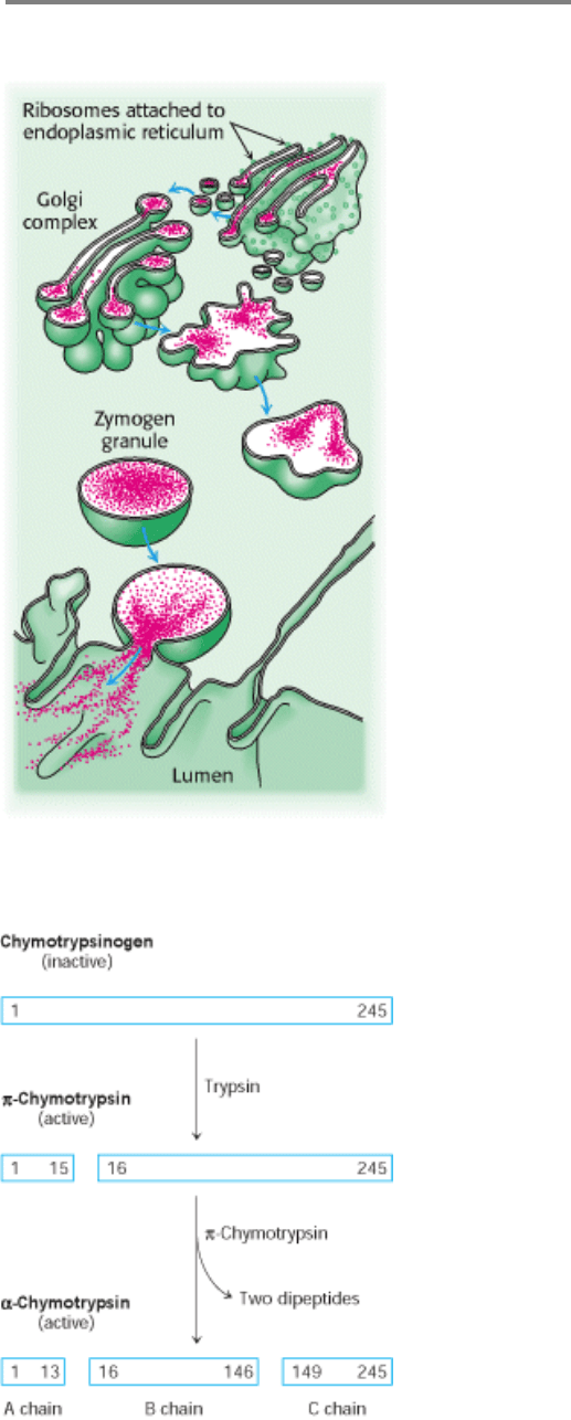
Figure 10.31. Secretion of Zymogens by an Acinar Cell of the Pancreas.
I. The Molecular Design of Life 10. Regulatory Strategies: Enzymes and Hemoglobin 10.5. Many Enzymes Are Activated by Specific Proteolytic Cleavage
Figure 10.32. Proteolytic Activation of Chymotrypsinogen. The three chains of α-chymotrypsin are linked by two
interchain disulfide bonds (A to B, and B to C).
I. The Molecular Design of Life 10. Regulatory Strategies: Enzymes and Hemoglobin 10.5. Many Enzymes Are Activated by Specific Proteolytic Cleavage
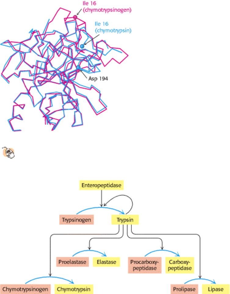
Figure 10.33. Conformations of Chymotrypsinogen (Red) and Chymotrypsin (Blue).
The electrostatic interaction
between the carboxylate of aspartate 194 and the α-amino group of isoleucine 16, essential for the structure of
active chymotrypsin, is possible only in chymotrypsin.
I. The Molecular Design of Life 10. Regulatory Strategies: Enzymes and Hemoglobin 10.5. Many Enzymes Are Activated by Specific Proteolytic Cleavage
Figure 10.34. Zymogen Activation by Proteolytic Cleavage. Enteropeptidase initiates the activation of the pancreatic
zymogens by activating trypsin, which then activates other zymogens. Active enzymes are shown in yellow; zymogens
are shown in orange.
I. The Molecular Design of Life 10. Regulatory Strategies: Enzymes and Hemoglobin 10.5. Many Enzymes Are Activated by Specific Proteolytic Cleavage
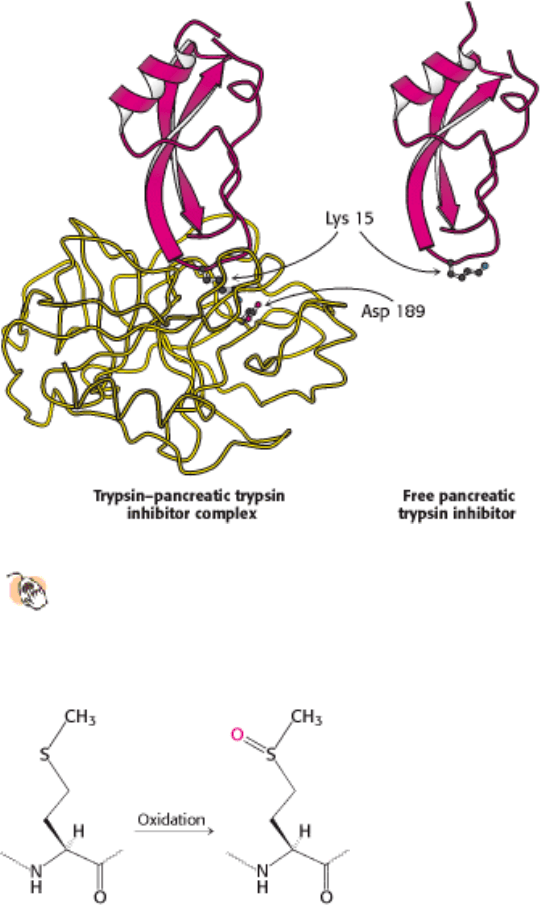
Figure 10.35. Interaction of Trypsin with Its Inhibitor.
Structure of a complex of trypsin (yellow) and pancreatic
trypsin inhibitor (red). Lysine 15 of the inhibitor penetrates into the active site of the enzyme and forms a salt
bridge with aspartate 189 in the active site. The bound inhibitor and the free inhibitor are almost identical in
structure.
I. The Molecular Design of Life 10. Regulatory Strategies: Enzymes and Hemoglobin 10.5. Many Enzymes Are Activated by Specific Proteolytic Cleavage
Figure 10.36. Oxidation of Methionine to the Sulfoxide.
I. The Molecular Design of Life 10. Regulatory Strategies: Enzymes and Hemoglobin 10.5. Many Enzymes Are Activated by Specific Proteolytic Cleavage
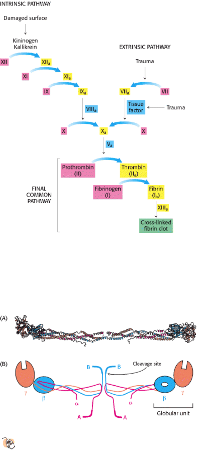
Figure 10.37. Blood-Clotting Cascade. A fibrin clot is formed by the interplay of the intrinsic, extrinsic, and final
common pathways. The intrinsic pathway begins with the activation of factor XII (Hageman factor) by contact with
abnormal surfaces produced by injury. The extrinsic pathway is triggered by trauma, which activates factor VII and
releases a lipoprotein, called tissue factor, from blood vessels. Inactive forms of clotting factors are shown in red; their
activated counterparts (indicated by the subscript "a") are in yellow. Stimulatory proteins that are not themselves
enzymes are shown in blue. A striking feature of this process is that the activated form of one clotting factor catalyzes
the activation of the next factor.
I. The Molecular Design of Life 10. Regulatory Strategies: Enzymes and Hemoglobin 10.5. Many Enzymes Are Activated by Specific Proteolytic Cleavage
Figure 10.38. Structure of a Fibrinogen Molecule.
(A) A ribbon diagram. The two rod regions are α-helical coiled
coils, connected to a globular region at each end. (B) A schematic representation showing the positions of the
fibrinopeptides A and B.
I. The Molecular Design of Life 10. Regulatory Strategies: Enzymes and Hemoglobin 10.5. Many Enzymes Are Activated by Specific Proteolytic Cleavage
Figure 10.39. Electron Micrograph of Fibrin. The 23-nm period along the fiber axis is half the length of a fibrinogen
molecule. [Courtesy of Dr. Henry Slayter.]
I. The Molecular Design of Life 10. Regulatory Strategies: Enzymes and Hemoglobin 10.5. Many Enzymes Are Activated by Specific Proteolytic Cleavage
Figure 10.40. Formation of a Fibrin Clot. (1) Thrombin cleaves fibrinopeptides A and B from the central globule of
fibrinogen. (2) Globular domains at the carboxyl-terminal ends of the β and γ chains interact with "knobs" exposed at the
amino-terminal ends of the β and α chains to form clots.
I. The Molecular Design of Life 10. Regulatory Strategies: Enzymes and Hemoglobin 10.5. Many Enzymes Are Activated by Specific Proteolytic Cleavage
Figure 10.41. Modular Structure of Prothrombin. Cleavage of two peptide bonds yields thrombin. All the γ-
carboxyglutamate residues are in the gla domain.
I. The Molecular Design of Life 10. Regulatory Strategies: Enzymes and Hemoglobin 10.5. Many Enzymes Are Activated by Specific Proteolytic Cleavage