Berg J.M., Tymoczko J.L., Stryer L. Biochemistry
Подождите немного. Документ загружается.

II. Transducing and Storing Energy 18. Oxidative Phosphorylation 18.5. Many Shuttles Allow Movement Across the Mitochondrial Membranes
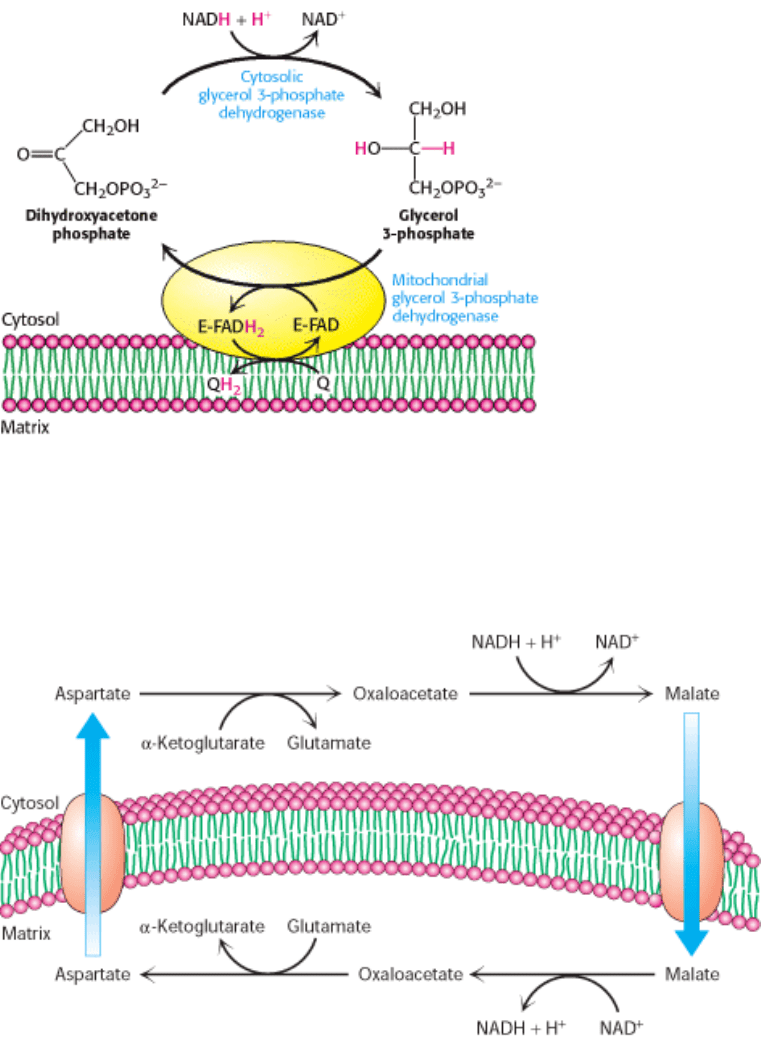
Figure 18.37. Glycerol 3-Phosphate Shuttle. Electrons from NADH can enter the mitochondrial electron transport
chain by being used to reduce dihydroxyacetone phosphate to glycerol 3-phosphate. Glycerol 3-phosphate is reoxidized
by electron transfer to an FAD prosthetic group in a membrane-bound glycerol 3-phosphate dehydrogenase. Subsequent
electron transfer to Q to form QH
2
allows these electrons to enter the electron-transport chain.
II. Transducing and Storing Energy 18. Oxidative Phosphorylation 18.5. Many Shuttles Allow Movement Across the Mitochondrial Membranes
Figure 18.38. Malate-Aspartate Shuttle.
II. Transducing and Storing Energy 18. Oxidative Phosphorylation 18.5. Many Shuttles Allow Movement Across the Mitochondrial Membranes
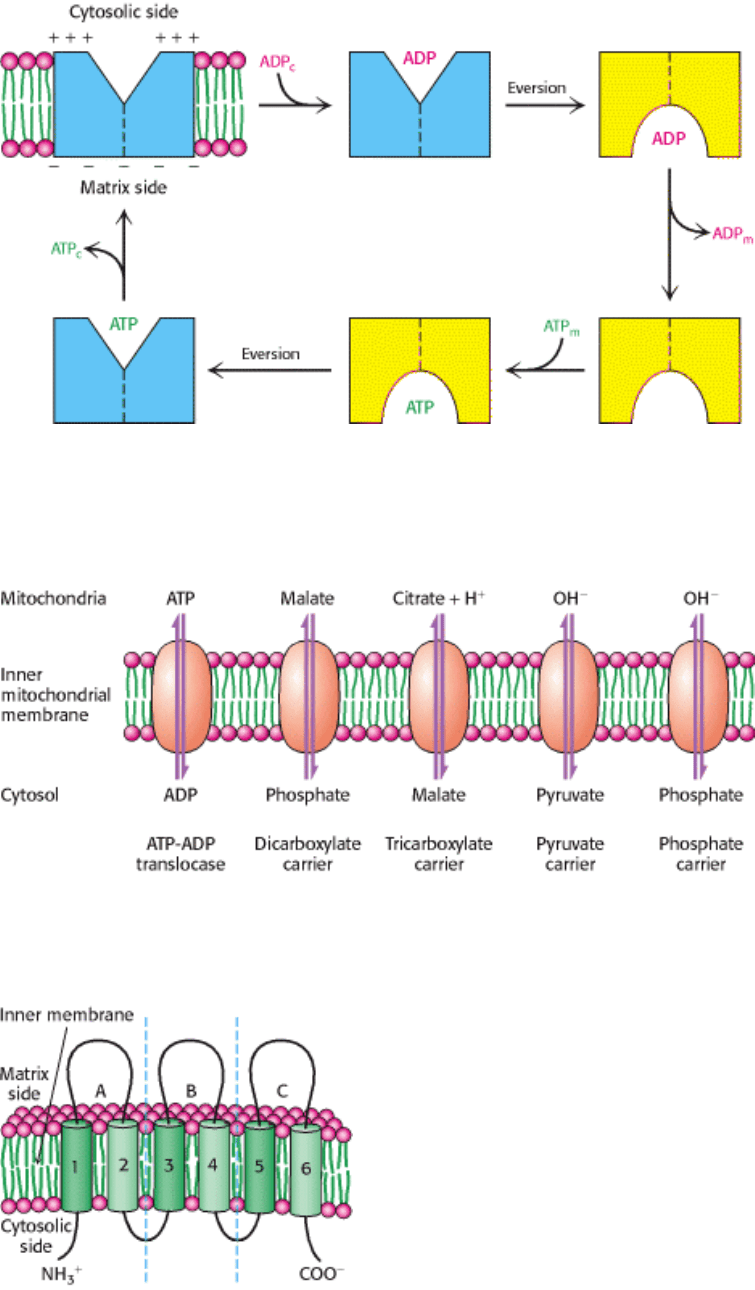
Figure 18.39. Mechanism of Mitochondrial ATP-ADP Translocase. The translocase catalyzes the coupled entry of
ADP and exit of ATP into and from the matrix. The reaction cycle is driven by membrane potential. The actual
conformational change corresponding to eversion of the binding site could be quite small.
II. Transducing and Storing Energy 18. Oxidative Phosphorylation 18.5. Many Shuttles Allow Movement Across the Mitochondrial Membranes
Figure 18.40. Mitochondrial Transporters. Transporters (also called carriers) are transmembrane proteins that move
ions and charged metabolites across the inner mitochondrial membrane.
II. Transducing and Storing Energy 18. Oxidative Phosphorylation 18.5. Many Shuttles Allow Movement Across the Mitochondrial Membranes
Figure 18.41. Structure of Mitochondrial Transporters. Many mitochondrial transporters consist of three similar 100-
residue units. These proteins contain six putative membrane-spanning segments. [After J. E. Walker. Curr. Opin. Struct.
Biol. 2(1992):519.]

II. Transducing and Storing Energy 18. Oxidative Phosphorylation
18.6. The Regulation of Cellular Respiration Is Governed Primarily by the Need for
ATP
Because ATP is the end product of cellular respiration, its concentration is the ultimate determinant of the rate of all of
the components of respiratory pathways.
18.6.1. The Complete Oxidation of Glucose Yields About 30 Molecules of ATP
We can now estimate how many molecules of ATP are formed when glucose is completely oxidized to CO
2
. The
number of ATP (or GTP) molecules formed in glycolysis and the citric acid cycle is unequivocally known because it is
determined by the stoichiometries of chemical reactions. In contrast, the ATP yield of oxidative phosphorylation is less
certain because the stoichiometries of proton pumping, ATP synthesis, and metabolite transport processes need not be
integer numbers or even have fixed values. As discussed earlier, the best current estimates for the number of protons
pumped out of the matrix by NADH-Q oxidoreductase, Q-cytochrome c oxidoreductase, and cytochrome c oxidase per
electron pair are four, two, and four, respectively. The synthesis of a molecule of ATP is driven by the flow of about
three protons through ATP synthase. An additional proton is consumed in transporting ATP from the matrix to the
cytosol. Hence, about 2.5 molecules of cytosolic ATP are generated as a result of the flow of a pair of electrons from
NADH to O
2
. For electrons that enter at the level of Q-cytochrome c oxidoreductase, such as those from the oxidation of
succinate or cytosolic NADH, the yield is about 1.5 molecules of ATP per electron pair. Hence, as tallied in Table 18.4,
about 30 molecules of ATP are formed when glucose is completely oxidized to CO
2
; this value supersedes the
traditional estimate of 36 molecules of ATP. Most of the ATP, 26 of 30 molecules formed, is generated by oxidative
phosphorylation. Recall that the anaerobic metabolism of glucose yields only 2 molecules of ATP.
18.6.2. The Rate of Oxidative Phosphorylation Is Determined by the Need for ATP
How is the rate of the electron-transport chain controlled? Under most physiological conditions, electron transport is
tightly coupled to phosphory-lation. Electrons do not usually flow through the electron-transport chain to O
2
unless
ADP is simultaneously phosphorylated to ATP. Oxidative phosphorylation requires a supply of NADH (or other source
of electrons at high potential), O
2
, ADP, and P
i
. The most important factor in determining the rate of oxidative
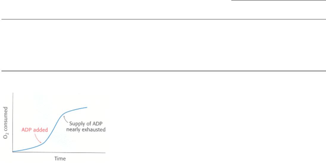
phosphorylation is the level of ADP. The rate of oxygen consumption by mitochondria increases markedly when ADP is
added and then returns to its initial value when the added ADP has been converted into ATP (Figure 18.42).
The regulation of the rate of oxidative phosphorylation by the ADP level is called respiratory control or acceptor
control. The level of ADP likewise affects the rate of the citric acid cycle because of its need for NAD
+
and FAD. The
physiological significance of this regulatory mechanism is evident. The ADP level increases when ATP is consumed,
and so oxidative phosphorylation is coupled to the utilization of ATP. Electrons do not flow from fuel molecules to O
2
unless ATP needs to be synthesized. We see here another example of the regulatory significance of the energy charge.
18.6.3. Oxidative Phosphorylation Can Be Inhibited at Many Stages
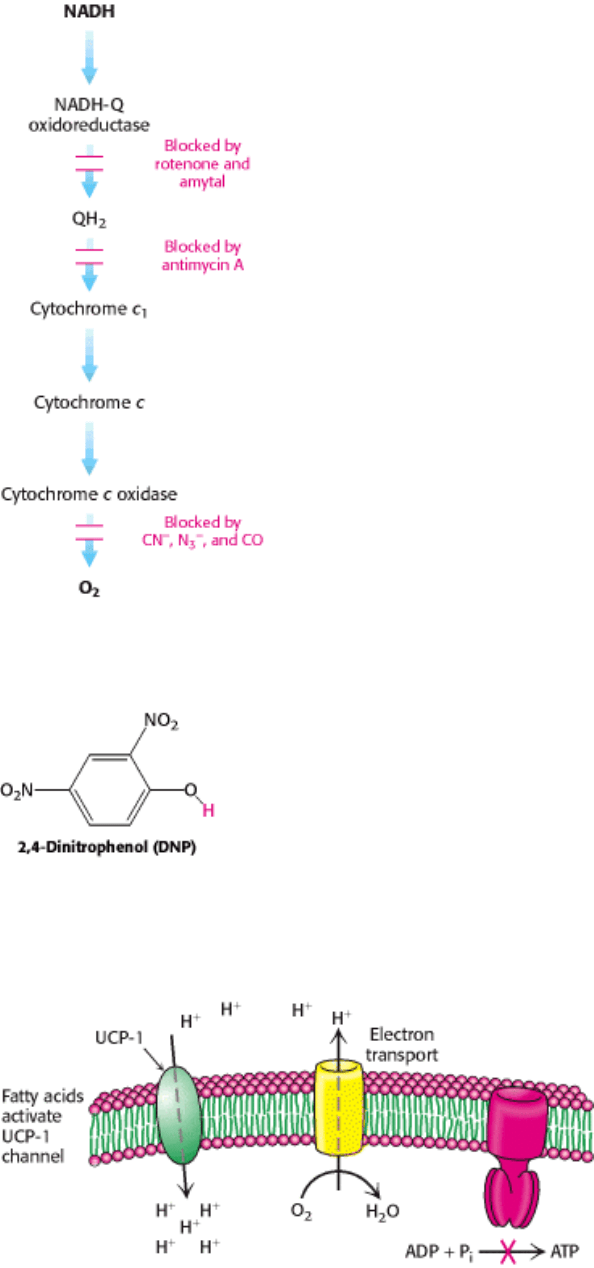
Oxidative phosphorylation is susceptible to inhibition at all stages of the process. Specific inhibitors of electron transport
were invaluable in revealing the sequence of electron carriers in the respiratory chain. For example, rotenone and amytal
block electron transfer in NADH-Q oxidoreductase and thereby prevent the utilization of NADH as a substrate (Figure
18.43). In contrast, electron flow resulting from the oxidation of succinate is unimpaired, because these electrons enter
through QH
2
, beyond the block. Antimycin A interferes with electron flow from cytochrome b
H
in Q-cytochrome c
oxidoreductase. Furthermore, electron flow in cytochrome c oxidase can be blocked by cyanide (CN
-
), azide (N
3
-
), and
carbon monoxide (CO). Cyanide and azide react with the ferric form of heme a
3
, whereas carbon monoxide inhibits the
ferrous form. Inhibition of the electron-transport chain also inhibits ATP synthesis because the proton-motive force can
no longer be generated.
ATP synthase also can be inhibited. Oligomycin and dicyclohexylcarbodiimide (DCCD) prevent the influx of protons
through ATP synthase. If actively respiring mitochondria are exposed to an inhibitor of ATP synthase, the electron-
transport chain ceases to operate. Indeed, this observation clearly illustrates that electron transport and ATP synthesis are
normally tightly coupled.
This tight coupling of electron transport and phosphorylation in mitochondria can be disrupted (uncoupled) by 2,4-
dinitrophenol (Figure 18.44) and certain other acidic aromatic compounds. These substances carry protons across the
inner mitochondrial membrane. In the presence of these uncouplers, electron transport from NADH to O
2
proceeds in a
normal fashion, but ATP is not formed by mitochondrial ATP synthase because the proton-motive force across the inner
mitochondrial membrane is dissipated. This loss of respiratory control leads to increased oxygen consumption and
oxidation of NADH. Indeed, in the accidental ingestion of uncouplers, large amounts of metabolic fuels are consumed,
but no energy is stored as ATP. Rather, energy is released as heat. DNP and other uncouplers are very useful in
metabolic studies because of their specific effect on oxidative phosphorylation. The regulated uncoupling of oxidative
phosphorylation is a biologically useful means of generating heat.
ATP-ADP translocase is specifically inhibited by very low concentrations of atractyloside (a plant glycoside) or
bongkrekic acid (an antibiotic from a mold). Atractyloside binds to the translocase when its nucleotide site faces the
cytosol, whereas bongkrekic acid binds when this site faces the mitochondrial matrix. Oxidative phosphorylation stops
soon after either inhibitor is added, showing that ATP-ADP translocase is essential.
18.6.4. Regulated Uncoupling Leads to the Generation of Heat
The uncoupling of oxidative phosphorylation is a means of generating heat to maintain body temperature in hibernating
animals, in some newborn animals (including human beings), and in mammals adapted to cold. Brown adipose tissue,
which is very rich in mitochondria (often referred to as brown fat mitochondria), is specialized for this process of
nonshivering thermogenesis. The inner mitochondrial membrane of these mitochondria contains a large amount of
uncoupling protein (UCP), here UCP-1, or thermogenin, a dimer of 33-kd subunits that resembles ATP-ADP
translocase. UCP-1 forms a pathway for the flow of protons from the cytosol to the matrix. In essence, UCP-1 generates
heat by short-circuiting the mitochondrial proton battery. This dissipative proton pathway is activated by free fatty acids
liberated from triacylglycerols in response to hormonal signals, such as β-adrenergic agonists (Figure 18.45).
In addition to UCP-1, two other uncoupling proteins have been identified. UCP-2, which is 56% identical in
sequence with UCP-1, is found in a wide variety of tissues. UCP-3 (57% identical with UCP-1 and 73% identical
with UCP-2) is localized to skeletal muscle and brown fat. This family of uncoupling proteins, especially UCP-2 and
UCP-3, may play a role in energy homeostasis. In fact, the genes for UCP-2 and UCP-3 map to regions of the human and
mouse chromosomes that have been linked to obesity, substantiating the notion that they function as a means of
regulating body weight. The use of uncoupling proteins is not limited to animals, however. The skunk cabbage uses an
analogous mechanism to heat its floral spikes, increasing the evaporation of odoriferous molecules that attract insects to
fertilize its flowers.
18.6.5. Mitochondrial Diseases Are Being Discovered
As befitting an organelle that is so central to energy metabolism, mitochondrial malfunction can lead to
pathological conditions. The number of diseases that can be attributed to mitochondrial mutations is steadily
growing in step with our growing understanding of the biochemistry and genetics of mitochondria. The first
mitochondrial disease to be understood was Leber hereditary optic neuropathy (LHON), a form of blindness that strikes
in midlife as a result of mutations to the NADH-Q oxidoreductase component of Complex I. Some of these mutations
impair NADH utilization, whereas others block electron transfer to Q. The accumulation of mutations in mitochondrial
genes in the course of several decades may contribute to aging, degenerative disorders, and cancer.

A human egg harbors several hundred thousand molecules of mitochondrial DNA, whereas a sperm contributes only a
few hundred and thus has little effect on the mitochondrial genotype. Because the maternally inherited mitochondria are
present in large numbers and not all of the mitochondria may be affected, the pathologies of mitochondrial mutants can
be quite complex. Even within a single family carrying an identical mutation, chance fluctuations in the percentage of
mitochondria with the mutation lead to large variations in the nature and severity of the symptoms of the pathological
condition as well as the time of onset. As the percentage of defective mitochondria increases, energy-generating capacity
diminishes until, at some threshold, the cell can no longer function properly. Defects in cellular respiration are doubly
dangerous. Not only does energy transduction decrease, but also the likelihood that reactive oxygen species will be
generated increases. Organs that are highly dependent on oxidative phosphorylation, such as the nervous system and the
heart, are most vulnerable to mutations in mitochondrial DNA.
18.6.6. Mitochondria Play a Key Role in Apoptosis
In the course of development or in cases of significant cell damage, individual cells within multicellular organisms
undergo programmed cell death, or apoptosis. Mitochondria act as control centers regulating this process. Although the
details have not yet been established, a pore called the mitochondrial permeability transition pore (mtPTP) forms in
damaged mitochondria. This pore appears to consist of VDAC (the adenine nucleotide translocator) and several other
mitochondrial proteins, including members of a family of proteins (Bcl family) that were initially discovered because of
their role in cancer. One of the most potent activators of apoptosis is cytochrome c. Its presence in the cytosol activates a
cascade of proteolytic enzymes called caspases. These cysteine proteases (Section 9.1.6) are conserved in evolution,
being found in organisms ranging from hydra to human beings. Cytochrome c, in conjunction with other proteins,
initiates the cascade by activating procaspase 9 to form caspase 9, which then activates other caspases. Activation of the
caspase cascade does not lead to generalized protein destruction. Rather, the caspases have particular targets. For
instance, the proteins that maintain cell structure are destroyed. Another example is the degradation of a protein that
inhibits an enzyme that destroys DNA (caspase-activated DNAse, CAD), freeing CAD to cleave the genetic material.
This cascade of proteolytic enzymes has been called "death by a thousand tiny cuts."
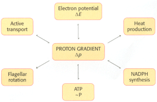
18.6.7. Power Transmission by Proton Gradients: A Central Motif of Bioenergetics
The main concept presented in this chapter is that mitochondrial electron transfer and ATP synthesis are linked by a
transmembrane proton gradient. ATP synthesis in bacteria and chloroplasts (Section 19.4) also is driven by proton
gradients. In fact, proton gradients power a variety of energy-requiring processes such as the active transport of calcium
ions by mitochondria, the entry of some amino acids and sugars into bacteria, the rotation of bacterial flagella, and the
transfer of electrons from NADP
+
to NADPH. Proton gradients can also be used to generate heat, as in hibernation. It is
evident that proton gradients are a central interconvertible currency of free energy in biological systems (Figure 18.46).
Mitchell noted that the proton-motive force is a marvelously simple and effective store of free energy because it requires
only a thin, closed lipid membrane between two aqueous phases.
II. Transducing and Storing Energy 18. Oxidative Phosphorylation 18.6. The Regulation of Cellular Respiration Is Governed Primarily by the Need for ATP
Table 18.4. ATP yield from the complete oxidation of glucose
Reaction sequence ATP yield per glucose molecule
Glycolysis: Conversion of glucose into pyruvate (in the cytosol)
Phosphorylation of glucose - 1
Phosphorylation of fructose 6-phosphate - 1
Dephosphorylation of 2 molecules of 1,3-BPG + 2
Dephosphorylation of 2 molecules of phosphoenolpyruvate + 2
2 molecules of NADH are formed in the oxidation of 2 molecules of
glyceraldehyde 3-phosphate
Conversion of pyruvate into acetyl CoA (inside mitochondria)
2 molecules of NADH are formed
Citric acid cycle (inside mitochondria)
2 molecules of guanosine triphosphate are formed from 2 molecules of succinyl
CoA
+ 2
6 molecules of NADH are formed in the oxidation of 2 molecules each of
isocitrate, α-ketoglutarate, and malate
2 molecules of FADH
2
are formed in the oxidation of 2 molecules of succinate
Oxidative phosphorylation (inside mitochondria)
2 molecules of NADH formed in glycolysis; each yields 1.5 molecules of ATP
(assuming transport of NADH by the glycerol 3-phosphate shuttle)
+ 3
2 molecules of NADH formed in the oxidative decarboxylation of pyruvate; each
yields 2.5 molecules of ATP
+ 5
2 molecules of FADH
2
formed in the citric acid cycle; each yields 1.5 molecules
of ATP
+ 3
6 molecules of NADH formed in the citric acid cycle; each yields 2.5 molecules of
ATP
+ 15
NET YIELD PER MOLECULE OF GLUCOSE
+ 30
Source: The ATP yield of oxidative phosphorylation is based on values given in P. C. Hinkle, M. A. Kumar, A. Resetar, and D. L.
Harris, Biochemistry 30(1991):3576.
Note: The current value of 30 molecules of ATP per molecule of glucose supersedes the earlier one of 36 molecules of ATP. The
stoichiometries of proton pumping, ATP synthesis, and metabolite transport should be regarded as estimates. About two more
molecules of ATP are formed per molecule of glucose oxidized when the malate-aspartate shuttle rather than the glycerol 3-
phosphate shuttle is used.
II. Transducing and Storing Energy 18. Oxidative Phosphorylation 18.6. The Regulation of Cellular Respiration Is Governed Primarily by the Need for ATP
Figure 18.42. Respiratory Control. Electrons are transferred to O
2
only if ADP is concomitantly phosphorylated to
ATP.
II. Transducing and Storing Energy 18. Oxidative Phosphorylation 18.6. The Regulation of Cellular Respiration Is Governed Primarily by the Need for ATP
Figure 18.43. Sites of Action of Some Inhibitors of Electron Transport.
II. Transducing and Storing Energy 18. Oxidative Phosphorylation 18.6. The Regulation of Cellular Respiration Is Governed Primarily by the Need for ATP
Figure 18.44. Uncoupler of Oxidative Phosphorylation. 2,4-Dinitrophenol, a lipid-soluble substance, can carry
protons across the inner mitochondrial membrane. The dissociable proton is shown in red.
II. Transducing and Storing Energy 18. Oxidative Phosphorylation 18.6. The Regulation of Cellular Respiration Is Governed Primarily by the Need for ATP
Figure 18.45. Action of an Uncoupling Protein. Uncoupling protein-1 (UCP-1) generates heat by permitting the influx
of protons into the mitochondria without the synthesis of ATP.
II. Transducing and Storing Energy 18. Oxidative Phosphorylation 18.6. The Regulation of Cellular Respiration Is Governed Primarily by the Need for ATP
Figure 18.46. The Proton Gradient Is an Interconvertible Form of Free Energy.
II. Transducing and Storing Energy 18. Oxidative Phosphorylation
Summary
Oxidative Phosphorylation in Eukaryotes Takes Place in Mitochondria
Mitochondria generate most of the ATP required by aerobic cells by a joint endeavor of the reactions of citric acid cycle,
which take place in the mitochondrial matrix, and oxidative phosphorylation, which takes place in the inner
mitochondrial membrane. Mitochondria are descendents of a free-living bacterium that established a symbiotic relation
with another cell.
Oxidative Phosphorylation Depends on Electron Transfer
In oxidative phosphorylation, the synthesis of ATP is coupled to the flow of electrons from NADH or FADH
2
to O
2
by a
proton gradient across the inner mitochondrial membrane. Electron flow through three asymmetrically oriented
transmembrane complexes results in the pumping of protons out of the mitochondrial matrix and the generation of a
membrane potential. ATP is synthesized when protons flow back to the matrix through a channel in an ATP-synthesizing
complex, called ATP synthase (also known as F
0
F
1
-ATPase). Oxidative phosphorylation exemplifies a fundamental
theme of bioenergetics: the transmission of free energy by proton gradients.
The Respiratory Chain Consists of Four Complexes: Three Proton Pumps and a
Physical Link to the Citric Acid Cycle
The electron carriers in the respiratory assembly of the inner mitochondrial membrane are quinones, flavins, iron-sulfur
complexes, heme groups of cytochromes, and copper ions. Electrons from NADH are transferred to the FMN prosthetic
group of NADH-Q oxidoreductase (Complex I), the first of four complexes. This oxidoreductase also contains Fe-S
centers. The electrons emerge in QH
2
, the reduced form of ubiquinone (Q). The citric acid cycle enzyme succinate
dehydrogenase is a component of the succinate-Q reductase complex (Complex II), which donates electrons from
FADH
2
to Q to form QH
2
.This highly mobile hydrophobic carrier transfers its electrons to Q-cytochrome c
oxidoreductase (Complex III), a complex that contains cytochromes b and c
1
and an Fe-S center. This complex reduces
cytochrome c, a water-soluble peripheral membrane protein. Cytochrome c, like Q, is a mobile carrier of electrons,
which it then transfers to cytochrome c oxidase (Complex IV). This complex contains cytochromes a and a
3
and three
copper ions. A heme iron ion and a copper ion in this oxidase transfer electrons to O
2
, the ultimate acceptor, to form
H
2
O.
A Proton Gradient Powers the Synthesis of ATP
The flow of electrons through Complexes I, III, and IV leads to the transfer of protons from the matrix side to the
cytosolic side of the inner mitochondrial membrane. A proton-motive force consisting of a pH gradient (matrix side
basic) and a membrane potential (matrix side negative) is generated. The flow of protons back to the matrix side through
ATP synthase drives ATP synthesis. The enzyme complex is a molecular motor made of two operational units: a rotating
component and a stationary component. The rotation of the γ subunit induces structural changes in the β subunit that
result in the synthesis and release of ATP from the enzyme. Proton influx provides the force for the rotation.
The flow of two electrons through NADH-Q oxidoreductase, Q-cytochrome c oxidoreductase, and cytochrome c oxidase
generates a gradient sufficient to synthesize 1, 0.5, and 1 molecule of ATP, respectively. Hence, 2.5 molecules of ATP
are formed per molecule of NADH oxidized in the mitochondrial matrix, whereas only 1.5 molecules of ATP are made
per molecule of FADH
2
oxidized because its electrons enter the chain at QH
2
, after the first proton-pumping site.
Many Shuttles Allow Movement Across the Mitochondrial Membranes
Mitochondria employ a host of carriers, or transporters, to move molecules across the inner mitochondrial membrane.
The electrons of cytoplasmic NADH are transferred into the mitochondria by the glycerol phosphate shuttle to form
FADH
2
from FAD. The entry of ADP into the mitochondrial matrix is coupled to the exit of ATP by ATP-ADP
translocase, a transporter driven by membrane potential.
The Regulation of Oxidative Phosphorylation Is Governed Primarily by the Need for
ATP
About 30 molecules of ATP are generated when a molecule of glucose is completely oxidized to CO
2
and H
2
O. Electron
transport is normally tightly coupled to phosphorylation. NADH and FADH
2
are oxidized only if ADP is simultaneously
phosphorylated to ATP, a form of regulation called acceptor or respiratory control. Uncouplers such as DNP can disrupt
this coupling; they dissipate the proton gradient by carrying protons across the inner mitochondrial membrane. Proteins
have been identified that uncouple electron transport and ATP synthesis for the generation of heat.
Key Terms
oxidative phosphorylation
proton-motive force
cellular respiration
electron-transport chain
reduction (redox, oxidation-reduction, E´
0
) potential
inverted region
coenzyme Q (Q, ubiquinone)
NADH-Q oxidoreductase (Complex I)
flavin mononucleotide (FMN)
iron-sulfur (nonheme iron) protein
succinate-Q reductase (Complex II)
Q-cytochrome c oxidoreductase (Complex III)
cytochrome c (cyt c)
Rieske center
Q cycle
cytochrome c oxidase (Complex IV)
superoxide dismutase
catalase
ATP synthase (Complex V, F
1
F
0
ATPase)
glycerol 3-phosphate shuttle
malate-aspartate shuttle
ATP-ADP translocase (adenine nucleotide translocase, ANT)
respiratory (acceptor) control
uncoupling protein (UCP)
programmed cell death (apoptosis)
caspase
II. Transducing and Storing Energy 18. Oxidative Phosphorylation
Problems
1.
Energy harvest. What is the yield of ATP when each of the following substrates is completely oxidized to CO
2
by a
mammalian cell homogenate? Assume that glycolysis, the citric acid cycle, and oxidative phosphorylation are fully
active.
(a) Pyruvate
(b) Lactate
(c) Fructose 1,6-bisphosphate
(d) Phosphoenolpyruvate