Egerton R.F. Electron Energy-Loss Spectroscopy in the Electron Microscope
Подождите немного. Документ загружается.

8 1 An Introduction to EELS
measure of the intensity of scattering from each atom (or molecule) and which can
be calculated by the use of either classical physics or quantum mechanics.
Inner-shell excitation gives rise to relatively low scattered intensity (due to the
low cross section) and therefore has a mean free path that is long compared to
the s pecimen thickness. The probability that a fast electron produces more than
one inner-shell excitation is therefore negligible. However, an electron that has
undergone inner-shell scattering may (with fair probability) also cause outer-shell
excitation. This “mixed” inelastic scattering again involves an energy loss that is
the sum of the two separate losses, and results in a broad peak above the ioniza-
tion threshold, displaced from the threshold by approximately the plasmon energy;
see Fig. 1.4. If necessary, this mixed-scattering intensity can be removed from the
spectrum by deconvolution.
1.3 The Development of Experimental Techniques
We will consider now how techniques evolved for recording and analyzing the
energy-loss spectrum of fast electrons, particularly in combination with electron
microscopy. More recent instrumental developments are dealt with in greater detail
in later chapters.
In his doctoral thesis, published in 1929, Rudberg reported measurements of the
kinetic energy of electrons after reflection from the surface of a metal such as copper
or silver. The kinetic energy was determined using a magnetic field spectrometer that
bent the electron trajectories through 180
◦
(in a 25-mm radius) and gave a resolu-
tion of about 1 part in 200, adequate for the low primary energies that were used
(40–900 eV). By measuring currents with a quadrant electrometer, the electron
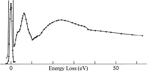
intensity could be plotted as a function of energy loss, as in Fig. 1.6. Rudberg
showed that the l oss spectrum was characteristic of the chemical composition of
the sample and independent of the primary energy and the angle of incidence. In
these experiments, oxidation of the surface was minimized by preparing the sample
Fig. 1.6 Energy-loss spectrum of 204-eV electrons reflected from the surface of an evaporated
film of copper (Rudberg, 1930). The zero-loss peak is shown on a reduced intensity scale. From
Rudberg (1930), with permission of The Royal Society
1.3 The Development of Experimental Techniques 9
in situ by evaporation onto a Mo or Ag substrate that was kept electrically heated.
Similar measurements were later carried out on a large number of elements by
Powell, Robins, and Best at the University of Western Australia. The reflection tech-
nique has since been refined to give energy resolution of a few milli electron volts
at incident energies of a few hundred electron volts (Ibach and Mills, 1982) and has
also been implemented in a field-emission scanning electron microscope (Cowley,
1982).
The first measurement of the energy spectrum of transmitted electrons was
reported by Ruthemann (1941), using higher incident energy (2–10 keV), an
improved magnetic spectrometer (bend radius = 175 mm, resolving power 1 in
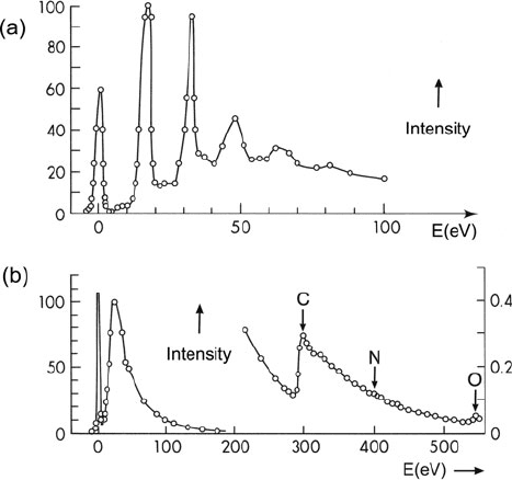
2000), and photographic recording. Figure 1.7a shows Ruthemann’s energy-loss
spectrum of a thin self-supporting film of Al; it displays a series of peaks at mul-
tiples of 16 eV, which were later interpreted in terms of plasma oscillation (Bohm
and Pines, 1951). In 1942, Ruthemann reported the observation of inner-shell losses
in a thin film of collodion, a form of nitrocellulose (Fig. 1.7b).
A first attempt to use inner-shell losses for elemental microanalysis was made by
Hillier and Baker (1944), who constructed an instrument that could focus 25–75 keV
electrons into a 20-nm probe and operate as either a microprobe or a shadow micro-
scope. Two condenser lenses were used to focus the electrons onto the specimen
Fig. 1.7 (a) Energy-loss spectrum of 5.3-keV electrons transmitted through a thin foil of alu-
minum (Ruthemann, 1941), exhibiting plasmon peaks at multiples of 16 eV loss. (b) Energy-loss
spectrum of 7.5-keV electrons transmitted through a thin film of collodion, showing K-ionization
edges arising from carbon, nitrogen, and oxygen. Reprinted from Ruthemann (1941), copyright
SpringerLink
10 1 An Introduction to EELS
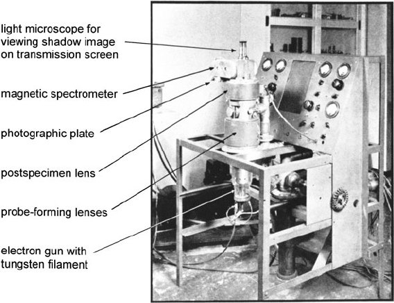
Fig. 1.8 Photograph of the first electron microanalyzer (Hillier and Baker, 1944). Two magnetic
lenses focused electrons onto the specimen, which was located within the bore of the second lens.
A third lens focused the transmitted beam into a 180
◦
spectrometer or produced a shadow image
of the specimen when the spectrometer field was turned off
and a third lens served to couple the transmitted electrons into a 180
◦
magnetic
spectrometer (Fig. 1.8). Because of the poor vacuum and resulting hydrocarbon con-
tamination, the 20-nm probe caused specimens to become opaque in a few seconds.
Therefore an incident beam diameter of 200 nm was used, corresponding to the
analysis of 10
−16
–10
−14
g of material. K-ionization edges were recorded from sev-
eral elements (including Si), as well as L- and M-edges of iron. Spectra of collodion
showed K-edges of carbon and oxygen; the nitrogen edge was usually absent, most
likely due to the preferential removal of nitrogen by the electron beam (Egerton,
1980f).
In 1949, Möllenstedt published the design of an electrostatic energy analyzer
in which electrons were slowed down by passing them between two cylindrical
electrodes connected to the electron-source voltage. This deceleration results in
high off-axis chromatic aberration (large dispersion) and an energy resolution of
about 1 part in 50,000, allowing high-resolution spectra to be recorded on photo-
graphic plates. The Möllenstedt analyzer was subsequently added to conventional
electron microscopes (CTEMs) in several laboratories, for example, by Marton (at
NBS, Washington), Boersch (Berlin), and Watanabe (Tokyo), and at the Cavendish
Laboratory (Cambridge). It was usually attached to the bottom of the TEM column,
allowing the microscope to retain its full range of magnification and diffraction
facilities. Because this analyzer is nonfocusing in a direction parallel to the elec-
trodes, a long (but narrow) entrance slit was used, enabling the spectrum to be
1.3 The Development of Experimental Techniques 11
recorded as a function of position in the specimen or, with a diffraction pattern
present on the TEM screen, as a function of electron scattering angle.
An alternative use of the deceleration principle was employed by Blackstock
et al. (1955), Klemperer and Shepherd (1963), Kincaid et al. (1978), and Ritsko
(1981). The electron source and analyzer were at ground potential, the electrons
being accelerated toward the specimen and decelerated afterward. This design pro-
vides good energy resolution but is difficult to apply to an electron microscope,
where the specimen cannot easily be raised to a high potential. Fink (1989)used
a spectrometer system with the sample at ground potential, the electron source,
monochromator, analyzer, and detector being at –170 keV. A retarding-field spec-
trometer was also used by Raether and colleagues in Hamburg, who conducted
in-depth studies of the angular distribution and dispersion of bulk and surface
plasmons in a wide variety of materials (Raether, 1980).
A combination of electric and magnetic fields (Wien filter) was first used
for transmission energy-loss measurements by Boersch et al. (1962) in Berlin.
For a given energy resolution, the entrance slit can be much wider than for t he
Möllenstedt analyzer (Curtis and Silcox, 1971), allowing the angular distribution
of both strong and weak energy-loss peaks to be studied in detail (Silcox, 1977,
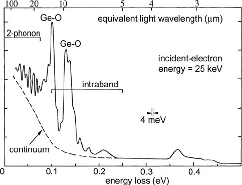
1979). By using a second Wien filter as a pre-specimen monochromator, Boersch
and colleagues eventually achieved an energy resolution of 4–6 meV at 25 keV
incident energy (Geiger et al., 1970). This degree of resolution is sufficient to reveal
phonon-loss peaks as well as vibrational modes and intraband electronic excita-
tions; see Fig. 1.9. The Berlin apparatus was also used to study the composition
of beam-induced contamination layers and weak energy gains (vibrational peaks at
23, 43, and 52 meV) could sometimes be detected (Katterwe, 1972). Since there
Fig. 1.9 Energy-loss spectrum of a 25-nm germanium film showing phonon and vibrational
modes, as well as intraband electronic transitions (Schröder, 1972)
12 1 An Introduction to EELS
was no strong focusing lens, the instrument lacked spatial resolution (the beam
diameter at the specimen was about 10 μm) and was not used extensively because
similar spatial resolution and better energy resolution could be obtained by infrared
spectroscopy.
1.3.1 Energy-Selecting (Energy-Filtering) Electron Microscopes
Instead of recording the energy-loss spectrum from a particular region of spec-
imen, it is sometimes preferable to display a magnified image of the specimen
(or its diffraction pattern) at a selected energy loss. This can be done by utilizing
the imaging properties of a magnetic field produced between prism-shaped pole-
pieces, as first demonstrated by Castaing and Henry (1962) at the University of
Paris. Like the normal unfiltered TEM image, a plasmon-loss image was found
to contain diffraction contrast due to differences in elastic scattering, but in suit-
able specimens it also conveyed “chemical contrast” that was useful for identifying
different crystallographic phases (Castaing, 1975). Installed in various laborato-
ries, the Castaing–Henry filter was also used to record spectra and images from
inner-shell energy losses (Colliex and Jouffrey, 1972; Henkelman and Ottensmeyer,
1974a; Egerton et al., 1974). At the University of Toronto, Ottensmeyer reduced the
aberrations of his filter by curving the prism edges, a design that was eventually
incorporated into a TEM by the Zeiss company.
In order to maintain a straight electron-optical column, the Castaing–Henry filter
uses an electrostatic mirror electrode at the electron gun potential, an arrange-
ment not well suited to high-voltage microscopes. For their 1-MeV microscope at
Toulouse, Jouffrey and colleagues adopted the purely magnetic “omega filter,” based
on a design by Rose and Plies (1974). In Berlin, Zeitler’s group improved this sys-
tem by correcting various aberrations, resulting in a commercial product: the Zeiss
EM-912 energy-filtering microscope (Bihr et al., 1991). The omega filter has since
been incorporated into TEM designs by other manufacturers.
An alternative method of energy filtering is based on the scanning transmis-
sion electron microscope (STEM). In 1968, Crewe and co-workers in Chicago built
the first high-resolution STEM and later used an energy analyzer to improve their
images of single heavy atoms on a thin substrate. At the same laboratory, Isaacson,
Johnson, and Lin recorded fine structure present in the energy-loss spectra of amino
acids and nucleic acid bases and used fading of this structure as a means of assessing
electron irradiation damage to these biologically important compounds.
In 1974, the Vacuum Generators Company marketed a field-emission STEM
(the HB5), on the prototype of which Burge and colleagues at London University
installed an electrostatic energy analyzer (Browne, 1979). Ferrier’s group at
Glasgow University investigated the practical advantages and disadvantages of
adding post-specimen lenses to the STEM, including their effect on spectrometer
performance. Isaacson designed an improved magnetic spectrometer for the HB5,
while Batson showed that superior energy resolution and stability could be obtained
by using a retarding-field Wien filter. Various groups (for example, Colliex and
1.3 The Development of Experimental Techniques 13
co-workers in Paris, Brown and Howie in Cambridge, Spence and colleagues in
Arizona) used the HB5 and its descendants to explore the high-resolution possi-
bilities of EELS. Leapman and co-workers at NIH (Washington) employed STEM
energy-selected imaging to record elemental maps of biological specimens at 10-nm
resolution and showed that energy-loss spectra can reveal concentrations (of lan-
thanides and transition metals) down to 10 ppm within 50-nm diameter areas of
an inorganic test specimen. Using a STEM with a high-excitation objective lens,
Pennycook and colleagues demonstrated that atomic columns in a suitably ori-
ented crystalline specimen can be imaged using high-angle elastic scattering, the
low-angle energy-loss signal being simultaneously available to extract chemical
information at atomic resolution. Correction of the objective lens spherical aber-
ration, as achieved in the NiOn STEM instrument, makes atom column imaging and
spectroscopy easier and has led to the identification and imaging of single atoms
(Krivanek et al., 2010).
1.3.2 Spectrometers as Attachments to a TEM
Starting in the mid-1970s, EELS attracted the attention of electron microscopists as
a method of light element microanalysis. For this purpose a single-prism magnetic
spectrometer mounted beneath a conventional TEM is sufficient; Marton (1946)
appears to have been the first to assemble such a system. At the Cavendish labora-
tory, Wittry (1969) employed the electron optical arrangement that is now widely
used: the prism operates with the projector lens crossover as its object point and a
spectrometer-entrance aperture (just below the TEM screen) selects the region of
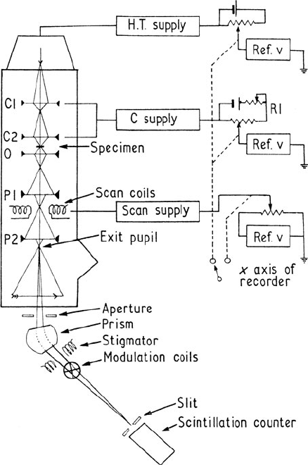
specimen (or its diffraction pattern) being analyzed (Fig. 1.10). Serial recording of
the spectrum was achieved by scanning it across an energy-selecting slit preced-
ing a single-channel electron detector (scintillator and photomultiplier tube). Based
on these principles, Krivanek constructed a magnetic spectrometer that was small
enough to fit below any conventional TEM, this design being marketed by the Gatan
company as their Model 607 serial recording EELS system. Joy and Maher (at Bell
Laboratories) and Egerton (in Alberta) developed data analysis systems and soft-
ware for electronic storage of energy-loss spectra and background subtraction at
core edges, as required for quantitative elemental analysis.
The energy-loss spectrum can be recorded simultaneously (rather than sequen-
tially) by means of a position-sensitive detector, such as a photodiode or charge-
coupled diode (CCD) array. Following development work in several university lab-
oratories, Gatan introduced in 1986 their Model 666 spectrometer, using quadrupole
lenses to project the spectrum onto a YAG transmission screen and photodiode
array. Parallel-recording spectrometers greatly reduce the time needed to record
inner-shell losses, resulting in less drift and electron irradiation of the specimen.
The Gatan Enfina spectrometer was a refinement of this design, with less light
spreading in the scintillator and the photodiode array replaced by a rectangular
(100 ×1340-element) CCD detector.
14 1 An Introduction to EELS
Fig. 1.10 TEM energy
analysis system with a
magnetic prism spectrometer
(45
◦
deflection angle) using
the projector lens crossover as
its object point (Wittry, 1969,
http://iopscience.iop.org/
0022-3727/2/12/317). The
spectrum was scanned across
the detection slit by applying
a ramp voltage to the
high-tension supply, a similar
ramp being applied to the
condenser lens supplies in
order to keep the diameter of
illumination constant.
Alternatively, scan coils could
be used to deflect the TEM
image or diffraction pattern
across the spectrometer
entrance aperture to obtain a
line scan at fixed energy loss
The Gatan imaging filter (GIF) used both quadrupole and sextupole lenses to
correct spectrometer aberrations, with a two-dimensional CCD array as the detec-
tor. Installed beneath a conventional TEM, it performed similar functions to an
in-column (e.g., omega) filter. The ability to produce an elemental map (by select-
ing an appropriate ionization edge and correcting for the pre-edge background)
increases the analytical power of EELS, allowing elemental segregation to be
imaged in a semiquantitative manner. The original GIF used a 1k × 1k detector,
increased to 2k × 2k in its successor, the GIF Tridiem. In 2009, Gatan introduced
the GIF Quantum incorporating fifth-order aberration correction (allowing a 9-mm
entrance aperture), faster CCD readout, and a 1-μs electrostatic shutter (developed
at Glasgow University) to allow near-simultaneous recording of the low-loss and
core-loss regions of a spectrum.
More complete information about a TEM specimen is contained in its spectrum
image, obtainable using a STEM with a parallel-recording detector to record an
entire energy-loss spectrum at each pixel or as a sequence of energy-selected images

1.4 Alternative Analytical Methods 15
recorded using a two-dimensional detector in an energy-filtering TEM. The Gatan
company developed software for spectrum image processing, making this technique
more powerful and easy to use.
For many years, the energy resolution of most TEM-EELS systems remained
around 1–2 eV, being limited by the energy width of thermionic (tungsten fila-
ment or LaB
6
) electron source. In the late 1990s, Schottky emission sources became
widely available, offering resolution down to 0.5 eV, and were followed by the com-
mercial development of gun monochromators, starting with a Wien filter design
from the FEI company (Tiemeijer et al., 2001). Following improvements to the sta-
bility of TEM high-voltage supplies and spectrometer power supplies, an energy
resolution down to 0.1 eV is now possible for a TEM located in a low-noise
environment.
1.4 Alternative Analytical Methods
Electron energy-loss spectroscopy is only one of the many techniques available
for determining the structure and/or chemical composition of a solid. Some of the
techniques that are capable of high spatial resolution are listed in Table 1.1.As
pointed out by Wittry (1980), each method employs a well-known physical principle
but attains usefulness as a microanalytical tool only when suitable instrumentation
becomes available.
Table 1.1 Imaging and analysis techniques employing electron, ion, and photon beams, with
estimates of the achievable spatial resolution
Incident
beam
Detected
signal Examples Resolution (nm)
Electron Electron Electron microscopy (TEM, STEM) 0.1
Electron diffraction (SAED, CBED) 10–1000
Electron energy-loss spectroscopy (EELS) <1
Auger electron spectroscopy (AES) ∼2
Photon X-ray emission spectroscopy (XES) 2–10
Cathodoluminescence (CL)
Ion Ion Rutherford backscattering spectroscopy (RBS) 1000
Secondary ion mass spectrometry (SIMS) 50
Local electrode atom probe (LEAP) 0.1
Photon Proton-induced x-ray emission (PIXE) 500
Photon Photon X-ray diffraction (XRD) 30
X-ray absorption spectroscopy (XAS) 20
X-ray fluorescence spectroscopy (XRF)
Electron X-ray photoelectron spectroscopy (XPS) 5–10
Ultraviolet photoelectron spectroscopy (UPS) 1000
Photoelectron microscopy (PEM or PEEM) 0.5
Ion Laser microprobe mass analysis (LAMMA) 1000
16 1 An Introduction to EELS
Some of these analytical techniques, such as Auger spectroscopy, are surface-
sensitive: they characterize the first monolayer (or few monolayers) of atoms.
Others, such as EELS or electron diffraction using many-keV electrons, probe
deeper into the bulk or (in the case of a thin specimen) provide information inte-
grated over specimen thickness. Which category of technique is preferable depends
on the kind of information required.
Analysis techniques might also be classified as destructive or nondestructive.
Secondary ion mass spectrometry (SIMS) and atom probe tomography are exam-
ples of techniques that are necessarily destructive (non-repeatable), a property that
may be a disadvantage but which can be utilized to give three-dimensional infor-
mation by “depth profiling.” Electron beam methods can also be destructive, since
inelastic scattering of the incident electrons can result in radiation damage. The
extent of this damage depends on the electron dose needed to give a useful sig-
nal. Elemental analysis by EELS, Auger, or x-ray emission spectroscopy relies on
inner-shell scattering, which is comparatively weak, so radiation damage can be a
serious problem. Transmission electron microscopy and electron diffraction utilize
elastic scattering, which is relatively strong. However, these latter techniques are
often used to determine structure down to the atomic level, so damage is sometimes
still a problem.
The different techniques can also be grouped according to the type of incident
and detected particle, as in Table 1.1. We now outline s ome of the important char-
acteristics of each technique, leaving electron energy-loss spectroscopy until the
end of the discussion. Not included is scanning tunneling spectroscopy, which can
attain atomic resolution when performed with a very sharp tip (in combination
with scanning tunneling microscopy). STS gives information about the density of
valence states and can achieve a degree of chemical specificity when combined with
vibrational-mode spectroscopy (Zandvliet and Van Houselt, 2009).
1.4.1 Ion Beam Methods
In secondary ion mass spectrometry (SIMS), a specimen is bombarded with
1–20 keV ions, causing surface atoms to be sputtered away, some as secondary
ions whose atomic number is determined by passing them through a mass
spectrometer. Since the surface is steadily eroded, elemental concentrations are
obtained as a function of depth. A spatial resolution of 1 μm is routine and
50–100 nm is possible with a liquid–metal source (Levi-Setti, 1983). All elements
are detectable, including hydrogen, which has been measured in silicon at con-
centrations below 1% (Magee, 1984). Imaging of the secondary ions is possible,
at sub-micrometer spatial resolution (Grivet and Septier, 1978). Quantification is
complicated by the fact that sputtering gives rise mainly to neutral atoms: the
yield of ions is low and highly dependent on the chemical composition of the
matrix. To avoid these difficulties, the sputtered atoms can be ionized (by an elec-
tron gun, laser, or radio-frequency cavity), the t echnique then being known as
SNMS.
1.4 Alternative Analytical Methods 17
If the incident beam consists of high-energy light ions (e.g., 1-MeV protons or He
ions), the sputtering rate is low but some of the ions are elastically scattered in the
backward direction. The energy-loss spectrum of these reflected primary ions con-
tains peaks or edges whose energy is characteristic of each backscattering element
and whose width gives an indication of the depth distribution, since ions that are
backscattered deep in the sample lose energy on their way out. Rutherford backscat-
tering spectroscopy (RBS) therefore offers a nondestructive method of performing
three-dimensional elemental analysis. However, the lateral spatial resolution is
limited by the current density available in the incident beam.
Very low elemental concentrations (0.1–10 ppm) can be determined from proton-
induced x-ray emission (PIXE). Most of the incident protons (typically of energy
1–5 MeV) are deflected only slightly by the nuclear field, so the bremsstrahlung
background to the characteristic x-ray peaks is lower t han when using incident
electrons. Spatial resolution of 1 μm has been demonstrated (Johansson, 1984).
Field-ion microscopy (FIM) relies on the field ionization of an image gas, typ-
ically neon or helium, above the apex of a sharp needle-shaped specimen. These
specimens are often electropolished from small bars cut from the bulk material.
Raising the field allows atoms to be field evaporated and elementally identified in
a time-of-flight mass spectrometer, the instrument then being known as an atom
probe (Miller, 2000). By accumulating data from many millions of field-evaporated
ions, one can obtain the three-dimensional distribution of each element at the atomic
scale. Although unsurpassed in terms of three-dimensional spatial resolution, atom
probe tomography (APT) has the disadvantage of examining only a small volume
(a truncated cone typically 100–1000 nm long and up to 200 nm in diameter) of the
specimen. However, focused ion beam (FIB) machines now make it possible to pre-
pare needle-shaped specimens from chosen regions of a bulk sample (Miller et al.,
2007).
Stender et al. (2009) have compared atom probe tomography with energy-
filtering TEM for analysis of Fe/Cr multilayers. For a compendium of detailed
information about ion beam techniques, see Wang and Nastasi (2010).
1.4.2 Incident Photons
X-ray diffraction is a convenient technique for determining the symmetry of crys-
tals and measuring lattice parameters to high accuracy. With laboratory x-ray
sources, a relatively large volume of s pecimen (many cubic micrometers) is nec-
essary to record diffraction spots that stand out above the instrumental background.
Synchrotron sources offer much higher intensity and improved reciprocal space res-
olution, since beam divergence can be made much smaller than for a laboratory
source. With a coherent beam, spatial resolution can also be obtained through lens-
less diffractive imaging, employing iterative Fourier techniques (Spence, 2005), and
imaging of viruses and proteins (at 0.8-nm resolution) has been achieved prior to
radiation damage by means of “diffract and destroy” techniques (Chapman, 2009).
Without such fast-pulse techniques, radiation damage to organic specimens by