Egerton R.F. Electron Energy-Loss Spectroscopy in the Electron Microscope
Подождите немного. Документ загружается.

18 1 An Introduction to EELS
x-rays has been estimated to be a factor of 10
3
to 10
4
larger than by electrons,
for the same diffraction information (Henderson, 1995). This factor arises from the
relatively weak elastic scattering of x-rays and the much greater energy (almost the
full photon energy) deposited by a photon during photoelectron excitation, com-
pared to about 40 eV per inelastic excitation (≈120 eV per elastic diffraction event)
for electrons.
In x-ray absorption spectroscopy (XAS), the intensity of a transmitted beam of
x-rays is measured as a function of incident wavelength (i.e., photon energy). To
obtain sufficient intensity and wavelength tunability, a high-brightness synchrotron
source is preferred. X-ray absorption edges occur at incident energies close to the
binding energy of each atomic shell and are a close analog to the ionization edges
seen in electron energy-loss spectra. Extended absorption fine structure (EXAFS)
occurs up to several hundred electron volts beyond the absorption edge, and can
be used to determine interatomic distances and other properties around a specific
atomic site, for both crystalline and amorphous specimens. X-ray absorption near-
edge structure (XANES or NEXAFS), covering a range –15 to +50 eV relative to
the binding energy, provides details of the electronic structure at a given atomic site,
and can be used for many different analytical purposes.
Soft x-rays from a synchrotron can be focused by means of zone plates to yield
an x-ray microscope with a resolution down to about 20 nm. Contrast due to differ-
ences in absorption coefficient can be used to map individual elements or even (via
XANES fine structure) different chemical environments of the same element (Ade
et al., 1992). Although spatial resolution is inferior to that of the electron micro-
scope, radiation damage may be less and the specimen need not be in vacuum. In
the case of thicker specimens, three-dimensional information is obtainable by the
use of tomographic techniques.
Photoelectron spectroscopy is carried out with incident x-rays (XPS) or ultravi-
olet radiation (UPS). In the former case, electrons are released from inner atomic
shells and enter an electron spectrometer that produces a spectrum containing peaks
at an energy directly related to the inner-shell binding energy of each element
present in the sample (Watts and Wolstenholme, 2005). Besides providing elemen-
tal analysis, the XPS peaks have chemical shifts that can reveal the oxidation state
of each element. In UPS, valence electrons are excited by the incident radiation,
and the electron spectrum is characteristic of the valence band states. Photoelectron
microscopy (PEEM) is possible by immersing the sample in a strong magnetic or
electrostatic field and imaging the photoelectrons, with the possibility of energy dis-
crimination (Beamson et al., 1981); a resolution of 5.4 nm has been demonstrated
(Könenkamp et al., 2010).
In laser microprobe mass analysis (LAMMA), light is focused into a small-
diameter probe (≥500 nm diameter) to volatilize a small region of a sample,
releasing ions that are analyzed in a mass spectrometer. All elements can be detected
and measured with a sensitivity of the order of 1 ppm. The analyzed volume is typ-
ically 0.1 μm
3
, so a sensitivity of 10
−19
g is feasible (Schmidt et al., 1980). As
in the case of SIMS, quantification is complicated by the fact that the ionization
probability is matrix dependent. If necessary, different isotopes can be distinguished.
1.4 Alternative Analytical Methods 19
1.4.3 Electron Beam Techniques
Transmission electron microscopy (TEM) is capable of atomic resolution, using
either a conventional (CTEM) or a scanning transmission (STEM) instrument.
In the case of crystalline specimens, CTEM “chemical lattice images” (phase-
contrast images obtained under particular conditions of specimen thickness and
defocus) allow columns of different elements t o be distinguished (Ourmadz et al.,
1990). However, a high-resolution STEM fitted with a high-angle annular dark-field
(HAADF) detector now enables atomic columns to be directly imaged, atomic num-
ber contrast arising from the fact that the high-angle scattering is almost proportional
to Z
2
.
Transmission electron diffraction also offers good spatial resolution. Selected
area electron diffraction (SAED) is limited by spherical aberration of the objective
lens (in an uncorrected instrument) but for convergent beam diffraction (CBED) the
analyzed region is defined by the diameter of the incident beam: dow n to about 1 nm
in the case of a field-emission gun, but broadened because of beam spreading in the
specimen unless the latter is very thin. Besides giving information about crystal
symmetry, CBED has been used to measure small (0.1%) changes in lattice param-
eter arising from compositional gradients. Some metal alloys contain precipitates
whose CBED pattern is sufficiently characteristic to enable their chemical composi-
tion to be identified through a fingerprinting procedure (Steeds, 1984) and the point
or space group symmetry in many cases, nicely complementing EELS technique
(Williams and Carter, 2009).
In general, TEM imaging and diffraction provide structural information that is
complementary to the structural and chemical information provided by EELS. All
three techniques can be combined in a single instrument without any sacrifice of
performance. In addition, the use of an electron spectrometer to produce energy-
filtered images and diffraction patterns can greatly increase the information available
(Auchterlonie et al., 1989; Spence and Zuo, 1992; Midgley et al., 1995).
Auger electron spectroscopy (AES) can be carried out with incident x-rays or
charged particles; however, the highest spatial resolution is obtained by using an
electron beam, which also permits scanning Auger microscopy (SAM) on suitable
specimens. Auger peak energies are characteristic of each element present at the
sample but AES is particularly sensitive to low-Z elements, which have high Auger
yields. Quantification is more complicated than for EELS and may need to rely
on the use of standards (Seah, 1983). The detected Auger electrons have energies
in the range 20–500 eV, where the escape depth is of the order 1 nm. The tech-
nique is therefore highly surface sensitive and requires ultrahigh vacuum. Bulk
specimens can be used, in which case backscattering limits the spatial resolution
to about 100 nm. Use of a thin specimen and improved electron optics allows a
spatial resolution of around 2 nm (Hembree and Venables, 1992) and the identifica-
tion of single atoms in a sufficiently radiation-resistant specimen might be possible
(Cazaux, 1983). In practice, a resolution of 50 nm is typical for a 35-keV, 1-nA inci-
dent beam (Rivière, 1982); the usable resolution is generally limited by statistical
noise present in the signal, a situation that is improved by use of a parallel-recording
20 1 An Introduction to EELS
electron detector. Even then, the measured signal will be less than for EELS because
only a fraction of the excited Auger electrons (those generated within an escape
depth of the surface) are detected.
X-ray emission spectroscopy (XES) can be performed on bulk specimens, for
example, using an electron-probe microanalyzer (EPMA) fitted with a wavelength-
dispersive (WDX) spectrometer. As a method of elemental analysis, t he EPMA
technique has been refined to give good accuracy, about 1–2% of the amount present
with appropriate standards and corrections for atomic number, absorption, and fluo-
rescence effects. The accuracy becomes 5–10% for biological specimens (Goldstein
et al., 2003) A mass fraction detection limit of 1000 ppm (0.1 wt%) is routine for
most elements, and 1 ppm is possible for certain materials and operating conditions
(Robinson and Graham, 1992), which corresponds to a detection limit of 10
−17
gin
an analyzed volume of a few cubic micrometers. The WDX spectrometer detects all
elements except H and He and has an energy resolution ≈10 eV. Alternatively, bulk
specimens can be analyzed in a scanning electron microscope fitted with an energy-
dispersive x-ray (EDX) spectrometer, offering shorter recording times but poorer
energy resolution (≈130 eV), which sometimes causes problems when analyzing
overlapping peaks.
Higher spatial resolution is available by using a thin specimen and a TEM fit-
ted with an EDX detector, particularly in STEM mode with an electron probe of
diameter below 10 nm. Characteristic x-rays are emitted isotropically from the spec-
imen, resulting in a geometrical collection efficiency of typically 1% (for 0.13-sr
collection solid angle). For a tungsten filament electron source, the detection limit
for medium-Z element in a 100-nm specimen was estimated to be about 10
−19
g
(Shuman et al., 1976; Joy and Maher, 1977). Estimates of the minimum detectable
concentration lie around 10 mmol/kg (0.04% by weight) for potassium in biological
tissue (Shuman et al., 1976). For materials science specimens, the detection limits
tend to be lower: metal catalyst particles of mass below 10
−20
g have been analyzed
using a field-emission source (Lyman et al., 1995), although radiation damage is a
potential problem (Dexpert et al., 1982). For medium-Z elements in a 100-nm-thick
Si matrix, mass fraction detection limits are in the range 0.05–3% (Joy and Maher,
1977; Williams, 1987). With a windowless or ultrathin window (UTW) detector,
elements down to boron can be detected, although the limited energy resolution of
the EDX detector can lead to peak-overlap problems at low photon energies; see
Fig. 1.11a. Quantitative analysis is usually carried out using the ratio methods of
Cliff and Lorimer (1975) or of Watanabe and Williams (2006) for thin inorganic
specimens or of Hall (1979) in the case of biological specimens. For the anal-
ysis of light elements, extensive absorption corrections are necessary (Chan and
Williams, 1985).
The ultimate spatial resolution of thin-film x-ray analysis is limited by elastic
scattering, which causes a broadening of the transmitted beam; see Fig. 1.12.For
a 100-nm-thick specimen and 100-keV incident electrons, this broadening is about
4 nm in carbon and increases with atomic number to 60 nm for a gold film of the
same thickness (Goldstein et al., 1977). Inelastic scattering also degrades the spatial
resolution, since it results in the production of fast secondary electrons that generate
1.4 Alternative Analytical Methods 21
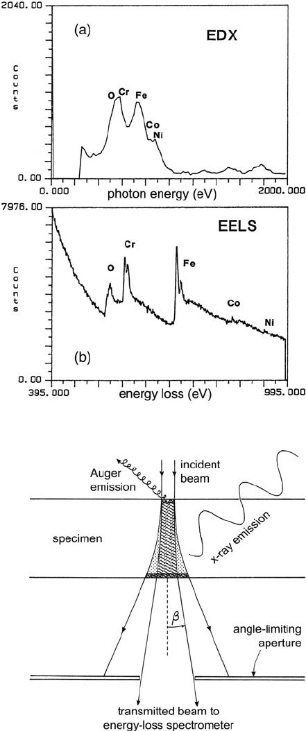
Fig. 1.11 (a) X-ray emission
spectrum recorded from an
oxidized region of stainless
steel, showing overlap of the
oxygen K-peak with the
L-peaks of chromium and
iron. (b) Ionization edges are
more clearly resolved in the
energy-loss spectrum, as a
result of the better energy
resolution of the electron
spectrometer (Zaluzec et al.,
1984). From Zaluzec et al.
(1984), copyright San
Francisco Press, Inc., with
permission
Fig. 1.12 Spreading of an
electron beam within a thin
specimen. X-rays are emitted
from the dotted region,
whereas the energy-loss
spectrum is recorded from the
hatched region,the
spectrometer entrance
aperture having a collimating
effect. Auger electrons are
emitted within a small depth
adjacent to each surface

22 1 An Introduction to EELS
characteristic x-rays, particularly from light elements. For energy losses between
1 and 10 keV, the fast secondaries are emitted almost perpendicular to the incident
beam direction and have a range of the order of 10–100 nm (Joy et al., 1982). An
x-ray signal is also generated at some distance from the incident beam by backscat-
tered electrons, by secondary fluorescence (Bentley et al., 1984), and by any stray
electrons or ions within the TEM column.
1.5 Comparison of EELS and EDX Spectroscopy
Originally, EDX detectors were protected from water vapor and hydrocarbons in the
microscope vacuum by a 10-μm-thick beryllium window, which strongly absorbed
photons of energy less than 1000 eV and precluded analysis of elements of atomic
number less than 11. The subsequent deployment of ultrathin (UTW) and atmo-
spheric pressure (ATW) windows allowed elements down to boron to be detected
routinely. Recently, windowless in-column silicon drift detectors have been devel-
oped (Schlossmacher et al., 2010) and offer a total solid angle as high as 0.9 sr,
allowing 7% of the emitted x-rays to be analyzed. Wavelength-dispersive spectrom-
eters with parallel-recording detectors are also available for the TEM and can detect
elements down to Li (Terauchi et al., 2010a) with an energy resolution of typically
1 eV but relatively low solid angle (Terauchi et al., 2010b). These developments
make EDX spectroscopy competitive with EELS for the detection of light elements
in a TEM specimen. Table 1.2 lists some of the factors relevant to a comparison of
these two techniques.
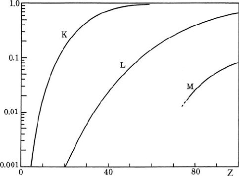
1.5.1 Detection Limits and Spatial Resolution
For the same incident beam current, the count rate of characteristic x-rays is less
than that of core-loss electrons (detectable by EELS) for two reasons. First, while
the K-line fluorescence yield is above 50% for Z > 32, it falls below 2% for Z <11
(see Fig. 1.13), reducing the generation rate for light elements. Second, character-
istic x-rays are emitted isotropically and the fraction recorded by an EDX detector
is below 10% (only 1% for a 0.13-sr detector), whereas the energy-loss electrons
are concentrated into a limited angular range, allowing a spectrometer collection
efficiency of typically 20–50%.
Table 1.2 Comparison of EELS with windowless EDX spectroscopy of a TEM specimen
Advantages of EELS Disadvantages of EELS
Higher core-loss signal Higher spectral background
Higher ultimate spatial resolution Very thin specimen needed
Absolute, standardless quantification Possible inaccuracy in crystals
Structural information available More operator intensive
1.5 Comparison of EELS and EDX Spectroscopy 23
Fig. 1.13 X-ray fluorescence yield for K-, L-, and M-shells, as a function of atomic number, from
Krause (1979)
Unfortunately, the EELS background, arising from inelastic scattering by all
atomic electrons whose binding energy is less than the edge energy, is generally
higher than the EDX background, which arises from stray radiation in the TEM
column and bremsstrahlung production. In addition, the characteristic features in
an energy-loss spectrum are not peaks but edges; the core-loss intensity is spread
over an extended energy range beyond the edge, making it less visible than the cor-
responding peak in the x-ray spectrum. It is possible to define a signal/noise ratio
(SNR) that takes account of the edge shape, and the minimum detectable concen-
tration of an element can be shown to depend on SNR, not signal/background ratio
(Section 5.6.3). On this basis, Leapman and Hunt (1991) compared the sensitivity
of EELS and EDX spectroscopy and showed EELS capable of detecting smaller
concentrations of elements of low atomic number; see Section 5.5.4. Using a field-
emission STEM and parallel-recording EELS, Leapman and Newbury (1993) could
detect concentrations down to 10 ppm for transition metals and lanthanides in pow-
dered glass samples. Shuman et al. (1984) reported a sensitivity of 20 ppm for Ca
in organic test specimens. I n some specimens, the detection limit is determined by
radiation damage and in this regard EELS is generally preferable to EDX spec-
troscopy because a larger fraction of the inner-shell excitations can be recorded by
the spectrometer.
EELS also offers slightly better spatial resolution than x-ray emission spec-
troscopy because the volume of specimen giving rise to the energy-loss signal can be
limited by means of an angle-limiting aperture, as shown in Fig. 1.12. The effects of
beam broadening (due to elastic scattering) and beam tails (if spherical aberration
of the probe-forming lens is not corrected) should therefore be less, as confirmed
experimentally (Collett et al., 1984; Titchmarsh, 1989; Genç et al., 2009). The
energy-loss signal is also unaffected by absorption, secondary fluorescence, and
24 1 An Introduction to EELS
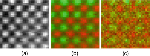
Fig. 1.14 (a) HAADF-STEM image of a [100]-projected GaAs specimen, showing bright Ga and
As atomic columns. (b) EELS image showing the Ga-L
23
intensity in green and As-L
23
intensity
in red.(c) EDX spectroscopy image with Ga-Kα intensity in green and As-Kα intensity in red.
Courtesy of M. Watanabe
the generation of fast secondary electrons within the specimen, making quantifi-
cation potentially more straightforward. With an aberration-corrected STEM and
efficient detectors, elemental maps showing individual atomic columns in a crystal
are now feasible, using either energy loss or EDX spectroscopy (Watanabe et al.,
2010a, b). As seen in Fig. 1.14, atomic columns are visible in the EDX image, but
the EELS map shows superior signal/noise ratio because of the larger number of
core excitations recorded.
Spatial resolution is a major factor determining the minimum detectable mass of
an element. Krivanek et al. (1991a, b) used a 1-nm probe with about 1 nA current to
identify clusters of one or two thorium atoms (on a thin carbon film) from the O
45
edge. More recently, Varela et al. (2004) reported the detection of single La atoms
inside a thin specimen of CaTiO
3
, using EELS and an aberration-corrected STEM.
The only alternative technique capable of single-atom identification is the field ion
atom probe (Section 1.4.1), whose applications have been limited by problems of
specimen preparation. However, focused ion beam (FIB) techniques ease the prepa-
ration of sharp needle-shaped tips, and the development of the local electrode atom
probe (LEAP) makes this technique a powerful competitor to EELS.
1.5.2 Specimen Requirements
If the specimen is too thick, plural scattering greatly increases the background to
ionization edges below 1000 eV, making these edges invisible for specimens thicker
than 100 or even 50 nm. This requirement places stringent demands on specimen
preparation, which can sometimes be met by ion milling (of inorganic materials) or
ultramicrotome preparation of ultrathin sections; see Section 1.6. The situation is
eased somewhat by the use of higher accelerating voltage, although in many materi-
als this introduces knock-on radiation damage by bulk- and surface-displacement
processes. EDX spectroscopy can tolerate thicker specimens (up to a few hun-
dred nanometers), although absorption corrections for light element quantification
become severe in specimens thicker than 100 nm.
1.5 Comparison of EELS and EDX Spectroscopy 25
1.5.3 Accuracy of Quantification
In EELS, the signal-intensity ratios depend only on the physics of the primary
excitation and are largely independent of the spectrometer. Quantification need not
involve the use of standards; measured core-loss intensities can be converted to ele-
mental ratios using cross sections that are calculated for the collection angle, range
of energy loss, and incident electron energy employed in the analysis. These cross
sections are known to within 5% for most K-edges and 15% for most L-edges, the
accuracy for other edges being highly variable (Egerton, 1993). EELS analysis of
45-nm NiO films distributed to four laboratories yielded elemental ratios within 10%
of stoichiometry (Bennett and Egerton, 1995); analysis of small areas of less-ideal
specimens would give more variable results.
In contrast, the relative intensities of EDX peaks depend on the properties of
the detector. For thin specimens, this problem is addressed within the k-factor and
ζ-factor methods, but because detector parameters are not precisely known, these
k-factors cannot be calculated with high accuracy. For the same reason, k-factors
measured in other instruments serve only as a rough guide. To achieve an accuracy
of better than 15%, the appropriate k-factors have usually been measured for each
analyzed element, using test specimens of known composition and the same x-ray
detector and microscope, operating at the same accelerating voltage (Williams and
Carter, 2009).
In the case of low-energy x-rays (e.g., K-peaks from light elements) the k-factors
are dependent on x-ray absorption in the protective window and front end of the
detector, and within the specimen itself. While it is possible to correct for s uch
absorption, the accuracy of the correction is dependent on the specimen geometry,
making the accuracy of light element EDX analysis generally worse than for heavier
elements.
Sometimes an overlap of peaks can prevent meaningful EDX analysis, as in the
case of light element quantification using K-peaks when there are heavier elements
whose L-peaks occur within 100 eV; see Fig. 1.11a. In the energy-loss spectrum,
these edges overlap but are more easily distinguished because of the better energy
resolution, close to 1 eV rather than 100 eV. Problems of background subtraction
(e.g., at the Cr edge in Fig. 1.11b) can often be overcome by fitting the energy-loss
spectrum to reference standards.
1.5.4 Ease of Use and Information Content
Changing from TEM or STEM imaging for recording an EDX spectrum typically
involves positioning the incident beam, ensuring that probe current is not excessive
(to avoid detector saturation), withdrawing any objective aperture, and inserting the
EDX detector. Once set up, the detector and electronics require little maintenance,
especially for detectors that do not require liquid-nitrogen cooling. EDX software
has been developed to the point where elemental ratios are predicted in a routine
fashion. Problems of peak overlap, absorption, and fluorescence can be important,
26 1 An Introduction to EELS
but not always. For these reasons, EDX spectroscopy remains the technique of
choice for most TEM/STEM elemental analysis.
Obtaining an energy-loss spectrum involves adjusting the spectrometer excitation
(positioning the zero-loss peak), choosing an energy dispersion and collection angle,
and verifying that the specimen is suitably thin. For some measurements, low spec-
trum drift is important and this condition may involve waiting for the microscope
high voltage and spectrometer power supplies to stabilize. Although improvements
in software have made spectral analysis more convenient, the success of basic opera-
tions such as the subtraction of instrumental and pre-edge backgrounds still depends
on the skill of the operator and some understanding of the physics involved.
To summarize, EELS is a more demanding technique than EDX spectroscopy
in terms of the equipment, expertise, and knowledge required. In return for this
investment, energy-loss spectroscopy offers greater elemental sensitivity for certain
specimens and the possibility of additional information, including an estimate of the
local thickness of a TEM specimen and information about its crystallographic and
electronic structure. In fact, EELS provides data similar to x-ray, ultraviolet, visible,
and (potentially) infrared spectroscopy, all carried out in the same instrument and
with the possibility of atomic-scale spatial resolution (Brown, 1997). Obtaining this
information is the subject of the remainder of this book; practical applications and
limitations are discussed in Chapter 5.
1.6 Further Reading
The following chapters assume some familiarity with the operation of a transmis-
sion electron microscope, a topic covered in many books, of which the 800-page
Williams and Carter (2009) is the most readable and comprehensive. Reimer and
Kohl (2008) give a thorough account of the physics and electron optics involved;
Egerton (2005) treats those topics at an introductory level. Hirsch et al. (1977)
remains a useful guide to diffraction-contrast imaging, while the text by De Graef
(2003) provides a more modern account of electron diffraction and crystallography.
Phase-contrast imaging is dealt with by Spence (2009) and by Buseck et al. (1988);
reflection imaging and diffraction are reviewed by Wang (1993, 1996).
The correction of electron lens aberrations has increased the performance of elec-
tron microscopes and some of the implications are described in Hawkes (2008).
A modern discussion of STEM techniques and achievements is contained in the
book edited by Nellist and Pennycook (2011). Progress in time-resolved (fem-
tosecond) TEM and EELS is well illustrated in Zewail and Thomas (2010).
Spence (2005) provides a lucid introduction to diffractive (lensless) imaging and
Chapman ( 2009) describe femtosecond diffractive imaging with a free-electron
laser.
Analytical electron microscopy is treated in volume 4 of Williams and Carter
(2009) and in several multi-author volumes: Hren et al. (1979), Joy et al. (1986),
and Lyman et al. (1990). A detailed review of alternative analytical techniques,
based on lectures from the 40th Scottish Universities Summer School in Physics
1.6 Further Reading 27
was published in book form (Fitzgerald et al., 1992) but the two-volume Science of
Microscopy edited by Hawkes and Spence (2008) is more recent and comprehensive.
TEM-EELS is outlined in the microscopy handbook of Brydson (2001). Recent
review articles dealing with basic principles of EELS include Colliex (2004), Spence
(2006), Egerton (2009), and Garcia de Abajo (2010), the latter from a more theo-
retical point of view. Materials science applications are described in some detail in
Disko et al. (1992); this multi-author book was revised and edited by Ahn (2004),
and now contains a digital CD version of the EELS Atlas (Ahn and Krivanek, 1983),
giving low-loss and core-loss spectra of many solid elements and some compounds.
Early progress in EELS is reviewed by Colliex (1984) and Marton et al. (1955).
Other review articles of historical and general interest include Silcox (1979), Joy
(1979), Joy and Maher (1980c), Isaacson (1981), Leapman (1984), Zaluzec (1988),
Egerton (1992b), and Colliex et al. (1976a).
The physics and spectroscopy of outer-shell excitation is well covered by Raether
(1965), Daniels et al. (1970), and Raether (1980). Basic theory of inelastic scattering
is given in Schattschneider (1986); magnetic (linear and chiral-dichroic) measure-
ments are described in Schattschneider (2011). The benefits of energy filtering in
TEM, as well as an account of the instrumentation and physics involved, are fully
covered in the multi-author volume edited by Reimer (1995). The effect of inelastic
scattering on TEM images and diffraction patterns is treated in depth by Spence and
Zuo (1992) and by Wang (1995).
EELS has been the subject of several workshops, which are represented by col-
lected papers i n Ultramicroscopy: vol. 110, no. 8 (EDGE 2009, Banff); vol. 106,
nos. 11 and 12 (EDGE 2005, Grundlsee), vol. 96, nos. 3 and 4 (SALSA 2002,
Guadeloupe), vol. 59, July 1995 (EELSI 1994, Leukerbad), and vol. 28, nos. 1–4
(Aussois); also in Microscopy, Microanalysis, Microstructures: vol. 6, no. 1, Feb.
1995 (Leukerbad) and vol. 2, nos. 2/3, April/June 1991 (Lake Tahoe). Workshops on
electron spectroscopic imaging (ESI) are documented in Ultramicroscopy (vol. 32,
no. 1, 1990: Tübingen), and Journal of Microscopy (April 1991: Dortmund; vol. 6,
Pt. 3, 1992: Munich).
Several web sites contain information useful to researchers using EELS,
including
http://www.TEM-EELS.ca/ (includes teaching material and the software
described in Appendix B)
http://www.felmi-zfe.tugraz.at/ (includes a collection of Digital Micrograph
scripts)
http://www.gatan.com/software/ (Digital Micrograph scripting resources)
http://www.public.asu.edu/~perkes/DMSUG.html (Digital Micrograph Scrip-
ting Users Group)
http://pc-web.cemes.fr/eelsdb/ (a database of energy-loss spectra),
http://people.ccmr.cornell.edu/~davidm/WEELS/ (includes spectral data).
http://unicorn.mcmaster.ca/corex/cedb-title.html (a database of core-loss spec-
tra recorded from gases by Hitchcock and colleagues at McMaster
University)