Givan A.L. Flow Cytometry. First Principles
Подождите немного. Документ загружается.

This increase is thought to be one of the early steps involved in
so-called signal transduction and can result in the activation of en-
zyme systems responsible for subsequent metabolic or developmental
changes. Lymphocytes show increases in intracellular calcium in
response to many kinds of speci®c and nonspeci®c surface ligand
binding, some of which lead to the cellular changes that we associate
with an immune response. Many other classes of cells also show cal-
cium changes in response to stimulation.
A range of dyes developed by Roger Tsien in California has been
useful in ¯ow cytometry because they can be loaded into living cells
where they will chelate calcium in a reversible equilibrium and ¯uo-
resce in proportion to their calcium load. A dye called ¯uo-3 absorbs
light from the 488 nm line of the argon laser and ¯uoresces little
in the absence of calcium but signi®cantly (at 530 nm) when binding
the ion. However, the use of ¯uo-3 is di½cult to standardize because
the amount of ¯uorescence varies with the amount of dye loaded
into the cells. A more useful dye is the related indo-1, which, al-
though it requires a light source with ultraviolet output for excitation
(a high-power argon laser or a mercury arc lamp, for example), per-
mits so-called ratiometric analysis of calcium. The term ratio in this
context simply means that indo-1 ¯uoresces at 485 nm (turquoise)
when free of calcium but at 405 nm (violet) in the chelated state. By
using the ratio between violet and turquoise ¯uorescence, we can get
a measure of the amount of calcium in a cell that is independent of
the amount of dye loaded. Some ¯ow cytometers can calculate the
ratio between any two of their parameters. For others, this calcula-
tion has to be done through post-acquisition software.
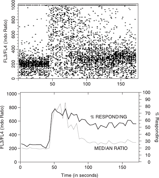
Figure 11.4 shows a time course of this ratio (related to internal
calcium) as it changes in response to the stimulation of lymphocytes
by a ligand bound to their surface receptors. Such a time course can
be performed on a ¯ow cytometer by loading cells with indo-1 in ad-
vance and then stimulating them with a ligand (in a calcium-containing
medium) immediately before placing the sample tube on the sample
manifold. Alternatively, the sample manifold of many research cy-
tometers can be modi®ed to permit rapid injection of reagents into the
sample tube while the sample is being drawn through the system. In
either case, measured cell parameters can then be acquired over the
subsequent minutes and their 405:485 ¯uorescence ratio determined.
Flow Cytometry200
Of course, to talk about kinetic measurements, we need to bring
in the parameter of time. Time is, in many ways, the hidden extra
parameter of every ¯ow system. Depending on the software being
used, it will be more or less easy to access this time parameter for use
in a kinetic pro®le. In the best case, each data ®le can have time as
an extra parameter for each cell. We would be able to plot any other
parameter(s) like turquoise and/or violet ¯uorescence against time
Fig. 11.4. The time course of a calcium response (measured by the indo-1 405/485
¯uorescence ratio) induced in lymphocytes by the addition of a stimulus. The upper
plot is a two-dimensional dot plot of the calcium ratio versus time. The lower plot
shows data binned in 5 s intervals and processed to give both the median ratio and
the percentage of cells above the base line as they change with time.
Research Frontiers 201
for each cell acquired into the computer memory, thus giving a time
course of the change of that parameter during the course of the
sample acquisition. It is also possible to use software to add a time
parameter to a data ®le. Knowing, from the ®le header information,
the start and ®nish time of the ®le, the software assumes constant cell
¯ow rate and assigns a time to the passage of each cell through the
laser beam.
This calcium ion procedure is an example of a class of protocols
that measure the kinetics of functional activity in living cells. They
depend on the ability to ®nd a compound able to enter living cells
and then alter its ¯uorescence in relation to a changing intracellular
environment. As we have seen, compounds are available that can
be used to assay calcium ion concentration. Other compounds will
alter their ¯uorescent properties in response to changes in pH or to
changes in membrane potential. Reduced ¯uorochromes such as di-
chloro¯uorescin diacetate, which ¯uoresce only when oxidized, have
been used to measure the production of peroxides during neutrophil
activation. Almost everyone starts out in ¯ow cytometry with work
on static systems. A shift of direction into the realm of function and
kinetic analysis requires a broadening of one's entrenched ideas of
what ¯ow cytometry is all about. The shift is almost certain to prove
both challenging and informative.
THE AQUATIC ENVIRONMENT
Blood has been an ideal object for ¯ow cytometric attention in large
part because it occurs naturally as a mixed population of single cells.
Lakes and oceans are also suspensions of mixed types of single cells.
As such, they would appear to present an obvious target for ¯ow
analysis. Indeed, ¯ow cytometers are now in place in many marine
laboratories and on board sea-going vessels.
Aquatic single cell organisms with a size range of about 0.02±
200 mm in diameter occur in nature in concentrations that range from
about 10
2
to 10
7
per ml. They include viruses, bacteria, cyanobacteria
(formerly known as blue-green algae), autotrophic phytoplankton
(unicellular plants), and heterotrophic zooplankton (unicellular ani-
mals). The analysis of aquatic organisms by ¯ow cytometry presents
Flow Cytometry202
some characteristic features that may serve to highlight the issues that,
to a greater or lesser extent, a¨ect all ¯ow analyses. In the ®rst place,
because of the presence of naturally occurring photosynthetic pig-
ments, the phytoplankton are highly auto¯uorescent (recall that some
of them contain phycoerythrin, peridinin-chlorophyll complexes, or
allophycocyanin). This auto¯uorescence leads to high background
intensity against which positive staining of low intensity may be di½-
cult to detect. The auto¯uorescence is also variable and may depend
on the environment or metabolic state of the cell. The auto¯uores-
cence can, however, be exploited and used to distinguish di¨erent
classes of organisms and di¨erent metabolic states.
Another characteristic of the aquatic environment is that the
abundance of organisms of di¨erent types is highly variable; aquatic
scientists do not have the benchmarks of a fairly tight ``normal
range'' that clinical scientists depend on. In addition, the abundance
of very small cells in the aquatic environment presents a challenge in
instrument tuning and sensitivity. Not all cytometers can distinguish
the forward scatter signal of nano- or picoplankton from optical
noise or from particulate matter in the sheath stream. Because of the
great size heterogeneity of plankton, a cytometer for aquatic analysis
must be able to cope with both small and large particles at the same
time. A ®nal problem is that the most common particles in aquatic
samples are not living; they represent decaying organic matter, silica-
or calcium-containing empty cell walls, and suspended sediment, which
are all di½cult for a ¯ow cytometer to distinguish from living cells.
Despite these problems, ¯ow cytometry has had some noted suc-
cess in aquatic research, particularly in relation to studies on the
phytoplankton. Because all phytoplankton possess chlorophyll, but
only the cyanobacteria possess the phycobiliproteins, auto¯uorescence
``signatures'' from water samples, based on the chlorophyll (¯uores-
cence >630 nm), phycoerythrin (¯uorescence <590 nm), and forward
scatter of particles, have been used to characterize the changes that
occur in plankton at di¨erent depths or at di¨erent locations (Figs.
11.5 and 11.6).
Figure 11.7 shows an example of the way in which ¯ow cytometric
analysis can distinguish six di¨erent species of plankton in culture
and de®ne which of these species are favored by grazing marine
scallops as a source of food. Results such as these have been used to
Research Frontiers 203
suggest modi®cations in the menu supplied to scallops being farmed
in aquaculture tanks.
Flow cytometry has also led to the notable discovery, reported by
Sallie Chisholm in 1988, of the existence of a novel group of small,
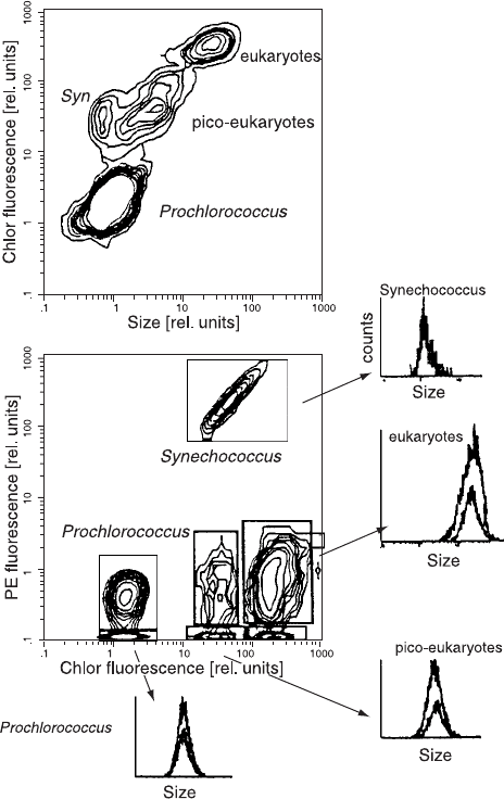
Fig. 11.5. The ¯ow cytometric signature of a seawater sample taken at 90 m depth
in the North Atlantic. Chlorophyll auto¯uorescence (>650 nm) has been plotted
against side scatter (``size'') and against phycoerythrin auto¯uorescence (530±590
nm). From Veldhuis and Kraay (2000).
Flow Cytometry204
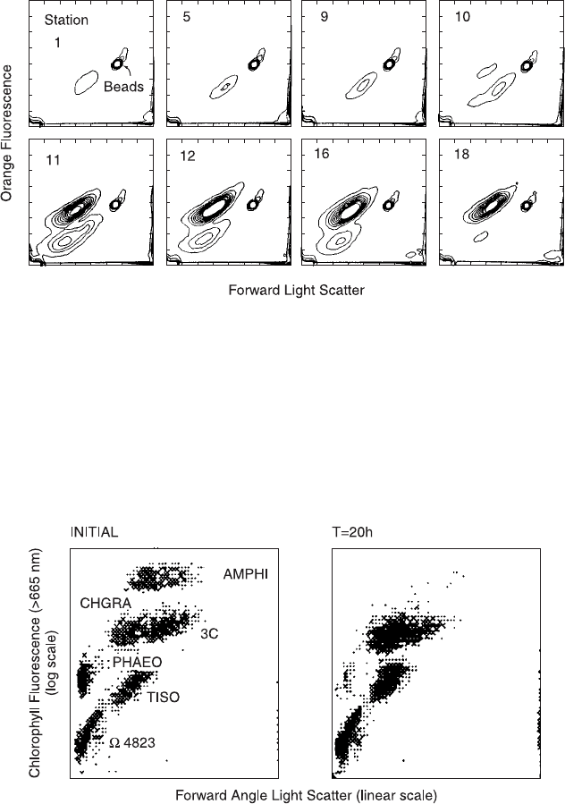
Fig. 11.6. Flow cytometric analysis of surface water from points at 1.5 mile intervals
o¨ shore from Cape Hatteras, North Carolina. Forward scatter and orange auto-
¯uorescence identify two Synechococcus populations with di¨erent phycoerythrin
content. Beads were used to calibrate the number of cells present. From Chisholm et
al. (1986).
Fig. 11.7. Small scallops were placed in tanks with six species of phytoplankton that
show distinctive ¯ow cytometric signatures. After 20 h of grazing, it was apparent
from the di¨erences between the ¯ow dot plots that the scallops had exhibited de®-
nite preference for two of the six species. Courtesy of Sandra Shumway.
Research Frontiers 205
prokaryotic phytoplankton; these free-living, marine prochlorophytes
(Prochlorococcus) are between 0.6 and 0.8 mm in size but possess
pigments more like those of eukaryotic plants than of other prokar-
yotes. The use of shipboard ¯ow cytometry during cruises o¨ south-
ern California, the Panama Basin, the Gulf of Mexico, the Carib-
bean, and the North Atlantic between Woods Hole, Massachusetts
and Dakar, Senegal (who said ¯ow cytometry isn't fun?) has found
these previously unknown prochlorophytes in remarkable abundance
and indicated that they may be responsible for a signi®cant portion
of the global photosynthetic productivity of the deep ocean. More
recently, Claude Courties (1994) has used ¯ow cytometry on samples
from the Thau lagoon in the Mediterranean o¨ the coast of France to
detect very small, eukaryotic picoplankton (about 1 mm) that have
been called ``the smallest eukaryotic organisms.''
The CytoBuoy project from the European Community Marine
Science and Technology Programme has been developing prototypes
for a small ¯ow cytometer (a 38 48 cm cylinder) that ®ts in ocean
buoys. Current implementation of the CytoBuoy includes a cyto-
meter inside a buoy, sampling just below the water surface. For-
ward scatter, side scatter, and orange ¯uorescence signals from par-
ticles are collected. It is projected that the CytoBuoy could take
samples in the ocean at depths up to 500 m. Data transmission for
continuous monitoring of ocean plankton could occur via short wave
over a distance of up to 50 km. At a rate of about 20 bytes per
second, this would give a sampling frequency of about one sample per
hour.
The problems presented by the heterogeneity of the aquatic envi-
ronment and the instrumental and conceptual developments made by
aquatic scientists toward handling these problems have led to ad-
vances that can enrich the work done in all ®elds of ¯ow analysis.
Cytometers that can deal with the instability of the ocean environ-
ment will be all the more dependable in a relatively stationary land-
based laboratory. Cytometers that are developed to handle both very
small and very large particles may allow ¯ow analysis to move more
decisively into the ®elds of microbiology, parasitology, mycology,
and botany. The general idea of studying auto¯uorescence instead of
trying to avoid or ignore it is one that may be pro®tably considered
by cytometrists in many areas of endeavor.
Flow Cytometry206
REPORTER MOLECULES
The Herzenbergs' group at Stanford has developed a ¯ow cytometric
method for assaying the presence of the enzyme b-galactosidase
(coded by the lacZ gene from Escherichia coli). The presence of this
enzyme can be detected by use of a so-called ¯uorogenic substrateÐ
in this case ¯uorescein digalactopyranoside (FDG), which is cleaved
by b-galactosidase to ¯uorescein monogalactoside and then to ¯uo-
rescein, which is ¯uorescent. The importance of assaying for the pres-
ence of b-galactosidase transcends any interest in regulation of expres-
sion of this enzyme in bacterial cells: The lacZ gene has been used
extensively in molecular biology as a reporter for the presence and/or
expression of recombinant genes in eukaryotic cells. Cloned genes can
be inserted, along with the lacZ bacterial gene, into eukaryotic cells;
if they are all under the control of the same promoter, expression of
the lacZ gene will then become a marker for the expression of the
cloned genes.
A creative use of this technique was developed by Nolan and
Krasnow at Stanford in a system they called whole animal cell sorting
(WACS). The system does not involve sorting of intact animals
(sheep to the left, goats to the right) but rather sorting of all the cells
from a whole animal, after they have been dissociated. Speci®cally,
the system has been used to study development of the fruit ¯y Dro-
sophila. Identi®able cell types in developing Drosophila embryos have
speci®c promoter regions in their genome that become activated in
the course of development to initiate the formation of gene products
typical of each cell type. Embryos can be transfected with lacZ into
chromosome positions driven by a cell-type-speci®c promoter. These
embryos (containing the introduced lacZ gene under the control of
a speci®c promoter) are then grown to a given developmental stage.
The cells expressing the reporter gene will contain b-galactosidase.
Depending on the promoter gene governing the lacZ gene, di¨erent
types of cells will therefore ¯uoresce when loaded with FDG. The
distribution of these ¯uorescent cells (and therefore the activity of the
speci®c promoter) can be visualized by looking at the intact embryo
(Fig. 11.8). The embryos can also, however, be dissociated and the
cells expressing the reporter gene then sorted by ¯ow cytometry,
based on ¯uorescein ¯uorescence intensity (Fig. 11.9). In this way,
Research Frontiers 207
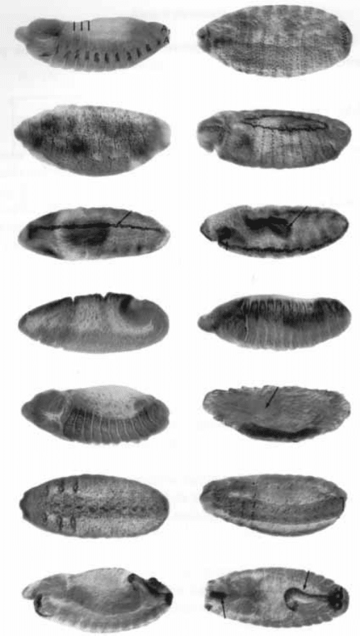
Fig. 11.8. Drosophila embryos transfected with the lacZ gene into association with
di¨erent cell-type-speci®c promoters. Depending on the promoter gene, cells in dif-
ferent patterns over the embryo surface will possess the enzyme b-galactosidase (in-
dicated by dark grains in this photograph). Courtesy of YN Jan from Bier et al.
(1989).
Flow Cytometry208
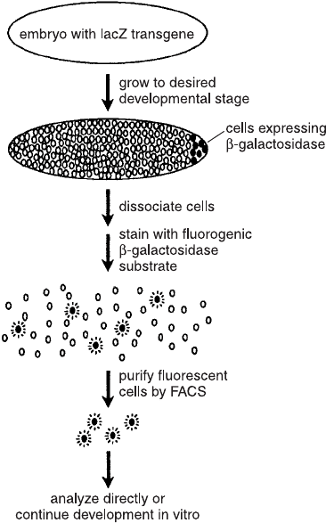
cells destined for di¨erent functions can be puri®ed and their subse-
quent development and interactions with other cells observed in cul-
ture (Fig. 11.10).
Over the past several years, the jelly®sh Aequorea victoria has had
remarkable impact on the ®eld of reporter molecules in biology.
When calcium ions bind to one of its proteins (aequorin), light is
emitted. In vitro, aequorin emits blue light when binding calcium.
The jelly®sh, however, produces green light because a second protein
(the imaginatively named ``green ¯uorescent protein'' or GFP) re-
ceives energy from aequorin and then emits ¯uorescence at a longer
wavelength (remember those tandem ¯uorochromes). In a landmark
paper by Chal®e et al. (1994), GFP was shown to remain ¯uorescent
when transfected into the bacterium Escherichia coli (under the con-
Fig. 11.9. The experimental protocol for whole animal cell sorting (WACS). Cour-
tesy of Mark Krasnow.
Research Frontiers 209