Lopes H.S., Cruz L.M. (eds.) Computational Biology and Applied Bioinformatics
Подождите немного. Документ загружается.


MicroArray Technology - Expression Profiling of MRNA and MicroRNA in Breast Cancer
97
experimentalists and bioinformaticians has contributed significantly to overcoming this
challenge (Ashburner et al, 2000). The GO Consortium was established with the aim of
producing a structured, precisely defined, common, controlled vocabulary for describing
the roles of genes and gene products in any organism. Initially a collaboration between three
organism databases: Flybase (The Flybase Consortium, 1999), Mouse Genome Informatics
(Blake et al, 2000) and the Saccharomyces Genome Database (Ball et al, 2000), the GO
Consortium has grown to include several of the world’s major repositories for plant, animal
and microbial genomes.
The Gene Ontology provides a structure that organizes genes into biologically related
groups according to three criteria. Genes and gene products are classified according to:
• Molecular Function: biochemical activity of gene products at the molecular level
• Biological Process: biological function of a gene product
• Cellular Component: location in the cell or extracellular environment where molecular
events occur
Every gene is described by a finite, uniform vocabulary. Each GO entry is defined by a
numeric ID in the format GO#######. These GO identifiers are fixed to the textual
definition of the term, which remains constant. A GO annotation is the specific association
between a GO identifier and a gene or protein and has a distinct evidence source that
supports the association. A gene product can take part in one or more biological process and
perform one or more molecular functions. Thus, a well characterized gene product can be
annotated to multiple GO terms in the three GO categories outlined above. GO terms are
related to each-other such that each term is placed in the context of all of the other terms in a
node-directed acyclic graph (DAC). The relationships used by the GO are: “is_a”, “part_of”,
“regulates”, “positively_regulates”, “negatively_regulates” and “disjoint_from”. Each term
in the DAC may have one or more parent terms and possibly one or more child nodes, and
the DAC gives a graphical representation of how GO terms relate to each other in a
hierarchical manner.
The development of Gene Ontology has facilitated analysis of microarray gene sets in the
context of the molecular functions and pathways in which they are involved (Blake &
Harris, 2002). GO-term analysis can be used to determine whether genetic “hits” show
enrichment for a particular group of biological processes, functions or cellular
compartments. One approach uses statistical analysis to determine whether a particular GO
is over or under-represented in the list of differentially expressed genes from a microarray
experiment. The statistical tests used for such analysis include hypergeometric, binomial or
Chi-square tests (Khatri et al, 2005).
An alternative approach known as “gene-set testing” has been described which involves
beginning with a known set of genes and testing whether this set as a whole is differentially
expressed in a microarray experiment (Lamb et al, 2003; Mootha et al, 2003). The results of
such analyses inform hypotheses regarding the biological significance of microarray
analyses.
Several tools have been developed to facilitate analysis of microarray data using GO, and a
list of these can be found at: http://www.geneontology.org/GO.tools.microarray.shtml
Analysing microarray datasets in combination with biological knowledge provided by GO
makes microarray data more accessible to the molecular biologist and can be a valuable
strategy for the selection of biomarkers and the determination of drug treatment effect in
breast cancer (Arciero et al, 2003; Cunliffe et al, 2003).

Computational Biology and Applied Bioinformatics
98
4.3 Microarray meta-analysis – combining datasets
Meta-analyses have confirmed that different prognostic signatures identify similar
biological subgroups of breast cancer patients (Fan et al, 2006) and have also shown that the
designation of tumours to a “good prognosis”/”low risk” group or a “poor
prognosis”/”high risk” group is largely dependent on the expression patterns of
proliferative genes. In fact, some of these signatures have been shown to have improved
performance when only the proliferative genes are used (Wirapati, 2008). Metanalyses of the
signatures have also proposed that the prognostic ability of the signatures is optimal in the
ER positive and HER2-negative subset of breast tumours (Desmedt, 2008; Wirapati, 2008),
the prognosis of this group of tumours being governed by proliferative activity.
Despite obvious clinical application, none of these prognostic assays are perfect, and they all
carry a false classification rate. The precise clinical value for these gene expression profiles
remains to be established by the MINDACT and TAILORx trials. In the interim the
performance of these assays is likely to be optimised by combining them with data from
traditional clinicopathological features, an approach which has been shown to increase
prognostic power (Sun et al, 2007).
Microarray technology has undoubtedly enhanced our understanding of the molecular
mechanisms underlying breast carcinogenesis; profiling studies have provided a myriad of
candidate genes that may be implicated in the cancer process and are potentially useful as
prognostic and predictive biomarkers or as therapeutic targets. However, as yet there is
little knowledge regarding the precise regulation of these genes and receptors, and further
molecular categories are likely to exist in addition to and within the molecular subtypes
already delineated. Accumulating data reveal the incredible and somewhat foreboding
complexity and variety of breast cancers and while mRNA expression profiling studies are
ongoing, a new player in breast cancer biology has come to the fore in recent years; a
recently discovered RNA species termed MiRNA (miRNA) which many scientists believe
may represent a crucial link in the cancer biology picture.
5. MicroRNA - a recently discovered layer of molecular complexity
It has been proposed that the discovery of miRNAs as regulators of gene expression represents
a paradigm changing event in biology and medicine. This discovery was made in 1993 by
researchers at the Ambros laboratory in Dartmouth Medical School, USA at which time it was
thought to be a biological entity specific to the nematode C. Elegans (Lee et al, 1993). In the
years following this discovery, hundreds of miRNAs were identified in animals and plants.
However it is only in the past 5 years that the field of miRNA research has really exploded
with the realisation that miRNAs are critical to the development of multicellular organisms
and the basic functions of cells (Bartel, 2004). MiRNAs are fundamental to genetic regulation,
and their aberrant expression and function have been linked to numerous diseases and
disorders (Bartel, 2004; Esquela-Kerscher & Slack, 2006). Importantly, miRNA have been
critically implicated in the pathogenesis of most human cancers, thus uncovering an entirely
new repertoire of molecular factors upstream of gene expression.
5.1 MicroRNA - novel cancer biomarkers
The first discovery of a link between miRNAs and malignancy was the identification of a
translocation-induced deletion at chromosome 13q14.3 in B-cell Chronic Lymphocytic

MicroArray Technology - Expression Profiling of MRNA and MicroRNA in Breast Cancer
99
Leukaemia (Calin et al, 2002). Loss of miR-15a and miR-16-1 from this locus results in
increased expression of the anti-apoptotic gene BCL2. Intensifying research in this field,
using a range of techniques including miRNA cloning, quantitative PCR, microarrays and
bead-based flow cytometric miRNA expression profiling has resulted in the identification
and confirmation of abnormal miRNA expression in a number of human malignancies
including breast cancer (Heneghan et al, 2010; Lowery et al, 2007). MiRNA expression has
been observed to be upregulated or downregulated in tumours compared with normal
tissue, supporting their dual role in carcinogenesis as either oncogenic miRNAs or tumour
suppressors respectively (Lu et al, 2005). The ability to profile miRNA expression in human
tumours has led to remarkable insight and knowledge regarding the developmental lineage
and differentiation states of tumours. It has been shown that distinct patterns of miRNA
expression are observed within a single developmental lineage, which reflect mechanisms of
transformation, and support the idea that miRNA expression patterns encode the
developmental history of human cancers. In contrast to mRNA profiles it is possible also to
successfully classify poorly differentiated tumours using miRNA expression profiles
(Volinia et al, 2006). In this manner, miRNA expression could potentially be used to
accurately diagnose poorly differentiated tissue samples of uncertain histological origin, e.g.
metastasis with an unknown primary tumour, thus facilitating treatment planning.
MicroRNAs exhibit unique, inherent characteristics which make them particularly attractive
for biomarker development. They are known to be dysregulated in cancer, with
pathognomonic or tissue specific expression profiles and even a modest number of miRNAs
is sufficient to classify human tumours, which is in contrast to the relatively large mRNA
signatures generated by microarray studies (Lu et al, 2005). Importantly, miRNA are
remarkably stable molecules. They undergo very little degradation even after processing
such as formalin fixation and remain largely intact in FFPE clinical tissues, lending
themselves well to the study of large archival cohorts with appropriate follow-up data (Li et
al, 2007; Xi et al, 2007). The exceptional stability of miRNAs in visceral tissue has stimulated
investigation into their possible preservation in the circulation and other bodily fluids
(urine, saliva etc.). The hypothesis is that circulating miRNAs, if detectable and quantifiable
would be the ideal biomarker accessible by minimally invasive approaches such as simple
phlebotomy (Cortez et al, 2009; Gilad et al, 2008; Mitchell et al, 2008).
5.2 MicroRNA microarray
The unique size and structure of miRNAs has necessitated the modification of existing
laboratory techniques, to facilitate their analysis. Due to the requirement for high quality
large RNA molecules, primarily for gene expression profiling, many laboratories adopted
column-based approaches to selectively isolate large RNA molecules, discarding small RNA
fractions which were believed to contain degradation products. Modifications to capture
miRNA have been made to existing protocols to facilitate analysis of the miRNA fraction.
Microarray technology has also been modified to facilitate miRNA expression profiling.
Labelling and probe design were initially problematic due to the small size of miRNA
molecules. Reduced specificity was also an issue due to the potential of pre-miRNA and pri-
miRNAs to produce signals in addition to active mature miRNA. Castoldi et al described a
novel miRNA microarray platform using locked nucleic acid (LNA)-modified capture
probes (Castoldi et al, 2006). LNA modification improved probe thermostability and
increased specificity, enabling miRNAs with single nucleotide differences to be

Computational Biology and Applied Bioinformatics
100
discriminated - an important consideration as sequence-related family members may be
involved in different physiological functions (Abbott et al, 2005). An alternative high
throughput miRNA profiling technique is the bead-based flow cytometric approach
developed by Lu et al.; individual polystyrene beads coupled to miRNA complementary
probes are marked with fluorescent tags (Lu et al, 2005). After hybridization with size-
fractioned RNAs and streptavidin-phycoerythrin staining, the beads are analysed using a
flow-cytometer to measure bead colour and pycoerythrin, denoting miRNA identity and
abundance respectively. This method offered high specificity for closely related miRNAs
because hybridization occurs in solution. The high-throughput capability of array-based
platforms make them an attractive option for miRNA studies compared to lower
throughput techniques such as northern blotting and cloning; which remain essential for the
validation of microarray data.
5.2.1 MicroRNA microarray - application to breast cancer
Microarray analysis of miRNA expression in breast cancer is in its’ infancy relative to
expression profiling of mRNA. However, there is increasing evidence to support the
potential for miRNAs as class predictors in breast cancer. The seminal report of aberrant
miRNA expression in breast cancer by Iorio et al. in 2005 identified 29 miRNAs that were
differentially expressed in breast cancer tissue compared to normal, a subset of which could
correctly discriminate between tumour and normal with 100% accuracy (Iorio et al, 2005).
Among the leading miRNAs differentially expressed; miR-10b, miR-125b and mR-145 were
downregulated whilst miR-21 and miR-155 were consistently over-expressed in breast
tumours. In addition, miRNA expression correlated with biopathological features such as
ER and PR expression (miR-30) and tumour stage (miR-213 and miR-203). Mattie et al.
subsequently identified unique sets of miRNAs associated with breast tumors defined by
their HER2/neu or ER/PR status (Mattie et al, 2006). We have described 3 miRNA
signatures predictive of ER, PR and Her2/neu receptor status, respectively, which were
identified by applying artificial neural network analysis to miRNA microarray expression
data (Lowery et al, 2009). Blenkiron et al used an integrated approach of both miRNA and
mRNA microarray expression profiling to classify tumours according to “intrinsic subtype”.
This approach identified a number of miRNAs that are differentially expressed according to
intrinsic breast cancer subtype and associated with clinicopathological factors including ER
status and tumour grade. Importantly, there was overlap between the differentially
expressed miRNAs identified in these studies.
There has been interest in assessing the prognostic value of miRNAs, and expression studies
in this regard have focused on detecting differences in miRNA expression between primary
breast tumours and metastatic lymph nodes. This approach has identified numerous
miRNA that are dysregulated in primary breast tumours compared to metastatic lymph
nodes (Baffa et al 2009; Huang et al, 2008). MiRNA have also been identified that are
differentially expressed in patients who had a “poor prognosis” or a short time to
development of distant metastasis (Foekens et al, 2008); miR-516-3p, miR-128a, miR-210, and
miR-7 were linked to aggressiveness of lymph node-negative, ER-positive human breast
cancer.
The potential predictive value of miRNA is also under investigation. Preclinical studies have
reported associations between miRNA expression and sensitivity to adjuvant breast cancer
therapy including chemotherapy, hormonal therapy and HER2/neu targeted therapy (Ma

MicroArray Technology - Expression Profiling of MRNA and MicroRNA in Breast Cancer
101
et al, 2010; Tessel et al, 2010; Wang et al, 2010), prompting analysis of tumour response in
clinical samples. Rodriguez-Gonzalez et al attempted to identify miRNAs related to
response to tamoxifen therapy by exploiting the Foekens dataset (Foekens, 2008) which
comprised miRNA expression levels of 249 miRNAs in 38 ER positive breast cancer patients.
Fifteen of these patients were hormone naive and experienced relapse, which was treated
with tamoxifen. Ten patients responded and five did not, progressing within 6 months. Five
miRNAs (miR-4221, miR-30a-3p, miR-187, miR-30c and miR-182) were the most
differentially expressed between patients who benefitted from tamoxifen and those who
failed therapy. The predictive value for these miRNAs was further assessed in 246 ER
positive primary tumours of hormone naive breast cancer patients who received tamoxifen
as monotherapy for metastatic disease. MiR-30a-3p, miR-30c and miR-182 were significantly
associated with response to tamoxifen, but only miR-30c remained an independent predictor
on multivariate analysis (Rodriguez-Gonzalez, 2010).
Microarray-based expression profiling has also been used to identify circulating miRNAs
which are differentially expressed in breast cancer patients and matched healthy controls.
Zhao et al profiled 1145 miRNAs in the plasma of 20 breast cancer patients and 20 controls,
identifying 26 miRNAs with at least two-fold differential expression which reasonably
separated the 20 cases from the 20 controls (Zhao et al, 2010). This is the first example of
genome-wide miRNA expression profiling in the circulation of breast cancer patients and
indicates potential for development of a signature of circulating miRNAs that may function
as a diagnostic biomarker of breast cancer.
At present diagnostic, prognostic and predictive miRNA signatures and markers remain
hypothesis generating. They require validation in larger, independent clinical cohorts prior
to any consideration for clinical application. Furthermore as additional short non-coding
RNAs are continuously identified through biomarker discovery programmes, the available
profiling technologies must adapt their platforms to incorporate newer potentially relevant
targets. MicroRNAs possess the additional attraction of potential for development as
therapeutic targets due to their ability to regulate gene expression. It is likely that future
microarray studies will adopt and integrated approach of miRNA and mRNA expression
analysis in an attempt to decipher regulatory pathways in addition to expression patterns.
6. Limitations of microarray technology & bioinformatic challenges
In addition to the great promises and opportunities held by microarray technologies, several
issues need to be borne in mind and appropriately addressed in order to perform reliable
and non-questionable experiments. As a result, several steps need to be addressed in order
to identify and validate reliable biomarkers in the scope of potential future clinical
application. This is one of the reasons why, despite the promises of using powerful high-
throughput technologies as such as microarray, only very few useful biomarkers have been
identified so far and/or have been translated to useful clinical assay or companion
diagnostics (Mammaprint
®
, Oncotype DX
®
). There still remains a lack of clinically relevant
biomarkers (Rifai et al, 2006). Amongst the limitations and pitfalls around the technology
and the use of microarrays, some of the most important are the reported lack of
reproducibility, as well as the massive amount of data generated, often extremely noisy and
with an increasing complexity. As for example, in the recent Affymetrix GeneChip 1.0 ST
microarray platform (designed to target all known and predicted exons in human, mouse
and rat genomes), where there is approximately 1.2 million exon clusters corresponding to

Computational Biology and Applied Bioinformatics
102
over 1.4 million probesets (Lancashire et al, 2009). As a result, it appears clearly that
extracting any relevant key component from such datasets requires robust mathematical
and/or statistical models running on efficient hardware to perform the appropriate
analyses.
With this in mind, it is clear that the identification of new biomarkers still requires a
concerted, multidisciplinary effort. It requires the expertise of the biologist or pathologist, to
extract the samples, the scientist to perform the analysis on the platform and then the
bioinformatician/biostatistician to analyse and interpret the output. The data-mining
required to cope with these types of data needs careful consideration and specific
computational tools, and as such remains a major challenge in bioinformatics.
6.1 Problems with the analysis of microarray data
6.1.1 Dimensionality and false discovery
The statistical analysis of mRNA or miRNA array data poses a number of challenges. This
type of data is of extremely high dimensionality i.e. has a large number of variables. Each of
these variables represents the relative expression of a mRNA or miRNA in a sample. Each of
these components contain noise, are non-linear may not follow a normal distribution
through a population and may be strongly correlated with other probes in the profile. These
characteristics mean that the data may violate many of the assumptions of conventional
statistical techniques, particularly with parametric tests.
The dimensionality of the data poses a significant problem, and remains as one of the most
critical when analysing microarray data. When one analyses this type of data, one has to
consider what is referred to as the curse of dimensionality, firstly described by Bellman in 1961
as the “exponential growth of the search space as a function of dimensionality” (Bellman, 1961;
Bishop, 1995). This occurs in highly dimensional systems where the number of dimensions
masks the true importance of an individual single dimension (variable). It is particularly
true in a microarray experiment when the number of probes representing the number of
miRNA/mRNA studied far exceeds the number of available samples. So there is the
potential for a probe that is in reality of high importance to be missed when considered with
a large number of other probes. This problem is overcome by breaking down the analysis
into single or small groups of variables and repeating the analysis rather than considering
the whole profile in one single analysis. Other methods consists of using pre-processing
methods and feature extraction algorithms in order to only analyse a subset of the data
supposed to hold the most relevant features (Bishop, 1995), as determined by the pre-
processing steps.
High dimensionality also creates problems due to false discovery. The false discovery rate
(FDR) introduced by Benjamini and Hochberg (Benjamini and Hochberg, 1995) is a measure
of the number of features incorrectly identified as “differential” and various approaches
have been suggested to accurately control the FDR. In this case if one has a high number of
dimensions and analyses each singly (as above) a proportion can appear to be of high
importance due to random chance considering the distribution, even when they are not. To
overcome this one has to examine a rank order of importance and when testing for
significance one has to correct the threshold for significance by dividing it by the number of
dimensions. So for example when analysing the significance of single probes from a profile
with 4,000 probes in it the threshold becomes P < 0.05 divided by 4,000 i.e. P < 0.0000125.

MicroArray Technology - Expression Profiling of MRNA and MicroRNA in Breast Cancer
103
6.1.2 Quality and noise
Noise also poses a problem in the analysis of mRNA or miRNA data. The inherent technical
and biological variability necessarily induces noise within the data, eventually leading to
biased results. The noise may lead to misinterpretation of sample groups that may actually
have no biological relevance. As a consequence extreme care needs to be taken to address
the problem of noise.
Noise may be random where it is applied to all parts of the miRNA equally or systematic
where particular probes inherently have more noise than others because of the nature of the
component miRNA or genomic code that they represent.
It is now widely acknowledged that the reported high level of noise found in microarray
data is the most critical pull-back of microarray-based studies, as it is pointed by the MAQC
Consortium (Shi et al, 2006; Klebanov and Yakovlev, 2007).
6.1.3 Complexity and non-normality
Because of the complex nature of the profile a particular mRNA or miRNA may be non-
normally distributed through a population. Such non-normality will immediately invalidate
any statistical test that uses parametric statistics i.e. depends on the assumption of a normal
distribution. Invalidated tests would include ANOVA and t-test. To overcome this, the data
would have to be transformed mathematically to follow a normal distribution or an
alternative non parametric test would have to be employed. Examples of non-parametric
tests include Kruskal-Wallis and Mann Whitney U which are ANOVA and unpaired T-Test
alternatives respectively. Generally non-parametric tests lack power compared to their
parametric alternatives and this may prove to be a problem in high dimensional space due
to the reasons described previously.
6.1.4 Reproducibility
Reproducibility has a marked effect on the accuracy of any analysis conducted. Furthermore
reproducibility has a profound effect on the impact of other issues such as dimensionality
and false detection. Robust scientific procedures requires that the results have to be
reproducible in order to reduce the within sample variability, the variability between
sample runs and the variability across multiple reading instruments. Aspects of variability
can be addressed using technical and experimental replicates. The averaging of samples
profiles can be used to increase the confidence in the profiles for comparison (Lancashire et
al., 2009). Technical replicates provide information on the variability associated with
instrumental variability whilst experimental (or biological) replicates give a measure of the
natural sample to sample variation. Problems in data analysis occur when the technical
variability is high. In this situation the problem in part can be resolved by increasing the
number of replicates. If however the technical variation is higher than the biological
variation then the sample cannot be analysed.
6.1.5 Auto-correlation or co-correlation
Auto correlation exists when two components within a system are strongly linearly
correlated with one another. In any complicated system there are likely to be a number of
components that are auto correlated. This is especially true in array profiling of biological
samples. Firstly due to biological processes one protein in a set of samples is likely to
interact or correlate with another through a population.

Computational Biology and Applied Bioinformatics
104
Auto correlation becomes a problem when using linear based regression approaches. This is
because one of the assumptions of regression using multiple components is that the
components are not auto correlated. If intensity for multiple miRNA probes are to be added
into a regression to develop a classifier these components should not be auto correlated.
Auto correlation can be tested for using the Durbin Watson test.
6.1.6 Generality
The whole purpose of biomarker (or set of biomarkers) identification, using high-
throughput technologies or any other, is to provide the clinicians with an accurate model in
order to assess a particular aspect. However, a model is only as good as its ability to
generalize to unseen real world data. A model only able to explain the population on which
it was developed would be purely useless for any application.
As a result, if one is to develop classifiers from mRNA or miRNA array data the features
identified should be generalised. That is they will predict for new cases in the general
population of cases. When analysing high dimensional data there is an increased risk of over
fitting, particularly when the analysis methods imply supervised training on a subset of the
population. So for example, when a large number of mRNA or miRNA are analysed there is
the potential for false detection to arise. If a random element identified through false
detection is included as a component of a classifier (model) then the generality of that
classifier will be reduce; i.e. it is not a feature that relates to the broader population but is a
feature specific to the primary set of data used to develop the classifier. Standards of
validation required to determine generality have been defined by Michiels et al, 2007.
Generality of classifiers can be increased by the application of bootstrapping or cross
validation approaches.
Some algorithms and approaches, that usually involve supervised training, suffer from
over-fitting (sometimes called memorisation). This is a process where a classifier is
developed for a primary dataset but models the noise within the data as well as the relevant
features. This means that the classifier will not accurately classify for new cases i.e. it does
not represent a general solution to the problem which is applicable to all cases. This is
analogous, for example, to one developing a classifier that predicts well the risk of
metastasis for breast cancer patients from Nottingham but will not predict well for a set of
cases from Denmark. Over fitted classifiers seldom represent the biology of the system being
investigated and the features identified are often falsely detected.
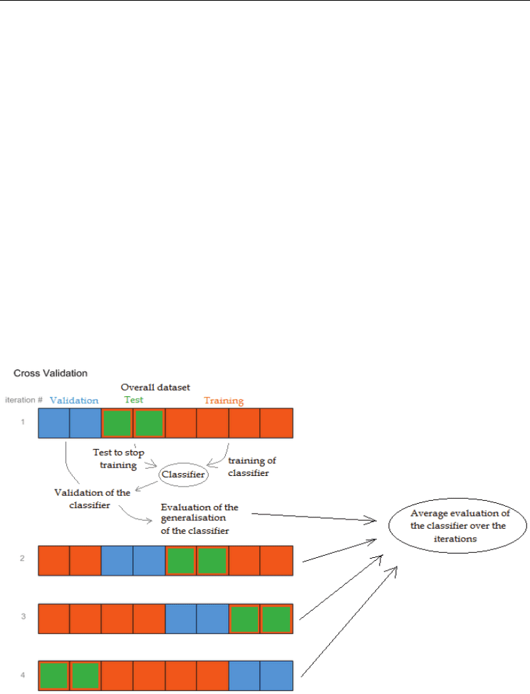
One of the most common solutions to avoid over-fitting is to apply a Cross Validation
technique in combination with the supervised training. Random sample cross validation is a
process of mixing data. Firstly the data are divided into two or three parts (figure 2); the first
part is used to develop the classifier and the second or second and third parts are used to
test the classifier. These parts are sometimes termed training, test and validation data sets
respectively. In certain classifiers such as Artificial Neural Network based classifiers the
second blind set is used for optimisation and to prevent over fitting. In random sample cross
validation the random selection and training process is repeated a number of times to create
a number of models each looking at the global dataset in a number of different ways (figure
2). Often the mean performance of these models is considered.
Leave one out cross validation is an approach also used to validate findings. In this case one
sample is left out of the analysis. Once training is complete the sample left out is tested. This
process is repeated a number of times to determine the ability of a classifier to predict
MicroArray Technology - Expression Profiling of MRNA and MicroRNA in Breast Cancer
105
unseen cases. This approach of random sample cross validation drives the classifier solution
to a generalised one by stopping the classifier from training too much on a seen dataset and
stopping the training earlier based on a blind dataset.
7. Methods used to analyse microarray data and their limitations
With the advent of cutting edge new technologies such as microarrays, the analysis tools for
the data produced need to be appropriately applied. Although expression arrays have
brought high hopes and expectations, they have brought tremendous challenges with them.
They have been proven to suffer from different limitations as previously discussed.
However, innovative computational analysis solutions have been developed and have been
proven efficient and successful at identifying markers of interest regarding particular
questions. This section presents some of the most common methods employed to overcome
the limitations discuss above, and to analyse expression array data.
7.1 Application of ordination techniques
If we are to utilise the mRNA or miRNA profile we have to identify robust features despite
its high dimensionality that are statistically valid for the general population not just for a
subset. Ordination techniques are used to map the variation in data. They are not directly
predictive and cannot classify directly unless combined with another classification
technique.
Fig. 2. Illustration of Cross Validation technique, here with three subsets: the training subset
used to train the classifier, the test subset used to stop the training when it has reached an
optimal performance on this subset, and a validation subset to evaluate the performance
(generalization ability) of the trained classifier.
Computational Biology and Applied Bioinformatics
106
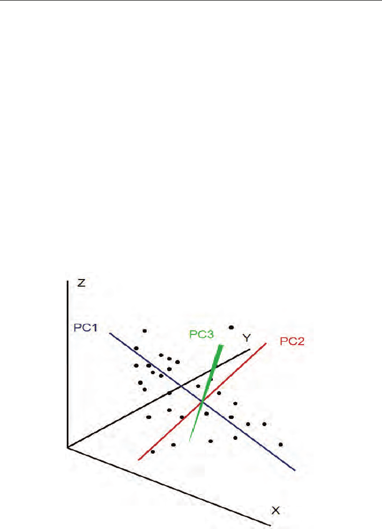
7.1.1 Principal components analysis
PCA is usually a method of choice for dimensionality reduction. It is a multivariate
exploratory technique used to simplify complex data space (Raychaudhuri et al, 2000) by
translating the data space into a new space defined by the principal components. It works
by identifying the main (principal) components that explain best the shape (variance) of a
data set. Each principal component is a vector (line) through the data set that explains a
proportion of the variance, it is the expression of a linear combination of the data. In PCA
the first component that is added is the one that explains the most variance the second
component added is then orthogonal to the first. Subsequent orthogonal components are
added until all of the variation is explained. The addition of vectors through a
multidimensional data set is difficult to visualise in print, we have tried to illustrate it with 3
dimensions in figure 3. In mRNA/miRNA profile data where thousands of dimensions
exist, PCA is a useful technique as it reduces the dimensionality to a manageable number of
principal components. If the majority of the variance is explained in 2 or 3 principal
components these can be used to visualise the structure of the population using 2 or 3
dimensional plots. A limited parameterisation can also be conducted to determine the
contribution of each parameter (miRNA) to each of the principal components. This however
suffers from the curse of dimensionality in high dimensional systems. Thus the main
limitation of using PCA for gene expression data is the inability to verify the association of a
principal component vector with the known experimental variables (Marengo et al, 2004).
This often makes it difficult to accurately identify the importance of the mRNA or miRNA in
the system, and make it a valuable tool only for data reduction.
Fig. 3. Example of a 3 dimension PCA with the 3 orthogonal PCs.