Lopes H.S., Cruz L.M. (eds.) Computational Biology and Applied Bioinformatics
Подождите немного. Документ загружается.

Functional Analysis of the Cervical Carcinoma
Transcriptome: Networks and New Genes Associated to Cancer
267
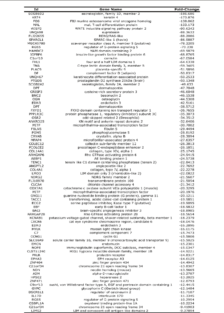
Table 2. List of genes down-regulated

Computational Biology and Applied Bioinformatics
268
4.1 Analyzing microarray data with novel software suite
If we want to display the data in just two dimensions, we want as much of the variation in
the data as possible captured in just two dimensions. Principal component analysis or PCA
has been developed for this purpose. Applying this PCA method in the cervical data we
observed some expected differences (Figure 3).
Fig. 3. Principal component analysis of cervical cancer samples. As expected, after perform
PCA analysis, the normal samples (red balls) were grouped out of CC Group (blue balls) in
a 3-D image.
In order to obtain a graphical representation of the differences between normal and tumor
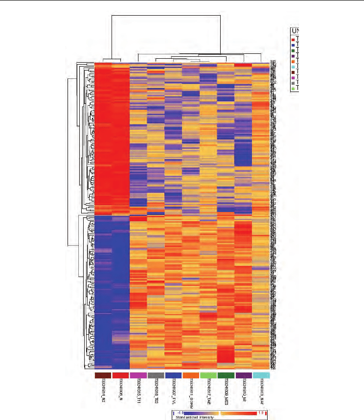
tissues, hierarchical cluster analysis was performed on all samples with a pseudo-color
visualization matrix of the 208 selected genes grouping with greater intra-group similarity
and differences between groups. The phylogenetic tree resulting from the hierarchical
complete linkage-clustering algorithm is shown in figure 4. The figure shows those genes
that are changing respect to health cervical tissue. In this method of clustering, allows to do
relationships among objects (genes or samples) and are represented by a tree whose branch
lengths reflect the degree of similarity between the objects, as assessed by a pairwise
similarity function. The computed trees can be used to arrange individual samples in the
original data table; this allows the samples or groups of samples with similar expression
patterns to be shown adjacent to each other.
In general, tumor samples showed heterogeneity among them compared with normal
samples, which had a more homogeneous gene expression profile. So, clustering analysis in
CC failed to show significant segregation of patients based on expression profiling possibly
Functional Analysis of the Cervical Carcinoma
Transcriptome: Networks and New Genes Associated to Cancer
269
Fig. 4. Hierarchical clustering of the gene expression data for cervical tissues. Clustering
analysis were performed for all tumours and normal samples. The data were clustered using
the standard hierarchical method with ward linkage and using the Perason correlation to
determine distance tumour. Before clustering, the data was filtered to remove genes that
were scored absent in 75% or more of the samples as they are likely to be measuring noise in
the system. The cluster of normal samples exhibit nearly identical patterns of gene
expression changes, on contrary, as expected the invasive samples grouped in a different
branch showing a heterogeneous gene expression. Blue color indentify downregulation and
red color an overexpression status.
Computational Biology and Applied Bioinformatics
270
due to the heterogeneous nature of the samples as well as the relatively small numbers of
samples in this study. Even when the samples were subjected to rigorous procedures of
analysis, the special selection of the patients, including age, clinical stage, HPV16 positive
and contraceptive oral status avoiding any bias, in this stage of the carcinogenesis process
(stage IIb) the pattern of gene expression is quite different between samples.
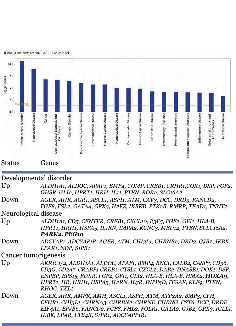
Table 3. Disease and Disorders functions in cervical cancer

Functional Analysis of the Cervical Carcinoma
Transcriptome: Networks and New Genes Associated to Cancer
271
We used IPA to investigate the biological relevant of the observed genome-wide expressed
gene changes by categorizing our data set into biological functions and/or diseases
Ingenuity Pathway analysis was applied. The 208 genes annotated list by PARTEK analysis
was submitted to the visualization IPA tool. This bioinformatics tool is employed for
visualizing expression data in the context of KEGG biological pathways; the importance IPA
is that retrieves an impact factor (IF) of genes that entire pathway involved, which can help
to obtain a clearer notion of the alteration level in each biological pathway, and understand
the complexity of these different process of the cancer cell. We imported a list of
significantly up and down regulated genes (with extension .txt) into the program to convert
the expression data into illustrations in an attempt to explore altered mechanisms in CC. To
overcome any possible incorrect IF in altered pathways due to different size of samples, we
submitted a similar quantity of up and down regulated genes. This allowed confirming that
genes involved in several metabolic pathways were altered in CC (see networks).
We were able to associate biological functions and diseases to the experimental results.
Fifteen pathways were obtained with a high score. Table 3 is showing the genes and the top
three disorders/disease of “small networks” based in the analysis of the data. As can be
seen, a clear route in cancer as it is known was not observed but some genes have been
previously associated; however, these data give important information involving “non
canonical” pathways in cancer.
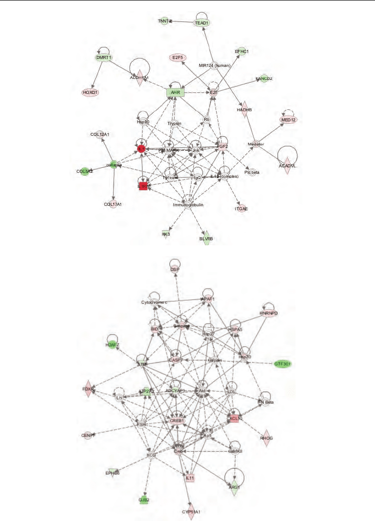
Finally, in the Figure 5 is showed a “hypothetical network in CC” based from the 15 small
networks. In addition to gene expression values, the proposed method uses Gene Ontology,
which is a reliable source of information on genes. The use of Gene Ontology can
compensate, in part, for the limitations of microarrays, such as having a small number of
samples and erroneous measurement results.
5. Discussion
In our results, non classical “cancer genes” were conserved, respect to expected genes as
MYC, FOS, RB, P53, HIF, etc. However, in the “strict sense of the word” when is considered
a cancer gene? By instance, over-expression, down-regulation, point mutation,
amplification, loss of heterozygosity, polymorphisms, epigenetic changes, etc. Thus, any
gene could be considered like cancer gene, if they are following special criteria as recently
was reported (27).
In this context, we decided to explore two non-related genes in cervical cancer PARK2 gene.
Interestingly, PARK2 gene mutations (point mutations and exonic deletions) were first
identified in autosomal recessive juvenile-onset parkinsonism. This gene is mapped to
6q25.2-q27 containing 12 small exons, and encodes parkin protein which functions as an E3
ligase, ubiquitinating proteins for destruction by the proteosome. Several substrates for
parkin have been identified, including a 22kD glycosolated form of synuclein, parkin-
associated endothelin receptor-like receptor (Pael-R), and CDCrel-1. Over-expression of
Pael-R causes it to become ubiquinated, insoluble, and unfolded, and lead to endoplasmic
reticulum stress and cell death (for review see 28). The location of Parkin is in a
chromosomal region that is frequently deleted in multiple tumor types, including
hepatocellular carcinoma (HCC), ovarian cancer, and breast cancer. The Parkin gene is
within FRA6E, the third most active common fragile site (29,30). Interestingly, all three
fragile sites regions were found consistently deleted in HCC (31) as well as in ovarian,
breast, and prostate cancers. Further PARKIN protein overexpression did not lead to
Computational Biology and Applied Bioinformatics
272
A
B
Functional Analysis of the Cervical Carcinoma
Transcriptome: Networks and New Genes Associated to Cancer
273
C
D
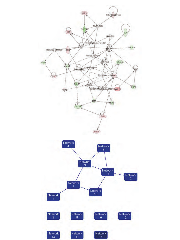
Fig. 5. Networks in cervical cancer. Top three networks diagrams generated as graphical
representations of the molecular relationship between genes and gene product. The gene

Computational Biology and Applied Bioinformatics
274
products are represented as nodes (shapes) and the biological relationship between two
nodes is represented as an edge (line). A) Network 1. skeletal and muscular system
development and function, embryonic development, tissue development, B) network 2;
dermatological diseases and conditions, cardiovascular disease, organismal injury and
abnormalities, C) network 3; cell cycle, organismal functions, organismal injury and
abnormalities, D) Ingenuity Pathways Analysis network of genes associated with CC. This
network diagram shows the biological association of 8 focus networks associated with
cervical cancer as graphical representation of the molecular relationship between
genes/gene produtcs.
increased sensitivity to all pro-apoptotic induction but may show specificity for a certain
type of cellular stress. At present, Parkin gene could be considered as new tumor suppressor
gene (32). In our case, per se Parkin expression in CC is interesting. It is widely described
that p53 master gene is constitutively expressed but under stress conditions as HPV
infection, DNA damage or point mutations its half-time life of the protein is increased.
Similar situation might be observed for Parkin gene due to increased expression in CC. This
could be supported because the 6q25 cytogenetic region in CC is not altered as happen for
TP53 gene (33). To this respect, we could hypothesize that a new overexpression of Parkin
gene could be involved in invasion cervical carcinogenesis. These findings could
demonstrate that the genetic context in which a mutation occurs can play a significant role
in determining the type of illness produced or associated.
It has been established that although we inherit two copies of all genes (except those that
reside on the sex chromosomes), there is a subset of these genes in which only the paternal
or maternal copy is functional. This phenomenon of monoallelic, parent-of-origin expression
of genes is termed genomic imprinting. Imprinted genes are normally involved in
embryonic growth and behavioral development, but occasionally they also function
inappropriately as oncogenes and tumor suppressor genes (34). Furthermore, it is well know
that a variety of genetic changes influence the development and progression of cancer.
These changes may result from inherited or spontaneous mutations that are not corrected by
repair mechanisms prior to DNA replication. It is increasingly clear that so called epigenetic
effects that do not affect the primary sequence of the genome also play an important role in
tumorigenesis (35).
Other gene overexpressed seen in this analysis was PEG10 gene. This gene is mapped in
chromosme 7q21. PEG10 protein prevents apoptosis in hepatocellular carcinoma cells
through interaction with SIAH1, a mediator of apoptosis. May also have a role in cell
growth promotion and hepatoma formation. Inhibits the TGF-beta signaling by interacting
with the TGF-beta receptor ALK1. This is a paternally expressed imprinted gene that
encodes transcripts containing two overlapping open reading frames (ORFs), RF1 and
RF1/RF2, as well as retroviral-like slippage and pseudoknot elements, which can induce a -1
nucleotide frame-shift. Increased expression of this gene is associated with hepatocellular
carcinomas. These findings link to cancer genetics and epigenetic by showing that a classic
proto-oncogene, MYC, acts directly upstream of a proliferation-positive imprinted gene,
PEG10 (36,37).
The HOX genes are a family of transcription factors that bind to specific sequences of DNA
in target genes regulating their expression. The role of HOX genes in adult cell
differentiation is still obscure, but growing evidence suggests that they may play an
important role in the development of cancer. We have previously reported that some HOX

Functional Analysis of the Cervical Carcinoma
Transcriptome: Networks and New Genes Associated to Cancer
275
genes could be related to CC. Specifically, HOXA9 was observed expressed in cervical
cancer by RT-PCR end point. In the present work, the data are showing that statistically
significative HOXA9 gene is differentially expressed in CC. Together to HOXB13, D9, D10,
and HOXC cluster (HOXC9, C11–C13) genes this family of genes might be an important
factor involved in CC (35).
It is clear that the most altered genes in CC are not commonly associated to cancer process.
This fact could suggest: 1) the “classic genes of cancer” are statistically significant altered
with tiny values, but there are some exceptions and specific tumor types as neuroblastomas
and N-MYC gene, Her2/neu in breast cancer. 2) At least in stage IIb of cervical carcinogenesis
could be involved genes related to “cellular economy” but not belonging to genes of cancer.
This is supported by recent reports showing molecular alterations in genes not previously
related to cancer (27). 3) The extreme values (high or low) in microarray analysis not always
represent strong candidates of markers in the models performed. What about the most
frequent?. (4) Integrative genomics, multicentre protocols in well selected samples, stratified
stages and clinical follow-up, will be the clue to get cancer hallmarks. In addition, the study
of the molecular function of selected genes strengthened the hypothesis that these genes are
involved in the process of cancer growth.
The data information obtained from microarray analysis should be validated because can
appear errors in positive and negative false due to nature of the massive assays. In this
context, in order to confirm the microarray data, additional molecular tool as end point
PCR, real time PCR, northern blot, immunohistochemistry should be performed and to
obtain results. (Mendez S. An Integrative microarray gene expression analysis, approach
identifies candidates’ array multi-experiments in Ovary Tumours, submitted to publication
2011).
6. Acknowledgements
This work was partially supported by CONACYT (México) grants 69719 AND 87244 from
FONDOS SECTORIALES. We appreciate the technical assistance of Laboratorio de
Oncología Genómica, CIS, HO-IMSS. Sergio JUAREZ and Mauricio SALCEDO made
similar efforts in the present work.
7. References
[1] Parkin DM, Bray F, Ferlay J, Pisani P. Global cancer statistics, 2002. CA Cancer J Clin.
2005, 55(2):74-108
[2] Secretaría de Salud (Méx). Registro Histopatológico de Neoplasias en México. México:
Secretaría de Salud, México D.F; 1999.
[3] Munoz N, Xavier Bosch F. Cervical cancer and human papillomavirus: epidemiological
evidence and perspectives for prevention. Salud Publica Mex 1997; 39:274–282.
[4] Boffetta P, Parkin DM. Cancer in developing countries. CA Cancer J Clin 1994 44:81-90
[5] Walboomers JM, Jacobs MV, Manos MM, Bosch FX, Kummer JA, Shah K, et al. Human
papillomavirus is a necessary cause of invasive cervical cancer worldwide. J Pathol
1999, 189: 12–19.

Computational Biology and Applied Bioinformatics
276
[6] Scheffner, M., B. A. Werness, J. M. Huibregtse, A. J. Levine, and P. M. Howley. The E6
oncoprotein encoded by human papillomavirus types 16 and 18 promotes the
degradation of p53. Cell 1990, 63:1129–1136.
[7] Boyer SN, Wazer DE, Band V. E7 protein of human papilloma virus-16 induces
degradation of retinoblastoma protein through the ubiquitin-proteasome pathway.
Cancer Res. 1996, 56(20):4620-4624.
[8] Duensing S, Munger K. The human papillomavirus type 16 E6 and E7 oncoproteins
independently induce numerical and structural chromosome instability. Cancer
Res. 2002, 62(23):7075-7082.
[9] Nees M, van Wijngaarden E, Bakos E, Schneider A, Dürst M. Identifcation of novel
molecular markers which correlate with HPVinduced tumor progression.
Oncogene 1998, 16:2447–2458.
[10] Brown P, Botstein R. Exploring the new world of the genome with DNA microarrays.
Nature Genet 1999, 21 (suppl):33-37.
[11] Lockhart DJ, Winzeler EA. Genomics, gene expression and DNA arrays. Nature, 2000,
405:827-836.
[12] Ruutu M, Peitsaro P, Johansson B, Syrjanen S: Transcriptional profiling of a human
papillomavirus 33-positive squamous epithelial cell line which acquired a selective
growth advantage after viral integration. Int J Cancer 2002, 100: 318-326.
[13] Duffy CL, Phillips SL, Klingelhutz AJ: Microarray analysis identifies differentiation-
associated genes regulated by human papillomavirus type 16 E6. Virol 2003, 314:
196-205.
[14] Thomas JT, Oh ST, Terhune SS, Laimins LA: Cellular changes induced by low-risk
human papillomavirus type 11 in keratinocytes that stably maintain viral episomes.
J Virol 2001, 75: 7564-7571.
[15] Garner-Hamrick PA, Fostel JM, Chien WM, Banerjee NS, Chow LT, Broker TR, Fisher
C: Global effects of human papillomavirus type 18 E6/E7 in an organotypic
keratinocyte culture system. J. Virol 2004, 78: 9041-9050.
[16] Toussaint-Smith E, Donner DB, Roman A: Expression of human papillomavirus type
16 E6 , E7 oncoproteins in primary foreskin keratinocytes is sufficient to alter the
expression of angiogenic factors. Oncogene 2004, 23: 2988-2995.
[17] Nees M, Geoghegan JM, Hyman T, Frank S, Miller L, Woodworth CD: Papillomavirus
type 16 oncogenes downregulate expression of interferon-responsive genes and
upregulate proliferation-associated, NF-kappaB-responsive genes in cervical
keratinocytes. J Virol 2001, 75: 4283-4296.
[18] Shim C, Zhang, Hun C, Lee. Profiling of differentially expressed genes in human
primary cervical cancer by complementary DNA expression array. Clin Cancer Res
1998, 4:3045-3050.
[19] Vazquez-Ortiz G, García JA, Ciudad CJ, Noé V, Peñuelas S, López-Romero R,
Mendoza-Lorenzo P, Piña-Sánchez P, Salcedo MDifferentially expressed genes
between high-risk human papillomavirus types in human cervical cancer cells. Int J
Gynecol Cancer. 2007, 17:484-491