Marshall L. Stoller, Maxwell V. Meng-Urinary Stone Disease
Подождите немного. Документ загружается.

390 Breiman and Coakley
(Fig. 11). Fat stranding represents edema, as well as engorged lymphatics, attempting to
drain increased renal interstitial fluid, secondary to post obstructive elevation of collect-
ing system pressure. The presence of both of these signs is associated with 99% positive
predictive value and 95% negative predictive value for ureteral obstruction (54).
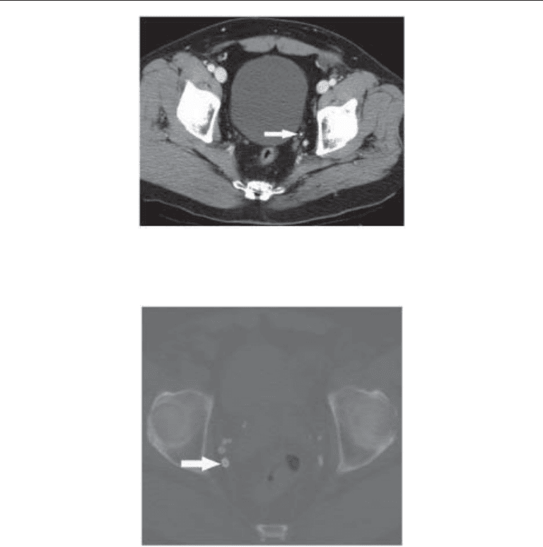
A calculus lodged in a ureter is often associated with thickening of the ureteral wall
resulting from edema, the ureteral rim sign, which may be helpful in differentiating a
ureteral calculus from a phlebolith (Fig. 19). A ureteral rim sign is noted in 76–77% of
calculi, but less than 10% of phleboliths (54,55). It is detected in as many as 91% of
ureteral stones 4 mm or less in size, but larger stones may obscure the ureteral rim sign.
C
OMPUTED-TOMOGRAPHY PITFALLS
Phleboliths are usually round with smooth contours and often contain a central lucency
(Fig. 20) (54). Phleboliths increase in number with age. Many phleboliths lie below a line
Fig. 19. CT image showing a small stone (arrow) in the distal left ureter. A thin surrounding rim
of tissue is visible, representing the edematous ureteral wall. This so-called ureteral rim sign
helps to differentiate a ureteral calculus from a phlebolith.
Fig. 20. Nonenhanced CT image (filmed at bone windows) demonstrating numerous pelvic
phleboliths. The characteristic central lucency is evident within the largest phlebolith (arrow).
Chapter 20 / Imaging of Urinary Stone Disease 391
drawn between the iliac spines, and are more lateral than the bladder and distal ureters.
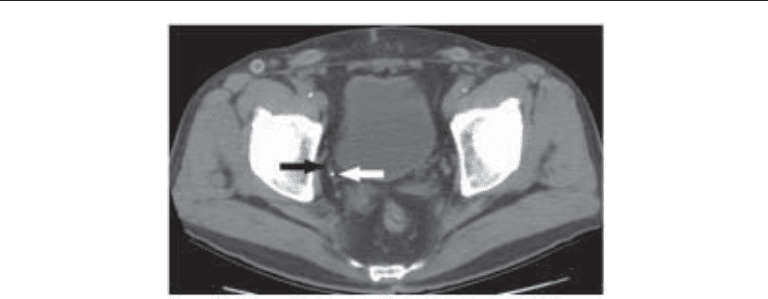
A pelvic phlebolith may be associated with a comet tail sign, representing a noncalcified
thrombus adjacent to a calcified component (55–58)(Fig. 21). Boridy et al demonstrated
a sensitivity of 65% and a specificity of 100% for a calcified phlebolith when the comet
tail sign was detected; no calculi were associated with a comet tail (57). Bell also dem-
onstrated 100% specificity for a phlebolith when a comet tail was noted (55). However,
Guest et al. demonstrated poor observer agreement on whether the comet tail sign was
present in patients with pelvic opacities, and the sign frequently was related to a ureteral
calculus (58). If problems persist in differentiating a phlebolith from a distal ureteral
calculus following a noncontrast CT, the use of intravenous contrast with excretory
phase images may be helpful for a definitive diagnosis. A gonadal vein phlebolith may
mimic a ureteral stone. It is important to carefully follow the course of the ureters and
periureteral vascular structures on each sequential image to avoid errors in interpreta-
tion. Arterial calcifications may lie in the vicinity of the renal collecting system or a
ureter and simulate a stone, particularly on X-rays.
I
NCIDENTAL NONSTONE PATHOLOGY DETECTED ON COMPUTED TOMOGRAPHY
Among the advantages of noncontrast CT as the primary modality in the assessment
of suspected urinary stone disease is the ability of CT to visualize extraurinary tract
abnormalities. These findings may be incidental, but in some cases may represent the
etiology of the patient’s clinical presentation. In one study, unsuspected findings that
could affect acute patient care were observed at 5.9% of unenhanced CT examinations
(59). Extraurinary tract conditions most often mimicking renal colic include; appen-
dicitis (Fig. 22), diverticulitis (Fig. 23), adnexal pathology including tubo-ovarian
abscess, retroperitoneal hemorrhage or infection, and retroperitoneal neoplasm, sec-
ondarily involving a ureter (Fig. 24). CT performed to assess suspected stone disease
may detect occult urinary and extra-urinary tract pathology, unrelated to the acute
clinical presentation, but potentially clinically significant. As with screening CT, these
findings include abdominal aortic aneurysm, gallstones, lymphadenopathy, orga-
nomegaly and occult malignancy (Fig. 25).
Fig. 21. Nonenhanced CT showing a pelvic phlebolith (white arrow). A linear strand of tissue
(black arrow) is seen extending from the phlebolith. This tissue strand is believed to represent the
atretic vessel within which the phlebolith formed, and this so-called “comet-tail” sign is another
finding that may help distinguish stones and phleboliths.
392 Breiman and Coakley
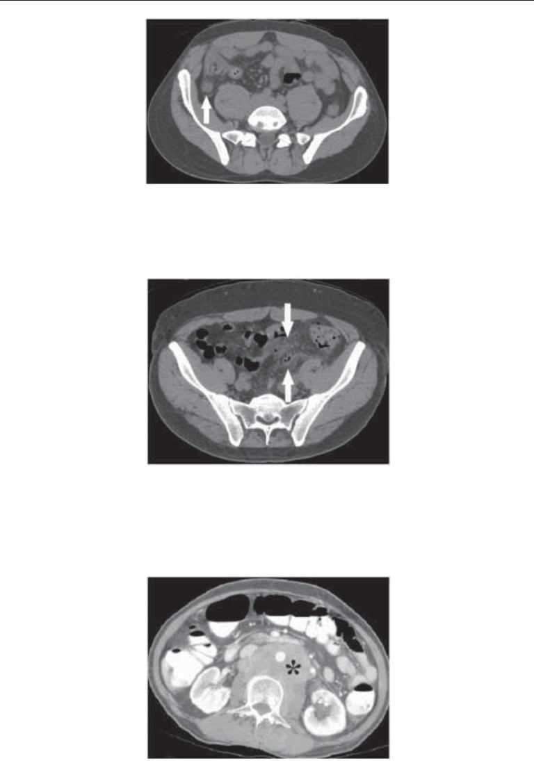
Fig. 24. Contrast-enhanced CT performed in a patient with left flank pain, after an initial
nonenhanced study (not shown) had failed to demonstrate urinary stones but did show a retro-
peritoneal mass. Enhancing soft tissue (asterisk) is seen encasing the aorta and displacing the
inferior vena cava. The left kidney is mildly hydronephrotic. A diagnosis of lymphoma was
established at biopsy.
Fig. 23. Nonenhanced CT image obtained in a patient with acute left sided flank pain. The scan
was requested by the emergency department to evaluate for possible urinary stone. Small bubbles
of free air and associated infiltration within the sigmoid mesocolon (arrows) were caused by
unsuspected perforated diverticulitis.
Fig. 22. Nonenhanced CT in a patient suspected of having a right ureteral calculus. The appendix
(arrow) is thickened with surrounding fat stranding. The radiological diagnosis of acute appen-
dicitis was confirmed at surgery.
Chapter 20 / Imaging of Urinary Stone Disease 393
Magnetic Resonance Imaging
Noncontrast CT is currently the modality of choice for the evaluation of acute flank
pain, but MR may be helpful in select patients, particularly in pregnancy when avoidance
of ionizing radiation is a priority. Most reports advocate the use of heavily T2-weighted
sequences for the MR assessment of the urinary tract. HASTE (half Fourier acquisition
single shot turbo spin echo) and RARE (rapid acquisition with relaxation enhancement)
sequences have been shown to be particularly useful in creating an MR urogram (60–63).
Maximum intensity projection (MIP) reconstructions in coronal and sagittal projections
may be helpful. Stones are identified as a filling defect (signal void) within the abundant
bright signal of the urine filled collecting system, ureter or bladder on a T2-weighted
sequence (Fig. 26) (60,64,65), although MR imaging is generally insensitive to urinary
stones (Fig. 27) and the primary use of this modality is in the demonstration of hydro-
nephrosis and hydroureter.
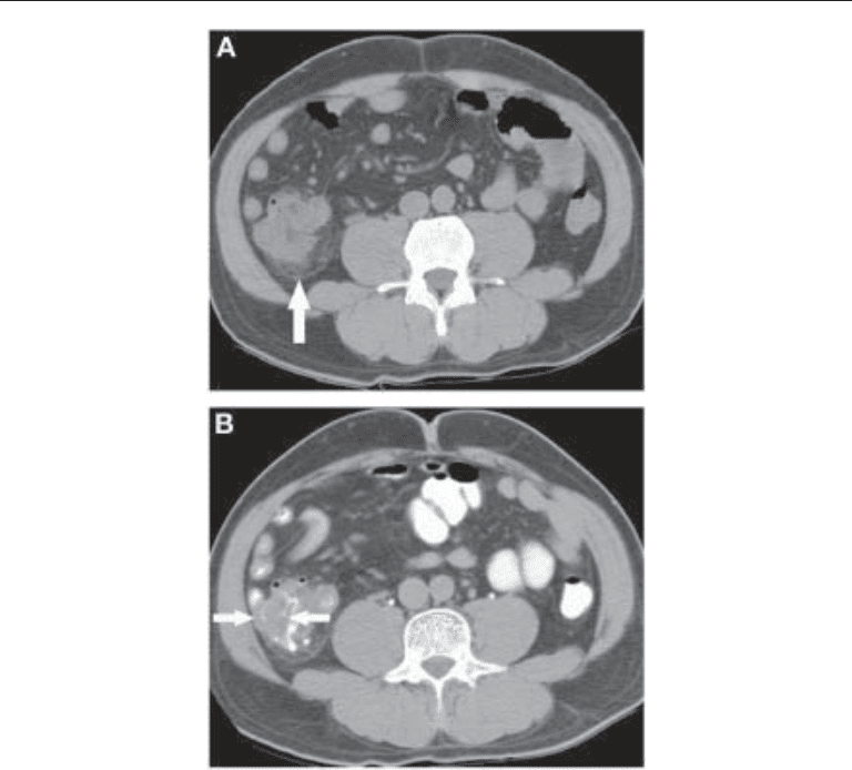
Fig. 25. (A) Nonenhanced urinary stone CT performed to assess right flank pain. Fat stranding
(arrow) is present around the cecum, and the cecal wall appears thickened. (B) Subsequent CT
performed after oral contrast confirms circumferential cecal wall thickening. Cecal carcinoma
was proven at resection.
394 Breiman and Coakley
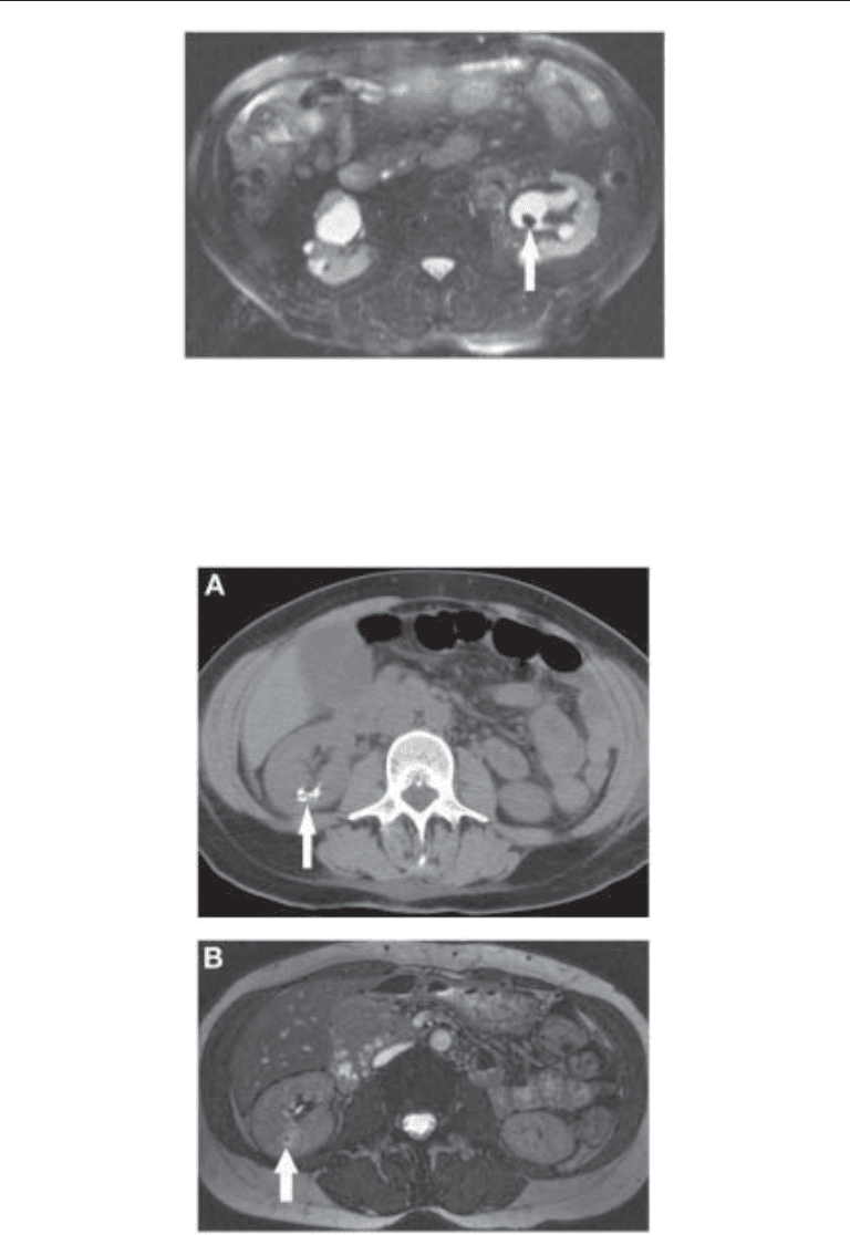
Fig. 26. Fat-saturated T2-weighted axial MR image demonstrates the characteristic appearance
of a urinary stone, which appears as a filling defect (arrow) within the fluid filled collecting
system. Fluid is bright on T2-weighted imaging, facilitating stone identification.
Fig. 27. (A) Nonenhanced axial CT image showing a cluster of small stones (arrow) within the
parenchyma of the lower pole of the right kidney. (B) Gradient echo axial T2-weighted MR image
at the same level. The cluster of stones (arrow) is difficult to appreciate. In general, MR imaging
is inferior to CT in the evaluation of stones, and is therefore rarely used for this indication.
Chapter 20 / Imaging of Urinary Stone Disease 395
Retrograde Pyelography
Retrograde pyelography may be helpful in azotemic patients and others in whom
opacification of the urinary tract is desirable but intravenous iodinated contrast is
contraindicated or produced insufficient luminal opacification (Fig. 28). For example,
a patient with multiple pelvic opacities in close proximity to the distal ureters in whom
a distal ureteral calculus can not be excluded by other examinations because of
contraindications for intravenous contrast or insufficient contrast excretion. A retro-
grade pyelogram may be helpful when other examinations are unsuccessful in detect-
ing a urinary stone in a patient with a strong clinical suspicion of stone disease.
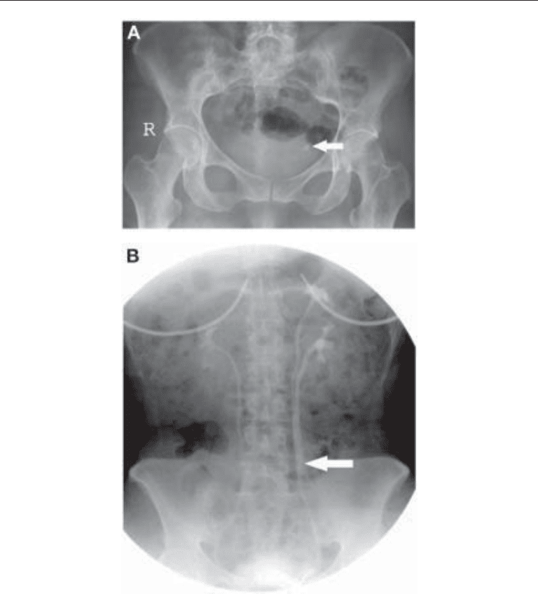
Fig. 28. (A) Plain radiograph showing a stone (arrow) in the distal left ureter. (B) Retrograde
pyelogram performed after laser lithotripsy of the stone shows the left ureter (arrow) to be stone
free. A duplicated left upper tract is also demonstrated.
396 Breiman and Coakley
Retrograde pyelography is occasionally used in association with SWL to locate a
faintly visible stone. The ureteral catheter used at retrograde pyelography may be
helpful as a reference for targeting a stone at lithotripsy. Dilute contrast is used with
fluoroscopy to prevent over distension and extravasation. If infection exists proximal
to obstruction the patient is at risk for sepsis, possibly life-threatening infection or
irreversible renal failure. Retrograde placement of a ureteral catheter above the level
of obstruction under fluoroscopic guidance, may provide adequate drainage of the
infected upper tract.
IMAGING BLADDER AND URETHRAL CALCULI
Primary bladder calculi are usually a manifestation of underlying pathology, includ-
ing voiding dysfunction, foreign bodies, infection, parasites, migrant renal calculi, blad-
der outlet obstruction, and neurogenic bladder (Fig. 29). Foreign bodies in the bladder
include chronic indwelling Foley catheters or double J ureteral catheters. Bladder infec-
tion may serve as a nidus for stone formation, as it does in the kidney. Post infection
bladder calculi are most common in males and are usually associated with a urea splitting
organism or a parasitic infection. Most bladder calculi are solitary, but multiple calculi
can be seen. Bladder calculi are most often composed of ammonium urate, uric acid, or
calcium oxalate. Uric acid stones are often radiolucent in the bladder on a radiograph,
as they are elsewhere, and may be identified as filling defects on an IVU. Bladder calculi
may be detected or confirmed with US as even radiolucent stones are echogenic on a
sonogram and usually cast an acoustic shadow. Stone mobility is easily demonstrated
with US when a patient changes position during scanning, aiding in the differentiation
of nonmobile calculi impacted at the ureterovesical junction, particularly the inter-
ureteric ridge portion of the distal ureter, from mobile bladder calculi.
Various structures may mimic bladder calculi on imaging studies, particularly X-ray
based procedures such as plain films and an IVU. These include: a punctate collection
of rectal contrast, dense impacted stool, phleboliths, an appendicolith, calcified fibroids
or adnexal masses, post inflammatory, hemorrhagic or infectious calcifications, and
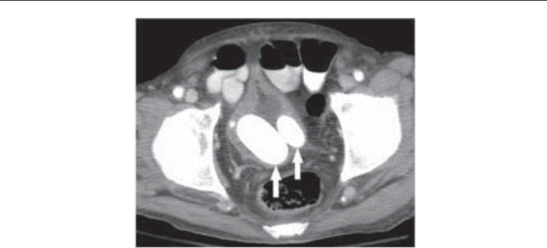
Fig. 29. Axial CT image in an elderly man with chronic bladder outlet obstruction. Large bladder
stones (arrows) are present within the bladder. Bladder wall thickening and a Foley catheter can
also be seen.
Chapter 20 / Imaging of Urinary Stone Disease 397
calcifications related to amyloidosis. Schistosomiasis is the most common cause of
bladder calcification (Fig. 30) where the disease is endemic in the Middle East and
Africa. Calcifications secondary to Schistosomiasis may also involve the distal ureters.
Calcifications related to tuberculosis may involve the bladder wall, seminal vesicles, vas
deferens and epididymis. However bladder calculi are rare in patients with tuberculosis.
Prostatic and seminal vesicle calcifications, including calcified amylacea of the pros-
tate in older males, may mimic bladder calculi on radiographs. Prostatic calcifications
are rarely associated with prostatitis. Seminal vesicle stones are often smooth, round and
may rarely be associated with hematospermia. They may be mistakenly attributed to
tuberculous infection.
Urethral calculi arise from the bladder and rarely from the upper tracts. Most ureteral
calculi pass unimpeded through the urethra. Urethral calculi may be secondary to urinary
stasis in a diverticulum or related to a stricture, possibly in the region of previous surgery.
In males, urethral stones are most commonly in the prostatic and bulbar portions and
are usually solitary. Urethral calculi are rare in females, secondary to a short urethra.
Urethral calculi may be secondary to diverticula, particularly in females (Fig. 31). Symp-
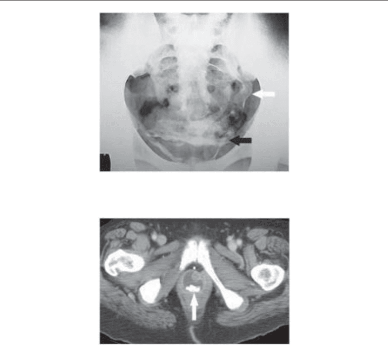
Fig. 31. Axial CT image in an elderly woman showing stones (arrow) within a urethral diverticu-
lum. The appearance of the diverticulum is characteristic.
Fig. 30. Plain abdominal radiograph in a patient with urinary schistosomiasis. Calcification of
the bladder (black arrow) and of the distal left ureter (white arrow) are evident.

398 Breiman and Coakley
toms secondary to urethral calculi are similar to those of bladder stones, including
intermittent interruption of the urinary stream, dribbling, terminal hematuria, infection
and pain.
IMAGING FLANK PAIN IN PREGNANCY
Ultrasound is often the initial modality used to search for suspected urinary stones in
a pregnant patient. If an obstructing calculus is identified, no further imaging may be
necessary. If US does not yield an explanation of the patient’s clinical presentation, a
KUB may be helpful. A KUB may be the initial examination when a ureteral calculus
is suspected in the second or third trimester of pregnancy. If an IVU is felt necessary
during the second or third trimester, a limited examination consisting of a single film 20
min post-IV contrast infusion may yield important additional information, while limit-
ing radiation. Most stones pass spontaneously in this population, but intervention may
be required for persistent pain, sepsis, progressive or high-grade hydronephrosis or
obstruction of a solitary kidney. If necessary, the workup may proceed to a retrograde
pyelogram with stent placement to relieve obstruction, leaving an offending stone to be
addressed postpartum. Hydronephrosis of pregnancy or over distention syndrome is
often encountered as an incidental finding on second and third trimester obstetric US, but
may be difficult to differentiate from an occult stone with ureteral distension
sonographically. Simple hydronephrosis of pregnancy is not usually associated with an
elevated resistive index, as occurs with obstruction secondary to a stone.
SUMMARY
The management of urinary stone disease benefits from a multidisciplinary approach,
combining traditional and high tech clinical and imaging approaches. Imaging plays a
pivotal role in the diagnosis, characterization and follow-up of calculi, often providing
crucial information contributing to the successful management of urinary stones. Goals
of imaging include not only the detection of stones but equally important, characteriza-
tion of stones and the urinary tract. This information contributes to a determination of
prognosis and to the selection of an appropriate management plan, selection and guid-
ance of therapy, as well as the assessment of the success of management and a determi-
nation of timing and selection of alternative therapies. Important imaging data includes
size, shape, composition, (hardness) of stones, as well as information relating to the
urinary tract, including: anatomic condition leading to stasis and stone formation, pres-
ence, level, and degree of obstruction, resultant presence and degree of complications,
such as atrophy or infection. Imaging is often important to the selection and design of
appropriate therapy, in monitoring the success of treatment and in detecting complica-
tions. Noncontrast CT has become the initial modality in the assessment of suspected
nephroureterolithiasis and urinary tract obstruction as it is accurate, readily available,
and does not usually require the use of intravenous iodinated contrast, avoiding the
potential risks of contrast in most patients. Other modalities still play a role in the
management of stone disease, particularly plain films and US.
ACKNOWLEDGMENT
The authors wish to acknowledge the contribution of Xia Wang, MD to the research
of the literature and selection and preparation of case material for this chapter.

Chapter 20 / Imaging of Urinary Stone Disease 399
REFERENCES
1. Smith RC, Varanelli M. Diagnosis and management of acute ureterolithiasis. AJR 2000; 175:
3–6.
2. Reiter WJ, Schon-Pernerstorfer H, Dorfinger K, Hofbauer J, Marberger M. Frequency of uroli-
thiasis in individuals seropositive for human immunodeficiency virus treated with indinavir is
higher than previously assumed. J Urol 1999; 161: 1082–1084.
3. Schwartz BF, Schenkman N, Armenakas NA, Stoller ML. Imaging characteristics of indinavir
calculi. J Urol 1999; 161: 1085–1087.
4. Bruce GR, Munch LC, Hoven AD, et al. Urolithiasis associated with the protease inhibitor
indinavir. Urology 1997; 50: 513–518.
5. Blake SP, McNicholas MM, Raptopoulos V. Nonopaque crystal deposition causing ureteric
obstruction in patients with HIV undergoing indinavir therapy. AJR 1998; 171: 717–720.
6. Leroy AJ. Diagnosis and treatment of nephrolithiasis: Current perspectives. AJR 1994; 163: 1309–
1313.
7. Amis ES, Newhouse JH. Urinary stone disease. In: Amises, Newhouse JH eds., Essentials of
Uroradiology. Boston, Little, Brown, 1991: 213–231.
8. Tublin ME, Murphy ME, Delong DM, Tessler FN, Kliewer MA. Conspicuity of renal calculi at
unenhanced CT: Effects of calculus composition and size and CT technique. Radiology 2002; 225:
91–96.
9. Motley G, Dalrymple N, Kessling C, Fischer J, Harmon W. Hounsfield unit density in the deter-
mination of urinary stone. Urology 2001; 58 (2): 170–173.
10. Williams JC Jr, Saw KC, Monga AG, Chua GT, Lingeman JE, McAteer JA. Correction of helical
CT attenuation values with wide beam collimation: in vitro test with urinary calculi. Acad Radiol
2001; 8 (6): 478–483.
11. Joseph P, Manda AK, Singh SK, Mandal P, Sankhwar SN, Sharma SK. Computerized tomography
attenuation value of renal calculus: can it predict successful fragmentation of the calculus by
extracorporeal shock wave lithotripsy? A preliminary study. J Urol 2002; 167: 1968–1971.
12. Low RK, Stoller ML. Endoscopic mapping of renal papillae for Randall’s plaques in patients with
urinary stone disease. J Urol 1997; 158: 2062–2064.
13. Evan AP, Lingeman JE, Coe FL, et al. Randall’s plaque of patients with nephrolithiasis begins in
basement membranes of thin loops of Henle. J Clin Invest 2003; 111: 607–616.
14. Coll DM, Varanelli MJ, Smith RC. Relationship of spontaneous passage of ureteral calculi to stone
size and location as revealed by unenhanced helical CT. AJR 2002; 178: 101–103.
15. Tuttle DN, Yeh BM, Meng MV, Breiman RS, Stoller ML, Coakley FV. Risk of injury to adjacent
organs at lower pole fluoroscopically-guided percutaneous nephrostomy: Evaluation by prone,
supine and multiplanar reformatted CT. J Vasc Interv Radiol. 2005; 16(11): 1489–1492.
16. Middleton AW Jr, Pfister RC. Stone containing pyelocaliceal diverticulum: Embroyogenic, ana-
tomic, radiologic and clinical characteristics. J Urol 1974; 111: 2–6.
17. Timmons JW, Malek RS, Hattery RR, Deweerd JH. Caliceal diverticulum. J Urol 1975; 114: 6–9.
18. Ginalski JM, Portmann L, Jaeger PH. Does medullary sponge kidney cause nephrolithiasis? AJR
1990; 155: 299–302.
19. Rose JG, Gillenwater JY. Pathophysiology of ureteral obstruction. Am J of Physiol 1973; 225:
830–837.
20. Michaelson G. Percutaneous puncture of the renal pelvis, intrapelvic pressure, and the concentrat-
ing capacity of the kidney in hydronephrosis. Acta Med Scand 1974; 559(Suppl): 1–26.
21. Gillenwater JY. The pathophysiology of urinary tract obstruction. In: Campbell’s Urology, 6th
Ed., (Walsh PC, Retik AB, Stamey TA, Vaughn ED, eds.). Saunders, Philadelphia, PA, 1992, p.
509.
22. Holmes MJ, O’Morchoe PJ, O’Morchoe CC. Morphology of the intrarenal lymphatic system:
capsular and hilar communications. Am J Anat 1977; 149: 333–337.
23. Heney NM, O’Morchoe PJ, O’Morchoe CC. The renal lymphatic system during obstructed urine
flow. J Urol 1971; 106: 455–462.
24. Leahy AL, Ryan PC, McEntee GM, Nelson AC, Fitzpatrick JM. Renal injury and recovery in
partial ureteric obstruction. J Urol 1989; 142: 199–203.