Marshall L. Stoller, Maxwell V. Meng-Urinary Stone Disease
Подождите немного. Документ загружается.


400 Breiman and Coakley
25. Kerr WS. Effect of complete ureteral obstruction for one week on kidney function. J Appl Physiol
1954; 6: 763.
26. Thornbury JR, Parker TW. Ureteral calculi. Semin Roentgenol 1982; 17: 133–138.
27. Schuster GA, Nazos D, Lewis GA. Preparation of outpatients for excretory urography: is bowel
preparation with laxatives and dietary restrictions necessary? AJR Am J Roentgenol 1995; 164:
1425–1428.
28. Boyd R, Gray AJ. Role of the plain radiograph and urinalysis in acute ureteric colic. J Accid Emerg
Med 1996; 13: 390, 391.
29. Svedstrom E, Alanen A, Nurmi N. Radiologic diagnosis of renal colic: The role of plain films,
excretory urography, and sonography. Eur J Radiol 1990; 11: 180–183.
30. Niall O, Russell J, MacGregor R, Duncan H, Mullins J. A comparison of noncontrast computerized
tomography with excretory urography in the assessment of acute flank pain. J Urol 1999; 161: 534–
537.
31. Assi Z, Platt JF, Francis IR, Cohan RH, Korobkin M. Sensitivity of CT scout radiography and
abdominal radiography for revealing ureteral calculi on helical CT: implications for radiologic
follow-up. AJR 2000; 175: 333–337.
32. Chu G, Rosenfield AT, Anderson K, Scout L, Smith RC. Sensitivity and value of digital CT scout
radiography for detecting ureteral stones in patients with Ureterolithiasis diagnosed on unenhanced
CT. AJR 1999; 173: 417–423.
33. Levine JA, Neitlich J, Verga M, Dalrymple N, Smith RC. Ureteral calculi in patients with flank
pain: correlation of plain radiography with unenhanced helical CT. Radiology 1997; 204: 27–31.
34. Jackman SV, Potter SR, Regan F, Jarrett TW. Plain abdominal X-ray versus computerized tomog-
raphy screening: sensitivity for localization after nonenhanced spiral computerized tomography.
J Urol 2000; 164: 308–310.
35. Goldwasser B, Cohan RH, Dunnick NR, Andriani CC, Carsen CC 3rd, Weinerth JL. Role of linear
tomography in evaluation of patients with nephrolithiasis. Urology 1989; 33: 253–256.
36. Ahn SH, Mayo-Smith WW, Murphy BL, Reinert SE, Cronan JJ. Acute nontraumatic abdominal
pain in adult patients: Abdominal radiography compared with CT evaluation. Radiology 2002;
225: 159–164.
37. Averch TD, O’Sullivan D, Breitenbach C, Beser N, Schulam PG, Moore RG, Kavoussi LR: Digital
radiographic imaging transfer: comparison with plain radiographs. J Endourol 1997; 11: 99–101.
38. Eisenberg RL, Berlin L When does malpractice become manslaughter? AJR 2002; 179: 331–335.
39. Juul N, Brons J, Torp-Pedersen S, Fredfeldt KE. Ultrasound versus intravenous urography in the
evaluation of patients with suspected obstructing urinary calculi. Scand J Urol Nephrol 1991
(Suppl); 137: 45–47.
40. Smith RC, Rosenfield AT, Choe KA, et al. Acute flank pain: Comparison of non contrast-enhanced
CT and intravenous urography. Radiology 1995; 194: 789–794.
41. Caro JJ, Trindade E, McGregor M. The risks of death and of severe nonfatal reactions with high-
vs low- osmolality contrast media: a meta-analysis. AJR 1991;156: 825–832.
42. Tepel M, van der Giet M, Schwarzfeld C, Laufer U, Liermann D, Zidek W. Prevention of radio-
graphic-contrast-agent-induced reductions in renal function by acetylcysteine. N Engl J Med
2000; 343: 180–184.
43. Aspelin P, Aubry P, Fransson SG, Strasser R, Willenbrock R, Berg KJ. Nephrotoxicity in High-
Risk Patients Study of Iso-Osmolar and Low-Osmolar Non-Ionic Contrast Media Study Investi-
gators. Nephrotoxic effects in high-risk patients undergoing angiography. N Engl J Med. 2003;
348: 491–499.
44. Fowler KAB, Locken JA, Duchesne JH, Williamson MR. US for detecting renal calculi with
nonenhanced CT as a reference standard. Radiology 2002; 222: 109–113.
45. Laing FC, Jeffrey RB, Wing VW. Ultrasound versus excretory urography in evaluating acute flank
pain. Radiology 1985; 154: 613–616.
46. Rodgers PM, Bates JA, Irving HC. Intrarenal Doppler ultrasound studies in normal and acutely
obstructed kidneys. Brit J Radiol 1992; 65: 207–212.
47. Mallek R, Bankier AA, Etele-Hainz A, Kletter K, Mostbeck GH. Distinction between obstructive
and nonobstructive hydronephrosis: Value of diuresis duplex Doppler sonography. AJR 1996;
166: 113–117.

Chapter 20 / Imaging of Urinary Stone Disease 401
48. Hill MC, Rich JI, Mardiat JG, Finder CA. Sonography versus excretory urography in acute flank
pain. AJR 1985; 144: 1235–1238.
49. Schwartz BF, Schenkman N, Armenakas NA, Stoller ML. Imaging characteristics of indinavir
calculi. J Urol 1999; 161: 1085–1087.
50. Olcott EW, Sommer FG, Napel S. Accuracy of detection and measurement of renal calculi: in vitro
comparison of three-dimensional spiral CT, radiography, and nephrotomography. Radiology 1997;
204: 19–25.
51. Smith RC, Verga M, McCarthy S, Rosenfield AT. Diagnosis of acute flank pain: Value of
unenhanced helical CT. AJR 1996; 166: 97–101.
52. Heneghan JP, McGuire KA, Leder RA, DeLong DM, Yoshizumi T, Nelson RC. Helical CT for
nephrolithiasis and ureterolithiasis: comparison of conventional and reduced radiation-dose tech-
niques. Radiology. 2003; 229: 575–580.
53. Levine J, Neitlich J, Smith RC. The value of prone scanning to distinguish ureterovesical junction
stones from ureteral stones that have passed into the bladder: leave no stone unturned. AJR 1999;
172: 977–981.
54. Smith RC, Verga M, Dalrymple N, McCarthy S, Rosenfield AT. Acute ureteral obstruction: value
of secondary signs of helical unenhanced CT. AJR 1996; 167: 1109–1113.
55. Bell TV, Fenlon HM, Davison BD, Ahari HK, Hussain S. Unenhanced helical CT criteria to
differentiate distal ureteral calculi from pelvic phleboliths. Radiology 1998; 207: 363–367.
56. Heneghan JP, Dalrymple NC, Verga M, Resenfield AT, Smith RC. Soft-tissue “rim” sign in the
diagnosis of ureteral calculi with use of nonenhanced helical CT. Radiology 1997; 202: 709–711.
57. Boridy IC, Nikolaidis P, Kawashima A, Goldman SM, Sandler CM. Ureterolithiasis: value of the
tail sign in differentiating phleboliths from ureteral calculi at nonenhanced helical CT. Radiology
1999; 211: 619–621.
58. Guest AR, Cohan RH, Korobkin M, et al. Assessment of the clinical utility of the rim and comet-
tail signs in differentiating ureteral stones from Phleboliths. AJR 2001; 177: 1285–1291.
59. Gottlieb RH, La TC, Erturk EN, et al. CT in detecting urinary tract calculi: influence on patient
imaging and clinical outcomes. Radiology 2002; 225: 441–449.
60. Sudah M, Vanninen R, Partanen K, Heino A, Vainio P, Ala-Opas M. MR urography in evaluation
of acute flank pain. AJR 2001; 176: 105–112.
61. O’Malley ME, Hahn PF, Yoder IC, Gazelle GS, McGovern FJ, Mueller PR. Comparison of excre-
tory phase, helical computed tomography with intravenous urography in patients with painless
haematuria. Clin Radiol 2003; 58: 294–300.
62. Sudah M, Vanninen RL, Partanen K, et al. Patients with acute flank pain: comparison of MR
urography with unenhanced helical CT. Radiology 2002; 223: 98–105.
63. Regan F, Bohlman ME, Khazan R, Rodriguez R, Schultze-Haakh H. MR urography using HASTE
imaging in the assessment of ureteric obstruction. AJR 1996; 167: 1115–1120.
64. Rothpearl D, Frager D, Subramanian A, et al. MR urography: technique and application. Radiol-
ogy 1995; 194: 125–130.
65. Roy C, Saussine C, Jahn C, et al. Fast imaging MR assessment of ureterohydronephrosis during
pregnancy. Magn Reson Imaging 1995; 13: 767–772.

Chapter 21 / Physician Safety 403
403
From: Current Clinical Urology, Urinary Stone Disease:
A Practical Guide to Medical and Surgical Management
Edited by: M. L. Stoller and M. V. Meng © Humana Press Inc., Totowa, NJ
21
Physician Safety
Ronald M. Yang, MD and Gary C. Bellman, MD
CONTENTS
INTRODUCTION
RADIATION SAFETY GUIDELINES
INFECTION-CONTROL PRACTICES
REFERENCES
Key Words: Radiation; fluoroscopy; safety; infection control.
INTRODUCTION
Percutaneous nephrolithotomy is a well-established procedure of proven safety and
efficacy. According to Bass et al., nearly 90% of target stones can be successfully
removed in the community center (1), whereas almost 100% of stones may be treated in
tertiary care centers (2–4). The benefit of low early morbidity (5,6) and early return to
work and recreational activities (7–10) has popularized this approach for treatment of
renal calculi. Central to the success of any percutaneous procedure is the establishment
of a safe and reliable access into the renal collecting systems. As with any interventional
procedure, the creation of a percutaneous access tract into the renal collecting system
requires imaging equipment for guidance. The availability of high-quality C-arm con-
figuration fluoroscopy equipment allows fluoroscopic monitoring essential for intro-
duction of complex intrarenal catheters and guidewire manipulation. Although the advent
of real-time diagnostic ultrasonography (10,11) and CT guidance (12) provides alterna-
tive guidance systems for urinary tract intervention, primary fluoroscopic guidance for
percutaneous nephrostomy placement is still the preferred technique for most percuta-
neous stone therapies. The use of fluoroscopic guidance has increased the exposure of
the urologists to the possible deleterious effects of radiation. In addition, risks of infec-
tion with deadly pathogens always exist in any surgical procedure. In this chapter, we
will discuss the aspects of physician safety in the treatment of urinary stone disease,
including radiation and infectious precautions.

404 Yang and Bellman
RADIATION SAFETY GUIDELINES
Radiation Effects
Ionizing radiation refers to forms of electromagnetic radiation and particulate matter
that can impart sufficient energy to break ionic and chemical bonds in the matter that
interact with them. X-rays and gamma rays are the most common agents used for diag-
nostic imaging. The standard international unit (SI) used to measure units of ionizing
radiation is the gray (Gy). Each gray is the quantity of energy (1 Joule) being absorbed
by 1 kg of mass. An older form of classification is the rad (radiation absorbed dose),
which is equivalent to .01 Gy (1 Gy = 100 rad).
To comprehensively describe the deleterious effects of ionizing radiation is beyond
the scope of the chapter. We will attempt to briefly review the biologic basis of radiation
effects and provide recommendations for methods the surgeon can use for self-protec-
tion. The mechanisms by which ionizing radiation produces biologic damage can be
broadly divided into either “direct action” effects vs “indirect action” effects. Direct
action effects refer to ionization of chemical bonds in essential biomolecules such as
DNA. The most deleterious effect of damage includes single- and double-strand break-
age, deletion of a base, and chemical crosslinking of two strands. This damage is ampli-
fied during DNA proliferation as the chromosome mismatches accumulate over multiple
cell replication cycles. These mistakes in the genome ultimately manifest clinically as
cell death or carcinoma. Other host factors can indirectly affect the clinical manifestation
of the DNA damage. Factors such as the cell mitotic rate, cell death rate, nutrient supply,
hormonal status, immunocompetence, DNA mismatch repair mechanisms, and presence
of other toxic substances such as chemicals or drugs can determine whether or not the
damage is clinically expressed (13). The whole body lethal dose of X-ray is approx 5 Gy,
and the yearly maximum permissible dose equivalent for occupation exposure in any one
year for the combined whole body is 5 rems as recommended by the National Commis-
sion on Radiation Protection (NCRP). Rem is equal to the number of rads multiplied by
a quality factor ranging from 1 to 20 that represents the degree of biologic insult for equal
amounts of difference types of ionizing radiation.
Radiation Protection Recommendations
With recent technological advancement and technical improvements, urologists are
using the help of different radiation imaging equipment for urologic procedures, includ-
ing percutaneous nephrolithotomies for renal stone extraction. Fluoroscopic devices are
central to the development of this endourologic technique. Despite this familiarity, the
urologist usually has little formal protective equipment other than the standard lead
aprons and thyroid shield.
The radiation dose necessary to produce skin injury is variable and depends on several
factors. Typical threshold doses include 300 rads for temporary depilation of skin and
600 rads for erythema to be visible in the skin. The consequences of receiving radiation
doses in the range of 1500–2000 rads include epidermal desquamation, dermal necrosis,
and secondary ulceration (14). Because the deleterious effects of radiation exposure is
well known (15), the endourologist should protect himself/herself and patients from
radiation exposure by using a dose that is as low as reasonably achievable (ALARA) with
no concurrent loss of image quality to minimize the radiation exposure.
Lead aprons and thyroid shields are the basic form of protection to the urologist. They
provide up to 99% of radiation protection to the thorax, abdominal, and genital region.

Chapter 21 / Physician Safety 405
Currently all hospitals in United States have lead aprons and shields available for all
operators of radiation equipment in the operating room. They are easy to use and provide
effective protection for the surgeon. One needs to remember to wear the lead aprons and
shields under the operative gown to maintain sterile continuity during the surgical pro-
cedure. Though lead aprons and thyroid shields provide effective protection to the thy-
roid, thorax, and genital regions, scatter radiation to the forehead of the urologist are
often not protected by this equipment. Radiation entering the patient is usually absorbed,
scattered, or transmitted as a function of the density of the tissue it encounters. Though
only 0.1% of incident beam is scattered in a 90° angle to the incident radiation, contri-
bution from tube leakage, scatter from machine components, and scatter from the patient
comprises the secondary radiation field (16). Though it is often difficult to objectively
compare the secondary radiation field exposure in different operative theater settings, in
our experience using fluoroscopic guidance for percutaneous access, the secondary
radiation field can measure as high as 187 mrad an hour 25 cm from the source of the
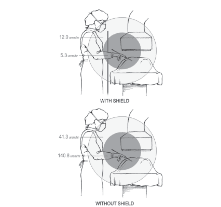
radiation and 51 mrad an hour 50 cm from the source (17). Numerous new radiation
shields that were originally used to shield urologists during cystoscopies to further
protect the urologist from the deleterious effect of radiation are now available for per-
cutaneous stone procedures (18).
In order for an effective radiation shield to be used in percutaneous procedures, the
shield should be able to be easily installed on the standard operative table and not
significantly hamper the surgeon’s view or access to the operative field (Fig. 1). Similar
shielding instruments have been described in the literature for use during gastrointestinal
(19) and interventional radiology procedures (20). Usually such shields are constructed
from 0.5 mm lead-equivalent vinyl-coated lead sheeting, and can be easily installed onto
a standard operating table using clamps that fasten onto steel supporting beams that hold
the shield onto the side of an operating table. The entire assembly takes less than 5 min
in experienced hands. The shield height can be adjusted to fit the urologist to provide an
optimal view and access to the operative field. These shields have been shown to reduce
an average of 96.1% of the radiation at a distance of 25 cm and 71.2% at a distance of
50 cm from the source of the radiation when used (17).
The NCRP states that the maximum permissible dose equivalent for occupation ex-
posure in any one year for the combined whole body is 5 rems. Given the experience of
the urologist, the number of cases performed per year, the difficulty in establishing the
percutaneous access, and the amount of intraoperative imaging required for each
endourologic procedure, an urologist may be exposed to a significant amount of radia-
tion. The radiation shield is an effective means of reducing that source of radiation.
The best method of reducing radiation exposure to the patient and urologist is to
reduce the amount of time spent near a radiation field and to shorten the time that a
fluoroscopy unit is used. As the deleterious effects of radiation is cumulative and results
in increase risk of DNA damage and cancer, the ALARA principal should be strictly
respected (15). In cases where the time is minimized and the maximal allowable distance
is employed, shielding is the most effective method of protection for the urologist. Lead
aprons and thyroid shields with an equivalent lead thickness of 0.5 mm should be used
by everyone exposed to the radiation field. In addition to the usage of lead aprons, the
urologist should also consider use of a mounted radiation shield such as the one used in
our study at the tableside to further minimize the radiation exposure owing to the close
proximity to the radiation source. The image intensifier should also be positioned above
the table with the radiation sources placed below the table to allow the table to act as a
shield and further reduce scattered radiation.
406 Yang and Bellman
As new generations of equipment, designed to reduce the dose of radiation delivered
to the patient and surgeon, such as the pulsed fluoroscopy and digital imaging fluoros-
copy unit becomes available, the combined method of shielding coupled with minimal
fluoroscopic usage time will minimize the deleterious effects of radiation to the urologist
and patient.
Conclusion
As technology advances, continuing efforts to reduce the radiation exposure of the
patient and urologist involved in endourologic procedures and improving imaging qual-
ity will be the ultimate goal for all parties involved. The use of basic, common sense
ALARA concepts, which include minimal fluoroscopic usage, maximal allowable dis-
tance from source of radiation, and implementation of effective shielding can provide a
significant reduction in radiation to both urologist and patient. The radiation shields can
also be one more step in the goal of reducing the amount of radiation exposure to the
urologist. Other methods such as the establishment of effective dose-minimizing imag-
ing protocols and the adaptation of equipment optimized in reduction of radiation expo-
Fig. 1. Exposure to radiation at 25 and 50 cm with and without radiation shield.

Chapter 21 / Physician Safety 407
sure are important steps toward the formation of a safer endourologic environment for
both the urologist and patient.
INFECTION-CONTROL PRACTICES
Introduction
With the advent of the AIDS epidemic, much attention has been focused on the
prevention of infectious disease transmission in the health-care setting. According to the
CDC, an exposure-prone invasive procedure is defined as “surgical entry into tissues,
cavities, or organs or repair of major traumatic injuries” associated with “an operating
or delivery room, emergency department, or outpatient setting, including both physi-
cian’ and dentists’ offices (21).” All procedures performed in endourology certainly
qualify as such and universal precautions should be strictly followed to minimize the risk
of exposure. In this chapter, we will briefly review the principles of universal precau-
tions, then embark on a discussion of the three most significant pathogens that can be
transmitted, namely HIV, hepatitis B, and hepatitis C. In the following discussion, let us
keep in mind that although the our discussion focuses on prevention of disease transmis-
sion from the patient to the health care worker (HCW), universal precautions are equally
effective in preventing transmission of disease from the HCW to the patient.
Universal Precautions
In 1987, the Center for Disease Control published an article in the Morbidity and
Mortality Weekly Report that was titled “Recommendations for prevention of HIV
transmission in health-care settings (1).” The recommendations included in the article
have since been known as “Universal Blood and Body Fluid Precautions.” By applying
the universal precaution principles, HCWs circumvent the need for extensive HIV test-
ing or specific HIV labeling of specimens or other materials (22). The basic tenant of
washing hands and wearing gloves, gowns, and masks is elementary to the practice of
modern sterile surgical technique. Eye protection using glasses, goggles, or face shields
should be stressed in all operative settings. Disposal of sharp instruments in special
puncture-resistant containers along with proper disposal of contaminated material and
sterilization techniques are other standard principles of the universal precautions. One
of the less implemented recommendations is to double-glove for all invasive procedures.
The benefit of this practice has consistently been shown to reduce the number of needle
sticks and cutaneous exposures during surgery (23). Given that a 25–60% occult-glove
perforation rate during surgery was documented in studies by vascular surgeons and
otolaryngologists, the additional layer offered by the second pair of gloves can be very
reassuring (24,25). It is also theorized that the amount of viral inoculum exposed to the
surgeon is also reduced to a minimum with the use of double gloves as the two layers of
latex literally wipes blood away from the penetrating instrument as it passes though the
layers of latex (26).
Whereas the practice of universal precautions focuses on what HCWs can do actively
to reduce infectious exposure risks, numerous innovations in equipment design have
been created to improve safety. Syringes that do not allow the user to recap the needle
and needles that have a recap guard, which can be slid on after usage, are a few of the
new safety measures employed on sharp instruments. Hospitals should actively encour-
age their material management departments to procure these instruments as allowed by
the budget.

408 Yang and Bellman
As for surgeons, one should weigh the benefit of replacing individual sharp instru-
ments with newly designed pieces that offer more protection. For example, the shielded
double-skin hook tenaculum can be used instead of the traditional double hook to pro-
vide exposure. Of course, the individual preference and ease of use for a particular
instrument in a particular situation can only be decided by the experience and preference
of each individual.
The operating room staff and scrub nurses should also adapt new techniques to
minimize risks. A no-pass technique should be used to avoid injury to either surgeon or
scrub nurse by avoiding direct handing off of instruments. Instead, instruments are
passed to and from the surgeon using a kidney basin as a neutral zone. Magnetic pads
can also be used to prevent instruments from slipping off the surgical field, reducing the
impulse to quickly grab for a potentially sharp instrument. Bovie and bipolar coagulators
instead of scalpels should be considered for soft tissue dissection to reduce possible
blade injury (7).
Among thoroughly educated health care professionals, universal precautions have
been demonstrated to show a decrease in body fluid exposure incidents. A study of 900
HCWs in a tertiary hospital showed that the blood contact rate was halved when univer-
sal precautions were implemented (27). It is simple and easy to implement. The mainstay
of infection prevention in endourology and any field of medicine should be focused on
adequate education of health care workers regarding universal precautions and actively
encouraging the strict adherence to them.
Blood-Borne Pathogens
To truly appreciate the infectious risk of any surgical procedure, one needs to under-
stand the nature of the disease. Of all the possible infectious pathogens that can be
transmitted during endourological cases, HIV, hepatitis B, and hepatitis C remain the
most feared. We will review each of the three diseases in detail and provide a more clear
understanding of the risks involved.
H
EPATITIS B VIRUS (HBV)
Of the three pathogens, hepatitis B is the only virus that health care workers can be
effectively vaccinated against. It is a double-stranded DNA virus that has been a known
to cause occupational infection since the 1940s (28). The method of transmission closely
parallels that of HIV: exposure to blood and other body fluids and sexual contact (29).
In the 1991 surveillance data published by the Center for Disease Control and Preven-
tion, 5100 health care workers who reported frequent blood contacts developed occupa-
tionally acquired HBV infection in the United States. Based on the natural history of the
disease, 255 (5%) of these individuals will eventually be hospitalized for their illness in
the acute period and 5 (0.1%) of those individuals would die of fulminant hepatic failure.
After the acute phase of the disease, 510 (10%) of the individuals would become chronic
carriers and 107 (21%) of the chronic carriers would succumb to cirrhosis or hepatocel-
lular carcinoma (30). Indeed, Hepatitis B is a deadly disease that will claim the lives of
a quarter of its victims.
One of the biggest risk factors associated with hepatitis B transmission from patients
to health care workers is the seroprevalence of the virus among the patients. Multiple
factors determine the seroprevalence of a particular disease, including ethnicity, country
of origin, intravenous drug use, and sexual contact with infected individuals. From a

Chapter 21 / Physician Safety 409
sample of four studies, the prevalence ranged between 0.9 and 6% in hospitalized and
emergency room patients (31–34). For health care workers, the mode of transmission of
hepatitis B virus is through blood contact from a sharp object injury. In any given case
of exposure, the risk of acquiring HBV infection is correlated to the e-antigen positivity
of the source patient. In other words, the higher the concentration of circulating HBV e
antigen in the blood, the higher the risk of infection. The risk of acquiring infection from
a single percutaneous exposure to e-antigen positive blood is approx 30% (30).
Vaccination for hepatitis B virus is an effective means of preventing infection with
HBV. A random testing of 3239 orthopedic surgeons in 1990 revealed that 90% of those
20–29 yr of age had been effectively vaccinated against the HBV, whereas only 35% of
those older than 60 yr reported vaccination (35). Another study of 770 hospital-based
surgeons in general surgery, gynecology, and orthopedics who were tested in 1992 sero-
survey in 21 hospitals showed that only 424 (55%) surgeons reported receiving at least
three doses of hepatitis B vaccine. In the same study, 225 (22%) of the 770 surgeons were
still susceptible to HBV infection based on their sero profile. Of these 225 individuals,
62 had reported receiving at least one dose of vaccine, but either were nonresponders or
had antibody levels that could not be detected (36). The findings above suggests that
although an effective means of prevention against HBV is available to most individuals,
many are still not utilizing them.
Hepatitis B prophylaxis after percutaneous injury centers on usage of hepatitis B
immunoglobulin (HBIG) and HBV vaccination sequence if the individual has not been
previously vaccinated. If the individual has been vaccinated and the antibody titer ad-
equate, no further precautions are required. If the titer is inadequate, the health care
worker should be given HBIG and a hepatitis B vaccine booster (29).
H
EPATITIS C VIRUS (HCV)
Of the three pathogens that are being discussed in detail, hepatitis C virus is probably
the least understood. It is a RNA virus that is transmitted the same way as hepatitis B
virus. Of the individuals infected, approx 20% will develop a clinically recognizable
acute infection. About half of those will progress to chronic active hepatitis and 20% of
those with chronic active hepatitis will develop cirrhosis (37). It is felt that owing to the
lower level of viremia associated with HCV infection (up to 10 trillion virion per mil-
liliter blood for HBV infection vs 100–1000 virions/mL blood for HCV infection) the
risk of transmission for each percutaneous exposure is approx 2.7–10% (38).
Data regarding the sero prevalence of HCV among patients is limited. The first-
generation tests for antibody to HCV has only been commercially available since 1990.
A random sample of 2523 patients from an urban hospital in Baltimore collected in 1991
showed that 454 (18%) individuals were seropositive (31). A separate Toronto region
study that included 3000 patients admitted to a teaching hospital revealed the
seroprevalence to be 0.5% (39). Thus the actual prevalence of HCV may vary greatly
based on the particular patient population that the surgeon interacts with.
Owing to the fact that no vaccination currently exists for HCV and treatment for the
virus is not 100% effective, it is considered a much deadlier disease compared to HBV.
Immediate post exposure prophylaxis requires topical disinfection with providone-
iodine solution. There is insufficient evidence for use of immunoglobulin after a needlestick
injury and it is currently not indicated for acute exposure. Antiviral agents are also not
recommended for lack of effectiveness (40). Treatment with interferon has been shown
to reduce the liver inflammation, but the drug is only effective in approx 50% of patients.

410 Yang and Bellman
In addition, the improvement is sustained only in 50% of those patients that responded
to interferon after cessation of treatment. Coupled with the poor effectiveness with the
multiple side effects that plague the patients during treatment, HCV should be consid-
ered a much more significant occupational hazard for surgeons.
H
UMAN IMMUNODEFICIENCY VIRUS (HIV)
Although the clinical syndrome caused by the HIV virus known as acquired immu-
nodeficiency syndrome (AIDS) has only been described in the literature within the past
20 yr, AIDS and HIV have captured the attention of the public like no other infectious
pathogens. HIV is a RNA retrovirus that uses its outer envelope protein gp-120 to bind
host cells that express the cell surface protein CD4. Once attached to the cell surface,
HIV invades the cell and integrates its DNA into the host cell chromosome via the
enzyme reverse transcriptase. After successful integration, the proviral DNA directs the
production and assembly of progeny virus, eventually leading to cell lysis and death (29).
Clinical latency averages 10 yr in the adult population. The CD4 cell population gradu-
ally declines over the course of the disease eventually leading to the immunocompro-
mised state manifested by AIDS-related complex and AIDS. HIV infection is a terminal
disease, which will eventually claim the lives of its victims. A full discussion of the
disease process is beyond the scope of this chapter, but we will focus on the important
risk factors for transmission and preventive measures to minimize exposure in the health
care setting.
Epidemiological studies have reported that approx 1.1–1.5 million Americans are
infected with HIV. The seroprevalence differs greatly depending on the population
surveyed (inner city vs suburban vs intravenous drug users) and generally range from
0.2% to 14% (41). The highest risk for HIV transmission for surgeons is due to percu-
taneous injury or mucocutaneous contact between breached skin barriers and blood. The
risk of percutaneous injury during operative procedures is related to the length of sur-
gery, amount of blood loss, and emergent nature of procedure (42). The rate for muco-
cutaneous exposure is not as well documented compared to the rate of percutaneous
exposure, but is believed to be higher compared to percutaneous exposure owing to the
higher incidence of exposure (29).
The rate of HIV infection per exposure to HIV-infected blood via hollow-bore needles
is estimated at about 0.3% (30). For solid-bore suture needles, the transmission rate is
significantly lower owing to the lower inoculum of blood delivered by the solid-bore
needles. Another important factor in infectivity rate is the titer of HIV in the patient’s
blood (43).
In addition to potential percutaneous exposure, a theoretical risk of aerosolized trans-
mission of HIV via the respiratory route was a concern in the last decade. However, no
current biologic or epidemiologic evidence support fears that transmission of aero-
solized HIV particles occurs through the respiratory route in vivo (29).
All health care institutions in the United States must have medications available to
health care workers within an hour of exposure to possible HIV infection based on CDC
guidelines. Institutions may provide “starter packs” through the institutional pharmacy
and may dispense them immediately upon approval by the exposure counselor. Before
releasing the medication, the exposure counselor should ascertain from the health care
worker whether there are underlying medical conditions, allergies to medications, or
other reasons the antiretroviral medication should not be administered. Most programs
have traditionally offered 4–6 wk of zidovudine prophylaxis based on the CDC’s recom-