Vaccari D.A., Strom P.F., Alleman J.E. Environmental Biology for Engineers and Scientists
Подождите немного. Документ загружается.

reversibly. Both have similar effects, but recovery from carbamate poisoning is faster,
making this a somewhat safer material to use. Another critical difference is that organo-
phosphate exposure, being irreversible, is cumulative. A series of small exposures succes-
sively depletes the useful supply of acetylcholinesterase until a level is reached that causes
detectable neurological effects. In fact, experiments with chickens have demonstrated that
the cumulative dose of an organophosphate pesticide that produces an effect can be less
than the single dose that would produce the same effect.
Membrane Changes A common target of industrial pollutants is the plasma membrane.
This is because many industrial chemicals, especially the solvents, are lipophilic. Once
absorbed into the organism, they preferentially partition into the lipid bilayer, changing
its physicochemical properties. These properties include its fluidity, permeability, and
physicochemical interactions with embedded proteins. The effect on proteins affects
their function, whi ch includes acting as receptors for normal biochemical functions or
as agents in active transport of ions and othe r substances across the plasma membrane.
These actions are the presumed cause of narcosis, one of the major symptoms of solvent
toxicity. In extreme cases, exposure to solvents can leach lipids out of the membrane, or
cause the membrane to rupture, destroying the cell or organelle.
Irritants Some of the more severe modes of action lead to irritation (damage to cells of
a tissue). These modes include oxidation, severe pH changes, dehydration, or precipitation
or denaturing of proteins. Dehydration can be caused by concentrated salt solutions.
Proteins can be precipitated or denatured not only by changes in pH or ionic strength,
but also by organics such as aldehydes or ketones. Examples of str ong oxidizers are
ozone and chlorine.
Free Radicals Oxidizers, ionizing radiation, and metabolic transformations of some
organic pollutants, can result in the formation of free radicals. Free radicals are com-
pounds with unpaired electrons. They are very reactive and electrophilic and therefore
tend to oxidize compounds. However, the reactions are nonspecific; that is, the radicals
tend to react with almost any compound with which they come into contact. Some
of the simpler radicals are the superoxide, peroxide, or hydroxyl free radicals:
HO
2
H
2
O
2
OH
The dot symbolizes the unpaired electron. Some toxins themselves react to become free
radicals, such as in the biotransformation of carbon tetrachloride:
CCl
4
)CCl
3
When radicals react with cellular compounds, they may either oxidize them, or they may
transform them into radicals, such as lipid or DNA free radicals, which undergo further
reactions. Several enzymes and the dietary antioxidants vitamin E, vitamin C, and glu-
tathione act to protect the cell by destroying radicals.
17.2 ABIOTIC FACTORS THAT AFFECT TOXICITY
A number of abiotic factors affect the toxicity of substances. Notable examples in aquatic
tests include the pH, temperature, dissolved oxygen content, salinity, alkalinity, and water
708 THE SCIENCE OF POISONS
hardness. The fact that all of these can have a simultaneous effect greatly complicates
the application of toxicity data to the field. In most of the following cases, the research
has been done on acute toxicity (i.e., those with rapid onset). Much less is known about
the effect of these factors on chronic toxicity (i.e., those with delayed onset).
Temperature affects organisms directly as well as by changing the response to toxins.
Thermal pollution of surface waters, often associated with cooling-wa ter discharge from
electric power plants, can affect fish behavior. By reducing the solubility of oxygen and
increasing microbial oxygen uptake rate, it can apply a stress to many aquatic organisms.
However, one cannot make generalizations about the effect of temperature on toxicity.
The threshold toxicity of zinc to Atlantic salmon was higher at 19
C than at 3
C, but
rainbow trout, which are in the same genus, were more tolerant to zinc at 3
C than at
20
C. The undissociated fraction of hydrogen cyanide shows one of the strongest tempe-
rature effects. Its LC
50
(the concentration that is lethal to 50% of test organisms) increases
with temperature in a log-linear relationship. The LC
50
increases (i.e., it becomes less
toxic) 12-fold, from 44 mg=L at 6.5
C to 530 mg=Lat25
C.
The effect of temperature can depend on other abiotic factors, thus forming a three-
way interaction between the toxin dosage, temper ature, and the other factor. In soft
water, napthalenic acids are twice as toxic to snails at 20
C than at 30
C. However, no
differences have been reported in hard water. One rule of thumb that can often be used is
that the time it take s for an organism exposed to a lethal concentration to succumb follows
a van’t Hoff relationship with temperature: survival time decreases by a factor of 2 or 3
with each 10
C increase. For this reason, shorter-term acute toxicity tests could show an
increasing toxicity with temperature, while the same test conducted for a longer time
could show decreasing toxicity.
Dissolved oxygen (DO) in water is saturated with respect to the atmosphere at concen-
trations ranging from 7.5 to 14:6mg=L, depending on temperature, salinity, and total
pressure (and therefore, altitude). Most fish will tolerate DO levels as low as 2:0mg=L,
although their activities may be reduced. DO can affect aquatic toxicity in several ways:
(1) by increasing exposure at a given activity level, since at low DO levels fish must cir-
culate more water through their gills; (2) indirectly by increasing organism activity; and
(3) by causing stress to organisms at low DO. Very commonly, there is no discernible
effect of DO, or the effects observed are due to the oxygen stress and not the toxin.
One case of strong interaction has been found in the effect of pulp mill effluent on young
salmon. Only 2% of effluent mixed with half-strength seawater caused the fish to gasp for
oxygen at the surface when the DO was reduced to 36% of saturation.
The pH range 6 .5 to 9.0 is considered harmless to fish. The pH interacts strongly with
the toxicity of weak acids and bases, such as cyanide, hydrogen sulfide, or ammonia, by
shifting the balance between ionized and nonionized forms (Sections 3.3 and 18.1).
The LC
50
of total sulfide is 64 mg=L at pH 6.5 and 800 mg=L at pH 8.7. Thus, the undis-
sociated form seems to be about 15 times as toxic as the ions. Metals are usually more
toxic in the ionic form. Although phenol is an acid, its toxicity does not change much with
pH. Alkalinity may have some effect. High-alkalinity waters are more buffered against
changes in pH. The CO
2
released by gills can cause a local decrease in pH, affecting
the toxicity of weak acids and bases. High-alkalinity waters are more buffered against
changes in pH, and therefore have less of an effect from CO
2
. Thus, at the same pH,
the toxicity of ammonia to fish would be expected to be less at lower alkalinities.
Water hardness is due primarily to dissolved calcium and magnesium. A soft water is
one with less than about 75 mg=L expressed in terms of CaCO
3
, and above 300 mg=Las
ABIOTIC FACTORS THAT AFFECT TOX ICITY 709
CaCO
3
, it is considered very hard. Surface waters tend to be soft; the world’s rivers have a
median hardness of 50 mg CaCO
3
=L. Groundwater from limestone regions range from
350 to 380 mg CaCO
3
=L.
Metal salts are less toxic in hard waters than in soft. For example, a very good correla-
tion was found between the LC
50
of copper in mg=L to salmonid fish vs. hardness (H)
in mg CaCO
3
=L:
LC
50
¼ 0:0034 H
0:91
ð17:1Þ
Based on such simple correlations, it was thought that membrane permeability was
being affected by calcium. However, if the tests w ere done in water with combina-
tions of pH and alkalinity different from those typically found in nature, the simple
relationship disappears. It seems that the re lationship was observed because in natural
waters the hardness and alkalinity vary together. Now it is thought that it is the alka-
linity that interacts with pH, a nd that t his is due to the various complex ions formed
by the equilibrium among the metal ion, hydroxide ions, and carbonate ions. For
example, copper forms at least seven species in solution, each of which may have its
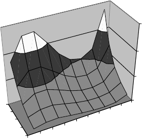
own toxicity. Figure 17.2 shows the effect of hardness and pH on the toxicity of copper
to rainbow trout. The curve represents a mathematically smoothed response sur-
face. The low toxicity at pH 8 is thought to be due to most of the copper being in
the form of carbonates and nonionized hydroxides, which may be less toxic than the
other forms.
Salinity is not an important variable in toxicity.
Temperature and humidity strongly affects the sensitivity of plants to toxins in the air.
An increase in either stimulates plants to open the leaf stomata, giving gaseous toxins
admittance to the interior of the leaves. Light intensity also affects plant response, but
the effect may be positive in some situations and negative in others.
Even time of day has been shown to have an effect on toxicity to animals. In rats and
mice the cytochrome P450 enzyme system, which metabolizes many toxins, is most active
just after dark. Factors relating to the care and handling of test animals, such as whether
they are caged individually or in groups, also affect the measured toxicity.
Figure 17-2. Hardness and pH interaction in c
“Hardness”. 478
9.0
8.0
7.0
6.0
5.0
30
100
360
0.0
200.0
400.0
600.0
pH
H (mg/L as
CaCO
3
)
96-hr LC
50
µg Cu/L
µ
Figure 17.2 Hardness and pH interaction in copper toxicity. (From Rand and Petrocelli, 1985.)
710
THE SCIENCE OF POISONS
17.3 INDIVIDUAL VARIABILITY
Even when given the same exposure to a toxin, individuals vary in their responses. The
causes of variation include genetic, nutritional, age and sex, metabolic activity level, life
stage, or exposure histor y leading to lesions, sensitization, or enzyme induction.
Several examples in humans will show that individuals of the same species can have
large genetic differences in responses to toxins. Many Orientals are genetically predis-
posed to a more rapid metabolism of ethanol to acetalydehyde. Since it is the metabolite
that is responsible for many of the symptoms of ethanol intoxication, these people react
strongly to even small doses, becoming flushed and uncomfortable. Another example is in
the inherited propensity that some people have toward specific cancers, such as retinoblas-
toma, or skin or colon cancer. These people have inherited one of the ‘‘hits’’ required to
convert a normal cell into a cancerous one. In a third example, milli ons of persons
throughout the world have red blood cells with defects in their respiration pathways,
such as a deficiency in glucose-6-phosphate dehydrogenase. These cells are not efficient
in maintaining glutathione, which protects against peroxide attacks. People with this type
of problem are especially sensitive to red blood cell hemolysis (breakdown) from certain
chemicals, such as aspirin and naphthalene, and from substances in some foods, such as
fava beans. This is an X-linked trait, exhibited only in males, who inherit it from their
mothers (Section 6.1.2).
The sex of an animal can have a great influence as well. Male mice are muc h more
sensitive than females to chloroform, possibly because males have a much higher concen-
tration of cytochrome P450. Female rats are more sensitive than males to certain organo-
phosphate pesticides. However, castration and hormone treatment render the males more
sensitive.
Young animals are typically 1.5 to 10 times as sensitive to toxins as adults, possibly
due to underdeveloped immunity or detoxification mechanisms. Malathion is about
28 times more toxic to newborn rats than to adults. However, this is not always the
case. DDT is about 20 times less toxic to newborns, and Dieldrin was about 4.5 times
less toxic. The young may absorb differently, and their blood–brain barrier is less efficient.
Preexisting disease can affect person’s response to a toxin in several ways. If the dis-
ease affects the kidney or liver, it may affect the half-life of a compound by changing the
rate of biotransformation or excretion. Of course, disease can also have an indirect effect
by rendering the person more vulnerable to any type of additional damage.
The relationship between nutrition and toxicity is now known to be a major factor
affecting toxic response. The effect occurs through altered absorption and renal function
and by affecting toxin distribution in tissues. Fasting or low-protein diets may reduce
cytochrome P450 act ivity. This can either increase toxicity (e.g., DDT) or decrease it
(e.g., chloroform). Lipids in the diet delay absorption of lipophobic substances and
enhance it for lipophilic substances. Essential fatty acids, such as linoleic acid, are impor-
tant to the cytochrome P450s. Fatty tissues can store lipophilic toxins away from
receptors. Thus, obesity actually protects against chronic toxicity of these compounds.
However, high-fat diets enhanc e absorption and retention of lead and fluoride.
Epidemiological evidence indicates that vitamin A is protective against lung cancer.
Exposing rats to PCB, DDT, and dieldrin significantly reduced the stores of vitamin A
in the liver. However, vitamin A can be toxic at high levels. On the other hand, b-carotene,
which is a precursor of vitamin A, is fairly nontoxic. Vitamins E and C are both important
antioxidants. Lipophilic vitamin E acts to protect the membranes from free radicals and
INDIVIDUAL VARIABILITY 711
other oxidizers; vitamin C does the same in the cytoplasm. In addition, in vitro experi-
ments suggest that vitamin C competes for nitrites, which are often found in preserved
foods. This blocks their reaction with amines that would otherwise form carcinogenic
nitroso compounds.
Zinc reduces the toxicity of lead and cadmium, and selenium is protective against
cadmium and mercury. Also, selenium is a coenzyme for glutathione peroxidase. Together
they destroy H
2
O
2
, which otherwise disrupts membranes by oxidation. Many metals are
enzyme cofactors (e.g., iron is essential for the cytochrome P450 system).
Enzymes may be induced by previous exposure to the toxin or to other substances. This
may persist for some time after the original exposure, affecting the response to a toxi n
until the induction effect dissipates.
17.4 TOXIC EFFECTS
The toxic modes of action result in numerous consequences, which are the toxic effects
observed. The effect of a toxic substance on an organism may be direct, induced, or
indirect. Indirect effects are those in which damage to an organism is caused by inter-
mediary effects, such as physical or chemical changes to the environment, or by loss of
food or shelter. Induced effects are those in which the toxicant renders the organism
vulnerable to other environmental forces, such as infectious disease or predation. Direct
effects are those that do not require such intermediate effects, such as toxins that produce
damage to tissues and organs, and may resu lt in its mortality.
The difference between direct and induced effects may be a matter of level of expo-
sure. At lower exposures an organism may be weakened and made vulnerable to preda-
tion. The same organism exposed to the same toxicant at a higher level may suffer
directly. Direct effects are easier to study under laboratory conditions. Often, it is difficult
to determine if an effect is direct or induced. For example, neoplasms (tumorlike growths)
have been observed in bivalve mollusks exposed to oil spills. However, experimental dif-
ficulties have prevented laboratory verification of a direct relationship.
Another way to classify toxic effects is as acute or chronic, or as lethal or nonlethal.
Acute effects are thos e that develop rapidly over a short time scale, usually within hours
to days, and usually from single or few exposures. Acute effects are usually associated
with mortality (death), but not necessarily so. Chronic effects are those that have a
long latency (time between exposure and occur rence of effect). They are often the result
of repeated exposures to lower doses than would produce acute effects. Because of the
long latency, chronic effects are inherently more difficult to study in the laboratory. It
may be necessary to maintain exposures for more than 10% of an organism’s lifetime
in order to observe them. Lethal effects are those that result in extinction of the organism.
Lethality is usually taken to mean morta lity but can also be prevention of reprodu-
ction. Sublethal effects include changes in biochemistry, physiology, histology (cell
structure), reproduction, or behavior. The latter includes changes in movement or aggres-
siveness. Behavioral changes can affect an individual’s chances of survival in the wild.
Table 17.1 summarizes a variety of sublethal toxicological endpoints.
Effects can be either reversible or nonreversible, depending on whether the damage
can be healed. A distinction is made between local effects and systemic effects. Local
effects occur where the toxicant was first contacted or absorbed, whereas systemic effects
are those that occur at other sites, therefore requiring absorption and transport. Systemic
712 THE SCIENCE OF POISONS

effects may be localize d in the sense that a particular organ is sensitive to a chemical. For
example, the air pollutant ozone has the local effect of causing damage to the linings of
the lungs, whereas the systemic toxin carbon monoxide does its damage by binding to the
hemoglobin of the red blood cells, preventing them from carrying oxygen to the tissues.
Another complicating factor is that the impact of a toxin may vary significantly accord-
ing to the life stage of the organism. For example, DDT is fairly nontoxic to adult
vertebrates at levels found in the environment. However, it causes the shells of eggs pro-
duced by birds with high body burdens to be so thin that the egg is unlikely to survive long
enough to hatch.
A toxin can also have effects at multiple levels of the biological hierarchy, from
biochemical and cellular to population and ecological .
17.4.1 Biochemical and Physiological Effects
Here the discussion is not on biochemical causes of toxic effects, as discussed in Sec-
tion 17.1, but on the secondary effects that are manifested biochemically. These are the
changes in the operation of biochemical pathways that do not involve the toxin directly.
They are the chemical response of the cell to toxin-induced damage to enzymes, reactants,
or cell structures involved in reactions. As mentioned above, the distinction is sometimes
arbitrary. For example, enzyme disruption by the complexation of metallic cofactors, or
production of free radicals by oxidants, could both be considered secondary effects. Some
TABLE 17.1 Factors Affected by Sublethal Toxic Exposures
Uptake, accumulation, and excretion
Complexation and storage, distribution within tissues and organs, kinetics of uptake and release,
bioconcentration, bioaccumulation.
Physiological effects
Photosynthesis and respiration, osmoregulation, feeding and nutrition, heartbeat rate, blood
circulation, body temperature, water balance.
Biochemical effects
Metabolism of carbohydrates, lipids, and proteins; pigmentation; enzyme activities; blood
chemistry; hormonal functions; oxygen uptake rate.
Behavioral effects (individual responses)
Sensory capacity, rhythmic activities, motor activity, appetite, equilibrium, motivation and
learning phenomena.
Behavioral effects (interindividual responses)
Migration, intraspecific attraction, aggregation, aggression, predation, vulnerability, mating.
Reproduction
Viability of eggs and sperm, breeding/mating behavior, fertilization and fertility, survival, life
stages, development of young.
Genetic
Chromosome damage, mutagenic and teratogenic effects.
Growth alterations and delays
Cell production, body and organ weights, developmental stages (e.g., larval and juvenile stages).
Histopathological effects
Abnormal growths, respiratory and sensory membrane changes, structural changes in tissue and
organ (e.g., reproductive organs).
Source: Connell and Miller (1984).
TOXIC EFFECTS
713
effects are more distinctly removed from the original chemical lesion. Examples include
mammalian effects such as allergic response, histamine production, hormone mimics, and
blood chemistry changes.
In mammals, many of the clinical measurements developed for medical use can also be
used as indicators of toxic stress. These include blood measurements, such as cell counts
and clotting times, and plasma chemical analysis such as chloride, cholesterol, glucose,
and protein. The advantage of using such measurements is that they could be more
sensitive than gross responses such as reduced growth or mortality, they could predict
long-term responses in short-term tests, and because they provide insight into the mecha-
nism of toxic action.
Many of the measurements developed for mammals have been applied to other ani-
mals, such as fish. However, the correlation between this type of data and environmental
impacts has not been sufficiently researched to be of broad use. Several biochemical
measurements have been developed specifically for fish. These include metabolic activity,
as indicated by oxygen consumption. For example, fish exposed to dieldrin required more
oxygen to swim against a current and had a reduced cruising speed.
17.4.2 Genotoxicity
Other irreversible changes include various forms of genotoxicity, or genetic damage.
These can cause three types of effects:
Mutagenesis is the formation of hereditable genetic changes.
Teratogenesis is the production of birth defects.
Carcinogenesis is the production of cancer.
17.4.3 Mutagenesis
Mutations were described in Section 6.2.3. Agents that can cause mutations are called
mutagens. Mutations can result in hereditable diseases, birth defects, or cancer. Accord-
ing to one study, 90% of carcinogens show mutagenic activity in a microbial assay (the
Ames test, Section 20.1.12). The same study found that few noncarcinogens are muta-
genic. Th e correlation between mutagenicity in laboratory tests and that in humans is
highly uncertain. Nevertheless, prudence dictates that substances known to be mutagenic
in appropriate laboratory models should be presumed to present a high risk to humans as
well.
Other types of genetic damage besides mutations can occur, including chromosomal
alterations such as broken chromosomes or change in number of chromosomes, or inexact
copying of the DNA during normal cellular replication. All of these can be caused by
toxic agents. Their results are typically more serious than mutations.
17.4.4 Teratogenesis
Birth defects are abnormal developments in embryos that are manifested either structu-
rally or functionally. Teratogenic agents act in a narrow range of doses between no obser-
vable effect levels and levels that are lethal to the embryo. Furthermore, they are
preferentially induced when the exposure occur s during a narrow time span during gesta-
tion, which corresponds to the time when organs are being formed. For humans this is
714 THE SCIENCE OF POISONS

approximately from day 26 to day 56 of gestation. The same dosage can cause different
birth defects, depending on whe n during this period the dose is administered. For exam-
ple, a cleft palate can be caused by exposure on one day, whereas heart defects may be
caused on another.
Although teratogenesis is classified here with genotoxic effects, its causes are not well
understood, and a number of other mechanisms probably also act. It seems that anything
that interferes with cell division can be a teratogen. This includes agents known to block
DNA expression, heavy metals such as lead and cadmium that inhibit enzymes, and even
substances that simply cause a delay in cell replication.
Other forms of toxicity do not correlate well with teratogenicity. Substance s that are
teratogenic may not be toxic to the mother, and vice versa. Surprisingly, many mutagens
are not teratogens. The sensitivity of the embryo can be decreased if the toxic substance
does not easily cross the placental barrier. On the other hand, the embryo may be more
sensitive than the mother to some compounds if the compounds act on cell division, since
embryos undergo this process at a high rate. Both deficiencies and excesses of some vita-
mins, such as vitamin A, are teratogenic. Other teratogens are listed in Table 17.2.
17.4.5 Carcinogenesis
Cancer is ‘‘possibly the most dreaded toxic event and probably the hardest for which to
provide reassuring safety precautions’’ (Williams and Burson, 1985). Normal cells
respond to the presence of adjacent cells by ceasing to replicate. In cancer, the mechan-
isms limiting the growth of cells become damaged. The damaged cells grow uncontrolla-
bly, forming tissue masses called tumors (except in some cases, such as leukemia, the
cancer of the white blood cells). This growth consumes the resources of the organism;
impinges on nearby tissues, causing them to atrophy; and ultimately results in mortality.
A great amount is understood about the causes and progresson of cancer, yet we are still
far from having a complete understanding, or even from knowing enough to treat the dis-
ease effectively.
The Stages of Cancer Cancer progresses in distinct stages. The first step, which pro-
duces no symptoms, is a first mutation that predisposes the cell to cancer. This step is
TABLE 17.2 Teratogenic Substances
Physical agents Hypothermia, hyperthermia, hypoxia, radiation
Agents causing hypoxia Carbon monoxide, carbon dioxide
Infections Rubella viruses, syphilis
Dietary deficiency or excess Vitamins A, D, and E, ascorbic acid, nicotinaminde, Zn, Mn,
Mg, Co
Hormone deficiency or excess Cortisone, insulin, androgens, estrogens
Natural toxins Aflatoxin B
1
, nicotine
Heavy metals Methyl mercury, lead, thallium, strontium, selenium
Solvents Benzene, carbon tetrachloride, 1,1-dichloroethane, dimethyl
sulfoxide, propylene glycol, xylene
Pesticides Insecticides, herbicides, fungicides
Other Azo dyes, antibiotics, sulfonamides, drugs (caffeine)
Source: Lu (1991).
TOXIC EFFECTS
715
called initiation. In the second step, promotion, the first clinical manifestations begin
with the formation of benign tumors. Finally, in progression, the tumors become malig-
nant, which in turn can spread to other tissues to form secondary tumors. In the nomen-
clature of cancer, the suffix -oma is appended to a tissue name to denote a benign tumor;
for example, hepatoma and osteoma are benign tumors of the liver and bone, respectively.
Malignant tumors are described using either carcinoma or sarcoma, for mesothelial or
epithelial tumors, respectively. Thus, names for the malignant tumors for liver and bone
are hepatocellular carcinoma and osteosarcoma.
Benign tumors exhibit cellular differentiation and grow by expansion, causing adja-
cent tissues to atrophy. The tumor shows some differentiation and has a clear boundary.
Benign tumor cells appear similar to normal cells under microscopic examination. Benign
tumors are usually not fatal, except when they impinge on critical tissues such as in the
brain. Brain tumors rarely become malignant because they are fatal before reaching that
stage. Benign tumors do not inevitably progress to the next stage, although their clinical
removal obviates that possibility.
Malignant tumors are undifferentiated. Instead of forming a discrete bounded struc-
ture as benign tumors do, they grow invasively into neighboring tissues. Malignant cells
appear obviously deranged. They are capable of the process of metastasis, in which
clumps of mali gnant cells migrate to other tissues through blood and lymph vessels, form-
ing secondary tumors. This rapidly increases the growth of cancerous tissue and accele-
rates the progression of clinical symptoms, especially weakness, a large amount of weight
loss, loss of various bodily functions, and pain.
What accounts for the observed stages of cancer? It is known that genetic damage
is the root cause of cancer. The damage can be either mutations or aberrations. The
latter include chromosome breakage, deletion of chromosome segments, or swapping
of segments between chromosomes. Other evidence, however, suggested involvement
of nongenotoxic agents. For example, when polynuclear aromatic hydrocarbons (PAHs)
are applied to the skin of a mouse, cancer does not occur until followed by application of
another chemical, such as phorbo l esters from croton oil. It does not even matter if the
second application is delayed for up to a year. Clearly, the PAH predisposes the skin
cells to cancer, and the esters stimulate progression to other stages. Furthermore, the
compounds that predispose were often found to be mutagenic, whereas the ones that only
stimulate progression often were not.
Types of Carcinogens The knowledge that carcinogens act by different mechanisms
led to a distinction in two types of carcinogens. The first type are called genotoxic car-
cinogens, which act either themselves or via metabolites to either damage DNA directly
or impair the processes of repair or transcription. This is initiation, as defined above, and
the chemicals are called initiators. Examples include nitrosamines, epoxides, and metals
such as cadmium, chromium, or nickel. The direct-acting genotoxins are often electro-
philic compounds that bind to DNA, similar to the action of mutagens. Others must be
biotransformed to be genotoxic and are called precarcinogens. Most genotoxic environ-
mental pollutants are in this category, including chlorinated hydrocarbons, aromatics such
as benzene, and PAHs. The mechanism for carcinogenic metals, such as arsenic, chro-
mium, and nickel, is not understood. They are thoug ht to impair DNA replication or
transcription by complexing with the DNA or associated proteins. Several nonchemical
carcinogens act by changing the cellular DNA and therefore may be classified as geno-
toxic. These include ionizing radiation and certain viruses.
716 THE SCIENCE OF POISONS
The second type are called epigenetic carcinogens or promoters. They do not affect
the DNA, but enhance the progress ion to cancer subsequent to initiation of genetic
damage by the genotoxic carcinogen. Epigenetic carcinogens act by (1) encouraging
cell division (promotion), (2) inhibiting intercellular communication, or (3) impeding
mechanisms for destroying aberrant cells. Tobacco smoke contains both initiators and
promoters.
Destruction of damaged cells is part of the function of the immune system, and immu-
nosuppressant drugs used in organ transplants are known to cause cancer by inhibiting
this function. One important group of pollutants that may act in this way is the dioxins.
They seem not to be genotoxic, but are both strong promoters and highly immunosup-
pressive. This is especially true for the form known as 2,3,7,8-tetrachlorodibenzodioxin
(2,3,7,8-TCDD).
Intercellular communication normally helps limit cell growth. Cells send each other
chemical signals that stop their division process when they are in contact. Cancer cells
do not respond to these signals.
Many environmental pollutants are promoters, not initiators. Cell division can be
stimulated a number of ways. Anything that kills or damages cells stimulates growth as
part of the healing process. This can include chemical toxins which act by other means,
and physical trauma such as burns, freezing, or possibly even mechanical injury. Even
implanted foreign solid materials, such as asbestos, plastics, metal, and glass, can promote
cancer. These are called solid-state carcinogens. A possible mechanism for this is that
their presence stimulates fibrosis, connective tissue cell growth, as the organism attempts
to encapsulate the foreign material. The more cell growth is stimulated, the greater the
chance that any cell, previously initiated by a genotoxic carcinogen, will be activated;
and the greater the chance of a transcription error causing an initiation.
Hormones are known to act as promoters. For example, estrogen administered to
menopausal females increases the risk of endometrial cancer. The synthetic hormone
diethylstilbestrol (DES) used to be given to pregnant women with high miscarriage risk
to improve their chances of carrying the pregnancy to full term. Tragically, it has been
found that daughters produced by those pregnancies are at a high risk of contracting
cervical cancer in their late teens or early 20s. Testosterone, or more precisely its meta-
bolite (dihydrotestosterone), promotes prostate cance r in men.
More important in an environmental context, many pollutants have been found to either
mimic or influence hormones. The herbicide Amitrole (aminotriazole) inhibi ts an enzyme
that uses iodine to form thyroxine. The pituitary responds to the low level of thyroxine by
stimulating thyroid growth. This in turn, can lead to cancer of the thyroid.
A variety of anthropogenic compounds found in nature have been shown to mimic the
hormone estrogen: xenoestrogens, endocrine disruptors,orenvironmental estrogens.
These include the chlorinated pesticides atrazine, chlordan e, DDT, endosulfan, kepone,
and methoxychlor, as well as dioxins and some polychlorinated biphenyl (PC B) con-
geners. Several compounds associated with plastics are xenoestrogens as well: Bisphenol
A is released by polycarbonates whe n heated. Nonylphenol is a softener for plastics used
in packaging and in flexible plastic tubing. Phthalates are also plastic softeners that are
commonly used in food packaging and which have been found in laboratory experiments
to cause reproductive disorders.The xenoestrogenic properties of some of these materials
were discovered when laboratory investigations were confounded by their presense. In
one case, cultures of breast cancer cells grew more rapidly than expected because of
contamination from laboratory plasticware. Possible xenoestrogenic effects have been
TOXIC EFFECTS 717