Watts J.F., Wolstenholme J. An Introduction to Surface Analysis by XPS and AES
Подождите немного. Документ загружается.


METALLURGY
2.0
.-
G
^
1.6
0.2
0.4 0.6
N/Ti ratio
0.8
1.0
Figure 5.13 Calibration curve
to
relate
the
convolution
of
nitrogen
and
titanium
features
to the
true N/Ti ratio which
has
been determined
by XPS
(reprinted
with permission
from
Baker,
M. A. et
al., "Auger electron spectroscopy/X-ray
photoelectron spectroscopy study
of
Ti-B-N
thin
films,"
Journal
of
Vacuum
Science
&
Technology
A,
13(3), 1995,
pp.
1633-1638,
Fig.
6.
Copyright 1995,
AVS)
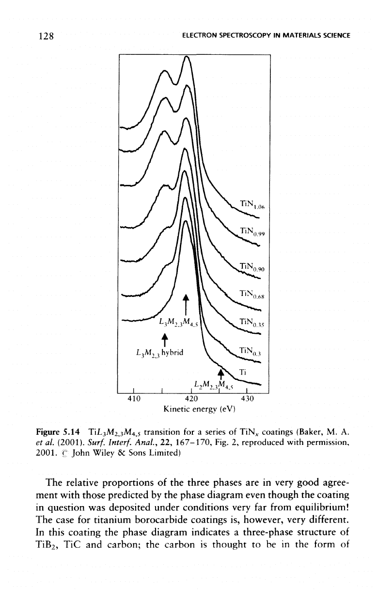
the
direct rather than
differential
form,
is
shown
in
Figure 5.14
for a
series
of
TiN
x
coatings,
and the
evolution
of a
TiL
3
M
2
,
3
hybrid
feature
at a
slightly lower kinetic energy
is
well resolved
in the
spectrum.
This hybrid
feature
is a
result
of
drawing
the Ti 3d
valence elec-
trons (close
to the
Fermi
Level)
towards
the N 2p
electron (sit-
uated below
the
Fermi
Level)
and the
formation
of a
hybrid bond.
The
involvement
of the
feature
in the
Auger
transition
is
then
seen
as
a
distinct feature
in the
Auger spectrum, which increases
in
intensity
with
increasing
nitrogen
content.
In a
similar manner
to the
previous
example
it is
possible
to
plot
a
calibration curve
of
peak area
ratio
TiL
3
M
23
hybrid/TiL
3
M
23
M
45
which
is
arguably more accurate than
the
method described using peak
to
peak heights
of the
differential
spectra, above.
The
slight drawback
is the
need
to
carry
out
careful
peak
fitting and
background removal
to
obtain
an
accurate estimation
of
the
ratio
of
components.
In
the
case
of the
boronitride
it is
possible
to
unravel
the
complexity
of
the
TiN, TiB
2
and BN
phases
in a
complex coating
by
study
of the
B
1s and N 1s XPS
spectra
as
shown
in
Figure 5.15.
128
ELECTRON SPECTROSCOPY
IN
MATERIALS
SCIENCE
410 420 430
Kinetic
energy (eV)
Figure 5.14 TiL
3
M
2,3
M
4,5
transition
for a
series
of
TiN
x
coatings (Baker,
M. A.
et
al.
(2001).
Surf.
Interf.
Anal.,
22,
167-170,
Fig.
2,
reproduced with permission,
2001.
C
John Wiley
&
Sons
Limited)
The
relative proportions
of the
three phases
are in
very good agree-
ment with those predicted
by the
phase diagram even though
the
coating
in
question
was
deposited under conditions very
far
from
equilibrium!
The
case
for
titanium borocarbide coatings
is,
however,
very
different.
In
this coating
the
phase diagram indicates
a
three-phase structure
of
TiB
2
,
TiC and
carbon;
the
carbon
is
thought
to be in the
form
of

METALLURGY
129
B
1s
TiB
1
TiB
195 190 185
Binding
energy (eV)
TiN
400
398 396
Binding
energy (eV)
Figure
5.15
B 1s and N 1s XPS
spectra
from
TiN,
TiB
2
,
BN, and
TiB
x
N
y
(reprinted
with permission from Baker,
M. A. et
al.,,
"Combined
X-ray photoelectron/Auger
electron spectroscopy glancing angle X-ray diffraction/extended X-ray absorption
on
fine
structure investigation
of
TiB
x
N
y
coatings,"
Journal
of
Vacuum Science
&
Technology
A,
15(2), 1997,
pp.
284-291,
Fig.
2.
Copyright 1997, AVS)
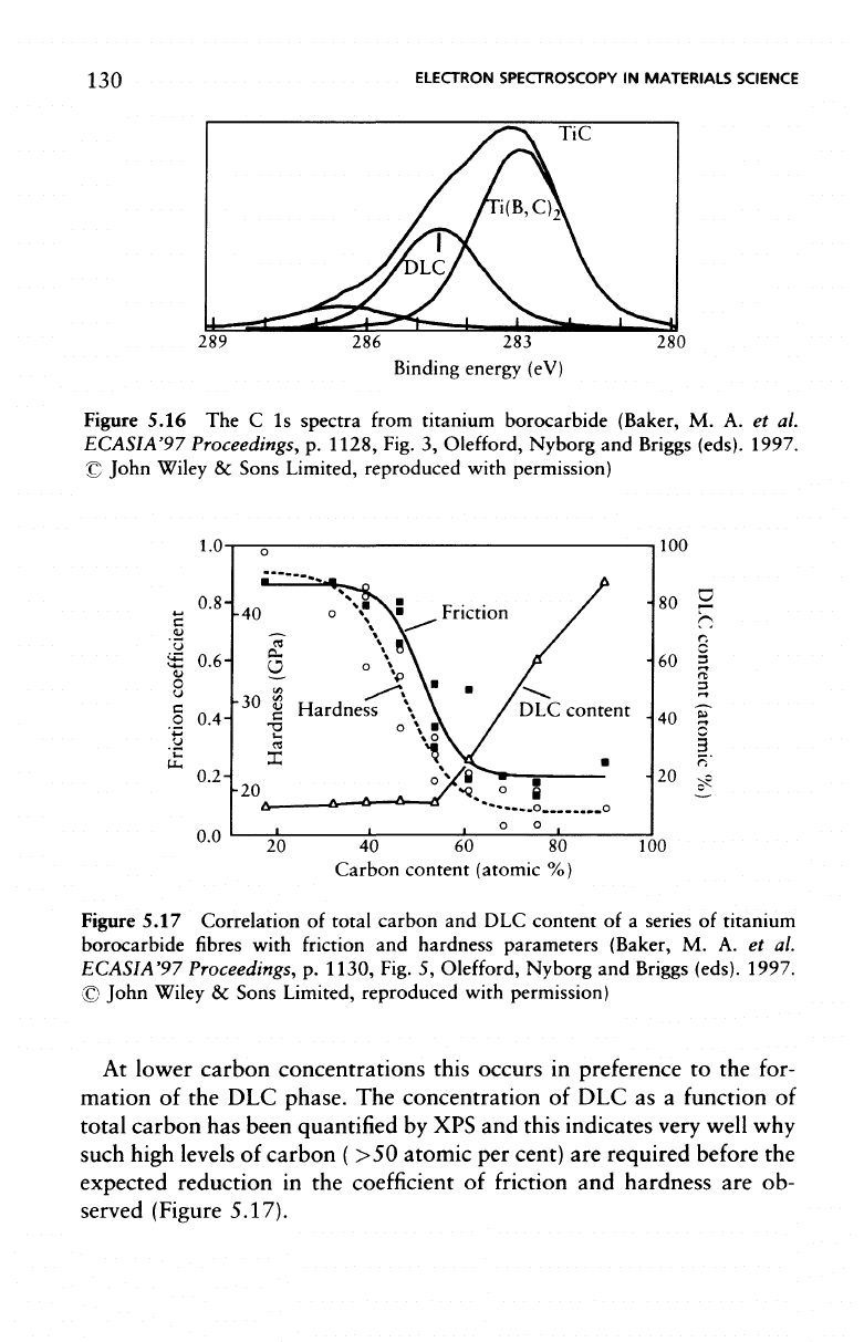
diamond-like carbon (DLC).
The C 1s
spectra indicate
that
there
is no
carbide
contribution
(Figure
5.16);
there
is,
however,
a
large component
at a
binding energy between that
of the
carbide
and
pure
carbon.
This
is
a
result
of
carbon substitution into
the
TiB2
phase instead
of
forming
the
two-phase carbide
and
boride structure.
130
ELECTRON
SPECTROSCOPY
IN
MATERIALS
SCIENCE
TiC
289
286 283
Binding
energy (eV)
280
Figure
5.16
The C 1s
spectra
from
titanium borocarbide
(Baker,
M. A. et al.
ECASIA'97
Proceedings,
p.
1128, Fig.
3,
Olefford,
Nyborg
and
Briggs
(eds).
1997.
©
John Wiley
&
Sons Limited, reproduced with
permission)
1.0
0.6-
0.4
0.2-
0.0
-40
DLC
content
-20
100
80
2
rs
r.
60
|
rs
3
40
1"
|
20 ?c
20
40 60 80
Carbon
content
(atomic
%)
100
Figure 5.17 Correlation
of
total carbon
and DLC
content
of a
series
of
titanium
borocarbide
fibres
with
friction
and
hardness parameters
(Baker,
M. A. et al.
ECASIA'97 Proceedings,
p.
1130, Fig.
5,
Olefford,
Nyborg
and
Briggs
(eds).
1997.
©
John Wiley
&
Sons Limited, reproduced with
permission)
At
lower carbon concentrations this occurs
in
preference
to the
for-
mation
of the DLC
phase.
The
concentration
of DLC as a
function
of
total
carbon
has
been
quantified
by XPS and
this indicates very well
why
such
high levels
of
carbon
( >50
atomic
per
cent)
are
required before
the
expected reduction
in the
coefficient
of
friction
and
hardness
are ob-
served
(Figure
5.17).
CORROSION
SCIENCE
These
two
examples serve
to
indicate
the
power
of XPS to
identify
and
quantify
surface
phases,
it is
then possible
to
relate such observa-
tions back
to the
phase diagram
to
establish whether equilibrium
or
metastable
phases exist
in the
coating.
5.3
Corrosion
Science
There
are two
areas within
corrosion
science
in
which electron
spectro-
scopy
has had a
dramatic impact:
the
interaction
of a
metal
surface
with
its
environment,
perhaps
to
form
a
passivating overlayer,
and the
break-
down
of the
surface
film by a
localized phenomenon such
as
pitting.
The
former
is
readily studied
by
XPS, where
the
ability
to
separate
the
spec-
trum
of the
underlying metal
from
that
of its
oxide
film
enables
the
definitive
identification
of
passivating
films on
alloys.
It
also enables
the
thickness
of
very
thin
films to be
estimated using
the
Beer-Lambert
equation
of
Chapter
1.
However, although
XPS
provides valuable information concerning
the
composition
and
perhaps growth kinetics
of
such
films,
their break-
down leading
to
major environmental
degradation
of the
metal
is a
localized
phenomenon requiring high spatial resolution surface analyti-
cal
methods, i.e., sub-micron scanning Auger
microscopy.
The
provision
of
compositional depth
profiles
of
corrosion
films is a
standard require-
ment
and may be
achieved
by
using
argon
ion
bombardment
in
con-
junction
with either
AES or
XPS.
In the
case
of
very
thin
films ( <4 nm)
angle
resolved
XPS can be
very informative.
Although
in
some cases
the
deconvolution
of the
metallic
and
cat-
ionic spectral information
is
straightforward, there
are
cases
of
great
importance
to the
corrosion scientist where
a
major research
effort
is
required
to
unravel
the
complexities
of two or
more valence states
along
with loss
features
present
in the
spectrum.
The
transition metals
in
general have presented
difficulties.
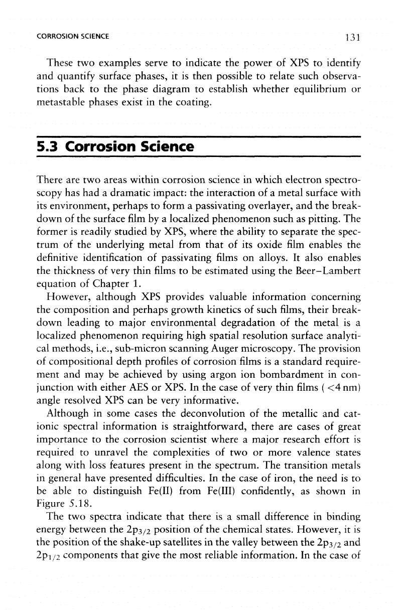
In the
case
of
iron,
the
need
is to
be
able
to
distinguish
Fe(II)
from
Fe(III)
confidently,
as
shown
in
Figure
5.18.
The
two
spectra
indicate
that
there
is a
small difference
in
binding
energy
between
the
2p
3/2
position
of the
chemical states. However,
it is
the
position
of the
shake-up
satellites
in the
valley between
the
2p
3/2
and
2p
1/2
components that
give
the
most reliable information.
In the
case
of
132
ELECTRON
SPECTROSCOPY
IN
MATERIALS
SCIENCE
Fe(III)
740
730 720
Binding
energy (eV)
710
700
Figure
5.18 Curve
fitting of
iron
2p
3/2
spectra
of (a)
Fe(III)
and (b)
Fe(II)
(the
satellites
(S)
provide
a
valuable
aid to
assignment
of
chemical state)
copper,
the
shake-up satellites provide
a
clear route
to the
identification
of
copper
(II),
as
seen
in
Figure 3.6,
but the
separation
of
metallic
copper
and the
monovalent
ion
requires
the use of the
X-ray induced
Auger
transition.
In
the
case
of the
Fe(II)
state,
these features give rise
to a
broadening
on
the
lower binding-energy side
of the
valley
between
the Fe
2p
3/2
and
Fe
2p
1/2
components
(Figure
5.18(a)),
for
Fe(III)
on the
higher-energy
side (Figure 5.18(b)).
The
curve
fitted
spectra
of
Figure 5.18 show
the
relative positions
and
intensities
of the
satellites.
In the
case
of a
mixed
phase,
all
these components, together with Fe(0)
(if
the film is
very
thin)
must
be
considered, preferably
for
both
2p
peaks
as a
check
for
self-
consistency.
The
backgrounds
of the
individual singlets will also
vary
with depth distribution.
It is
only
by
using sophisticated computer
curve-fitting
routines
in
combination with extensive knowledge
of
peak
shape
and
relative intensities, that this
has
become possible. This type
of
CORROSION SCIENCE
133
information
is
well documented
for all
elements
of
interest
to the
corro-
sion
scientist although,
in
some cases,
it is
necessary
to
consider
the
X-ray
induced
Auger peaks
in the XPS
spectrum
as
well. These
are
particularly
informative
for
magnesium, copper,
and
zinc, where
the
photoelectron
spectra alone
do not
provide unambiguous chemical
state
information.
A
little used,
but
nevertheless valuable, method
in
which surface anal-
ysis
can
assist
in the
determination
of
electrochemical history,
is by the
monitoring
of
cations
or
anions absorbed
from
aqueous solutions.
For
instance,
if a
metal electrode
is
polarized cathodically
in
MgCl2 solu-
tion,
it
will preferentially adsorb cations
on the
metal surface, seen
in
the
electron spectrum (XPS
or
AES)
as an
excess
of
magnesium
to
chlorine.
If the
electrode
has
been polarized anodically
the
reverse
is
true. This approach
is
useful
in
establishing
if a
pit,
or
other corrosion
site
is
active
or
benign,
it can
also
be
used
to
assess
the
potential
distribution
around
an
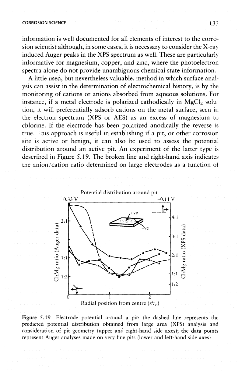
active pit.
An
experiment
of the
latter type
is
described
in
Figure 5.19.
The
broken
line
and
right-hand axis indicates
the
anion/cation ratio determined
on
large electrodes
as a
function
of
Potential distribution around
pit
0.33V
-0.11
2:1
•2
1:1
4:1
X
.2
£
OS
i-4
60
1:2
0 1 2
Radial
position
from
centre
(r/r
0
)
Figure
5.19 Electrode potential around
a
pit:
the
dashed line represents
the
predicted potential distribution obtained
from
large area (XPS) analysis
and
consideration
of pit
geometry (upper
and
right-hand side axes);
the
data points
represent
Auger
analyses
made
on
very
fine
pits
(lower
and
left-hand
side
axes)
134
ELECTRON
SPECTROSCOPY
IN
MATERIALS SCIENCE
electrode potential
for a
constant charge passed, plotted against
the
predicted potential distribution
for
small
pits. Microanalysis
of
three
pits
of
about
2 um in
diameter
by
Auger spectroscopy allows
the
elec-
trode
potential
distribution around
a pit to be
determined experimen-
tally,
and
shows excellent agreement with that predicted
by the
broken
line
(the combination
of XPS and
theory), indicating
an
active anodic
centre
to the pit
surrounded
by a
cathodic halo
as
illustrated
in the
schematic
diagram
of
Figure 5.19.
Such
pitting
may be
related
to
microstructural
features
of the
alloy,
and the
addition
of
X-ray analysis
facilities
to a
scanning Auger micro-
scope enables features such
as
inclusions
to be
identified
and
imaged
at
the
same time
as the
surface
phases.
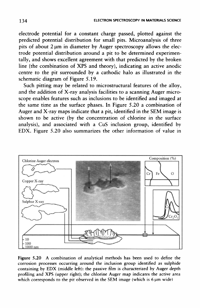
In
Figure 5.20
a
combination
of
Auger
and
X-ray maps indicate that
a
pit,
identified
in the SEM
image
is
shown
to be
active
(by the
concentration
of
chlorine
in the
surface
analysis),
and
associated with
a CuS
inclusion group,
identified
by
EDX.
Figure
5.20 also
summarizes
the
other
information
of
value
in
Figure 5.20
A
combination
of
analytical methods
has
been used
to
define
the
corrosion
processes
occurring around
the
inclusion group identified
as
sulphide
containing
by EDX
(middle
left):
the
passive
film is
characterized
by
Auger depth
profiling
and XPS
(upper right);
the
chlorine Auger
map
indicates
the
active area
which
corresponds
to the pit
observed
in the SEM
image
(which
is 6 urn
wide)
CORROSION
SCIENCE
135
corrosion
studies,
the
chemical state information
from
XPS,
and the
compositional change within
the
very
thin passive
film
obtained
by-
sputter
depth profiling.
The
versatility
of a
scanning Auger microscope
fitted
with
an
energy
dispersive
X-ray detector
has
been illustrated
by
studies
of the
initiation
of
pitting
corrosion
in a
stainless steel
at
oxide
and
manganese sulphide
inclusions.
A
system
of
this type
is
illustrated
in the
next
Chapter,
(Figure
6.3,
p.
169).
In
order
to
follow
the
corrosion process
as a
function
of
time
it
is
necessary
to
re-establish
the
specimen repeatedly
in the
Auger system
for
analysis, following exposure
to the
corrosive environment. This
is
best
achieved
by the
judicious
use of
microhardness indents around
the
inclusion
group
of
interest. Although inclusions
in
steels will have good
contrast
in
optical
microscopy this
is not the
case with
electron
micro-
scopy and, until
corrosion
features
are
evident
in the
secondary electron
image,
relocation
of the
specimen
is a
very uncertain process. Hence
the
need
for
physical markings such
as the
microhardness indents.
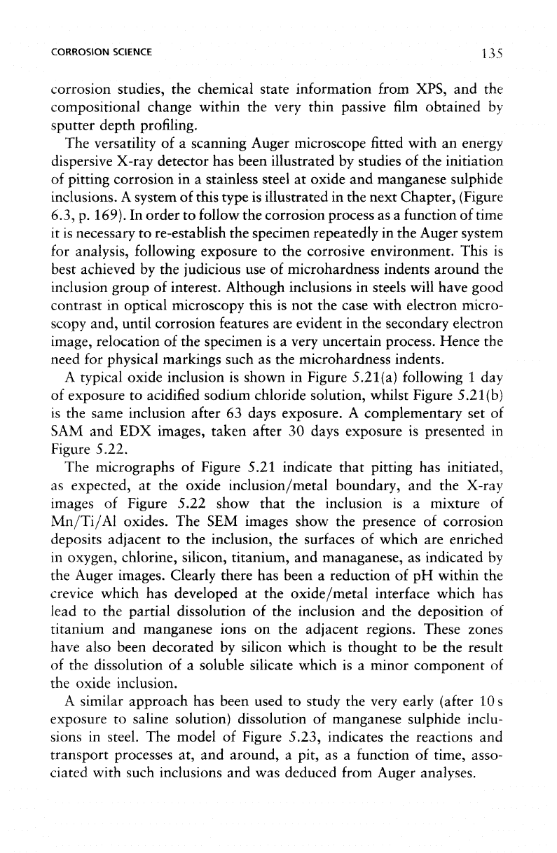
A
typical oxide inclusion
is
shown
in
Figure 5.21
(a)
following
1 day
of
exposure
to
acidified sodium chloride solution, whilst Figure 5.2l(b)
is
the
same inclusion
after
63
days exposure.
A
complementary
set of
SAM
and EDX
images, taken
after
30
days exposure
is
presented
in
Figure
5.22.
The
micrographs
of
Figure 5.21 indicate that pitting
has
initiated,
as
expected,
at the
oxide inclusion/metal boundary,
and the
X-ray
images
of
Figure 5.22 show that
the
inclusion
is a
mixture
of
Mn/Ti/Al
oxides.
The SEM
images show
the
presence
of
corrosion
deposits adjacent
to the
inclusion,
the
surfaces
of
which
are
enriched
in
oxygen,
chlorine,
silicon,
titanium,
and
managanese,
as
indicated
by
the
Auger images. Clearly there
has
been
a
reduction
of pH
within
the
crevice
which
has
developed
at the
oxide/metal interface which
has
lead
to the
partial dissolution
of the
inclusion
and the
deposition
of
titanium
and
manganese ions
on the
adjacent regions. These zones
have
also been decorated
by
silicon which
is
thought
to be the
result
of
the
dissolution
of a
soluble silicate which
is a
minor component
of
the
oxide inclusion.
A
similar approach
has
been used
to
study
the
very early
(after
10s
exposure
to
saline solution) dissolution
of
manganese sulphide
inclu-
sions
in
steel.
The
model
of
Figure 5.23, indicates
the
reactions
and
transport processes
at, and
around,
a
pit,
as a
function
of
time, asso-
ciated
with such inclusions
and was
deduced
from
Auger analyses.
136
ELECTRON SPECTROSCOPY
IN
MATERIALS SCIENCE
Figure
5.21
A
typical
oxide
inclusion
(a)
following
1 day of
exposure
to
acidified
sodium chloride
solution,
and (b)
after
63
days
exposure
(reprinted from Baker,
M. A. and
Castle,
J. E. The
initiation
and
pitting
corrosion
of
stainless steels
at
oxide
inclusions. Corrosion Science,
33, pp.
1295-1312,
Fig.
2.
Copyright 1992, with
permission
from
Elsevier Science)