Watts J.F., Wolstenholme J. An Introduction to Surface Analysis by XPS and AES
Подождите немного. Документ загружается.

MECHANICAL SECTIONING
43
Mechanical
Sectioning
To
analyse
to
greater depths than
is
practical using sputtering,
it is
necessary
to
resort
to an ex
situ,
mechanical process
for
removing
ma-
terial.
Two
related methods will
be
outlined here, angle lapping
and
ball
cratering.
4.3.1
Angle
lapping
When this method
is
employed, material
is
removed
by
polishing
the
specimen
at a
very shallow angle
(
<3°)
and
then introducing
the
sam-
ple,
with
any
buried interface
now
exposed, into
the
spectrometer.
A
brief
ion
etch
to
remove contamination
is all
that
is
needed prior
to
analysis.
By
carrying
out
Auger point analyses
in a
stepwise manner
the
variation
of
concentration with depth
is
established
and it is a
matter
of
simple geometry
to
convert
the
position
of the
analysis
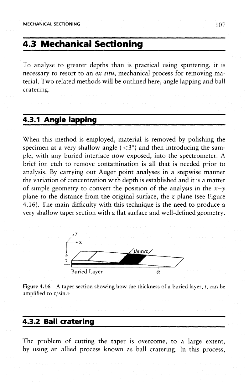
in the x-y
plane
to the
distance from
the
original surface,
the z
plane (see Figure
4.16).
The
main
difficulty
with this technique
is the
need
to
produce
a
very
shallow taper section with
a
flat
surface
and
well-defined geometry.
Buried
Layer
Figure
4.16
A
taper
section showing
how the
thickness
of a
buried layer,
t, can be
amplified
to
t/sina
4.3.2
Ball
cratering
The
problem
of
cutting
the
taper
is
overcome,
to a
large extent,
by
using
an
allied process known
as
ball cratering.
In
this
process,
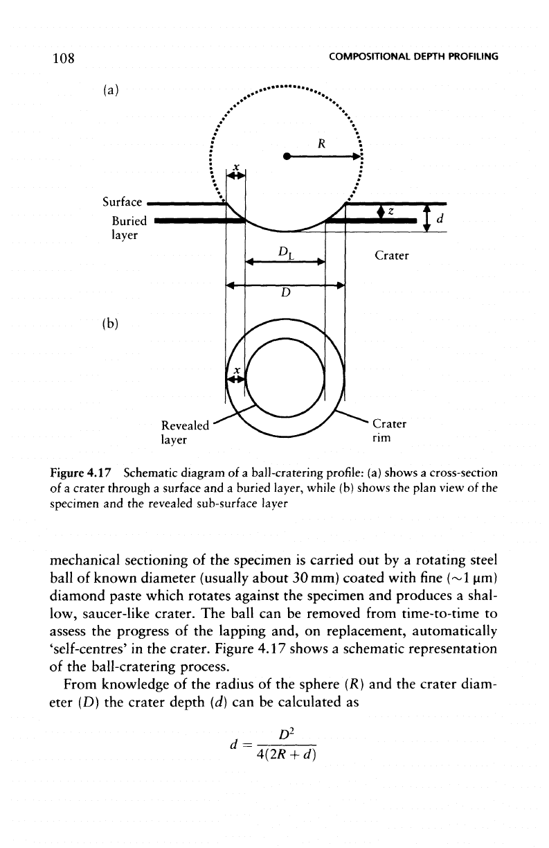
108
COMPOSITIONAL
DEPTH PROFILING
Revealed
layer
Figure
4.17 Schematic diagram
of a
ball-cratering profile:
(a)
shows
a
cross-section
of
a
crater through
a
surface
and a
buried layer, while
(b)
shows
the
plan view
of the
specimen
and the
revealed sub-surface
layer
mechanical sectioning
of the
specimen
is
carried
out by a
rotating steel
ball
of
known diameter
(usually
about
30 mm)
coated
with
fine (~1
urn)
diamond
paste which rotates against
the
specimen
and
produces
a
shal-
low,
saucer-like
crater.
The
ball
can be
removed
from
time-to-time
to
assess
the
progress
of the
lapping and,
on
replacement, automatically
'self-centres'
in the
crater. Figure 4.17 shows
a
schematic representation
of
the
ball-cratering process.
From
knowledge
of the
radius
of the
sphere
(R) and the
crater
diam-
eter
(D) the
crater depth
(d) can be
calculated
as
d
=
D
2
4(2R
+ d)
MECHANICAL SECTIONING
109
but,
as d is
very small compared with
R,
this approximates
to
d
=
D
2
/8R
If
x is the
radial distance
from
the
edge
of the
crater
to the
revealed
buried
layer then
the
depth
of the
layer,
z, can be
shown
to be:
By
recording Auger point analyses along
the
surface
of the
crater
a
compositional depth profile
can be
determined.
If
there
are
buried inter-
faces
of
special interest,
ion
sputtering
may be
used
at a
point
on the
crater close
to the
interface
to
obtain
better depth
resolution.
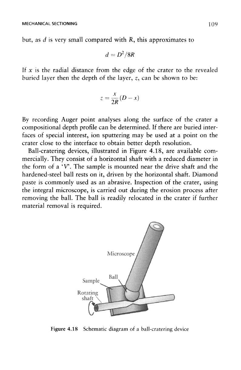
Ball-cratering devices, illustrated
in
Figure 4.18,
are
available com-
mercially. They
consist
of a
horizontal
shaft
with
a
reduced diameter
in
the
form
of a
'V'.
The
sample
is
mounted near
the
drive
shaft
and the
hardened-steel ball rests
on it,
driven
by the
horizontal shaft. Diamond
paste
is
commonly used
as an
abrasive. Inspection
of the
crater, using
the
integral microscope,
is
carried
out
during
the
erosion process
after
removing
the
ball.
The
ball
is
readily relocated
in the
crater
if
further
material
removal
is
required.
Figure
4.18 Schematic diagram
of a
ball-cratering device
COMPOSITIONAL
DEPTH
PROFILING
Ball
cratering works well
for
metals
and
oxides
but
there
are
problems
with
both
soft
and
certain brittle materials. Polymers
are
extremely
difficult
to
handle
but
some success
has
been obtained
by
using
a
ball-
cratering
machine equipped with
a
cryostage.
4.4
Conclusions
Sputter depth profiling
is by far the
most popular means
of
producing
a
compositional
depth
profile
in
surface analysis. Although
the
above
discussions suggest that
it is
fraught
with
difficulties,
it is
fair
to say
that
the
majority
of the
problems
can be
circumvented
or
reduced
to an
acceptable level
by
careful
experimental techniques.
It is for
this reason
that,
as a
method
for
depth profiling,
it is
widely used
in
studies
of
metals, oxides, ceramics,
and
semiconductors,
as we
shall
see in the
next
chapter.
The
analysis depth which
is
feasible varies with
the
sample
and the
system
employed
but the
upper limit
is of the
order
of a few
microns.
The
main
reason
for
this
is the
time
for the
experiment. Sputter profiling
becomes unsuitable when
the
layer
thicknesses become either
very
large
or
very small. When
the
thickness
is so
great that
the
time
for the
experiment
would
be
excessive then either angle lapping
or
ball crater-
ing
should
be
considered.
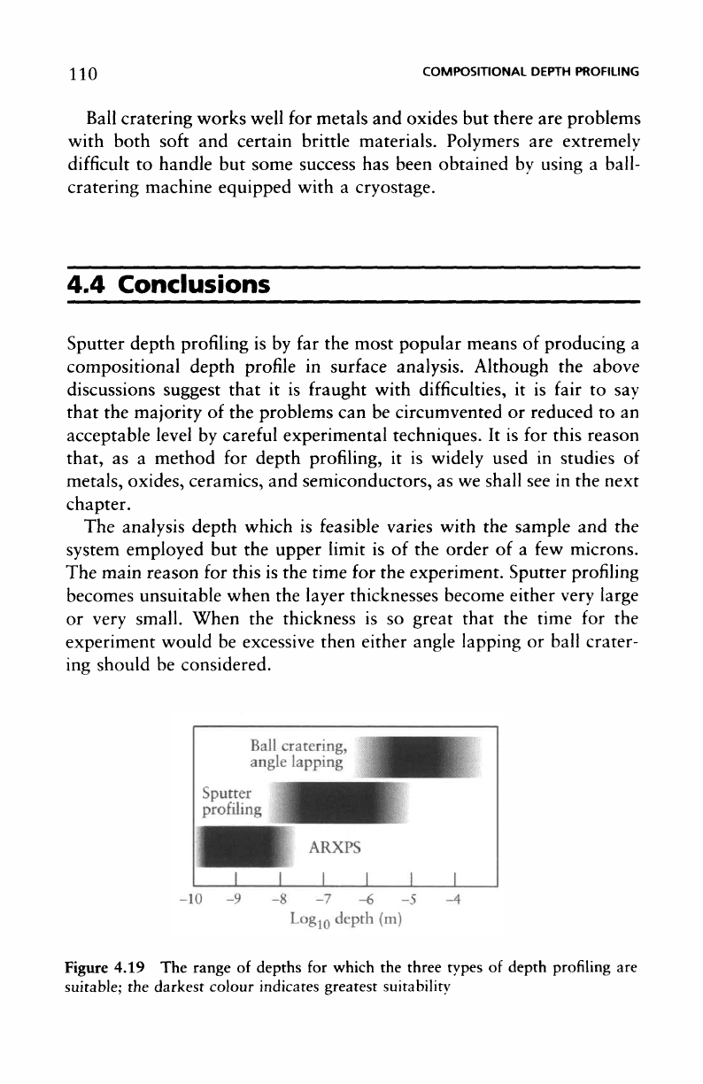
Figure
4.19
The
range
of
depths
for
which
the
three types
of
depth
profiling
are
suitable;
the
darkest colour indicates greatest suitability
CONCLUSIONS
1 I |
For
very
thin layers there
may be
problems associated with
the
attain-
ment
of the
so-called sputter equilibrium leading
to
uncertainties
in the
sputter
yield. Furthermore,
the
process
of
sputtering
can
lead
to
changes
in
the
chemical composition
of the
material. Under these circumstances
the use of
ARXPS
has
many advantages, bearing
in
mind that
it is
only
appropriate
for
layers
up to
about
10 nm.
Figure
4.19 shows
the
range
of
depths
for
which each
of the
three
types
of
profiling method
is
appropriate.
The
depth ranges will depend,
to
some
extent,
upon
the
material
and so
Figure 4.19 should
be
regarded
as a
guide
only.

This page intentionally left blank
5
Applications
of
Electron
Spectroscopy
in
Materials
Science
5.1
Introduction
So
far,
in
this text,
we
have been concerned with
the
practice
of
electron
Spectroscopy
and the
interpretation
of the
resultant spectra.
We
will
now
consider
the way in
which
it is
possible
to
make
use of
these
surface
analysis
techniques
to
provide information which
furthers
our
knowledge
in
a
particular discipline. Although
XPS and AES
together with
SAM are
used
widely
in all
branches
of
pure
and
applied sciences
- as
well
as for
trouble
shooting
and
quality assurance purposes
- the
only area
that
we
will
consider
in
this chapter
is
their
use in
materials science investigations.
If
we
subdivide this group,
it is
possible
to
identify
the
following applica-
tions headings: metallurgy (including surface engineering),
corrosion,
microelectronic materials
and
devices, polymers,
and
adhesion.
We
shall
consider each
of
these areas
in
turn; representative references
for
each
are
listed
in the
Bibliography which will provide interested readers with
further
examples
and
guidance
in
their particular
field.
5.2
Metallurgy
In
the field of
metallurgy
it is
Auger electron Spectroscopy which
has
proved
to be the
most popular technique,
and
with good reason.
The
majority
of
investigations
are
concerned with
the
diffusion
of
elements
ELECTRON
SPECTROSCOPY
IN
MATERIALS
SCIENCE
within metallic matrices. This
may
take
the
form
of
interdiffusion
of
metallic
coatings
with
the
substrate,
or the
surface
segregation
of
minor
alloying elements
on
heating
in
oxidizing
or
reducing atmospheres.
However,
the
major
contribution
of
Auger
electron
spectroscopy
to
metallurgy, especially
in the
early days
of the
development
of
surface
analysis,
was the
investigation
of
grain-boundary segregation
and
embrittlement
in
structural steels.
In
addition,
both
AES and XPS
have
been used
in
'quality assurance'
and
sometimes 'forensic' roles
to
ensure
(for
example) rolled-steel sheet
is of
adequate cleanliness,
or to
identify
surface
phases which lead
to
poor compaction
in
powder metallurgy
processing.
5.2.1
Grain-boundary
segregation
The
embrittlement
of
structural steels results from
the
aggregation
of
certain elements, present
in
very
low or
trace quantities
in the
bulk
material,
at the
prior austenite grain-boundaries.
The
grain boundaries
are
weakened
to
such
an
extent
that
they become
the
preferred fracture
path,
with catastrophic
effects
on the
material's mechanical integrity.
The
elements
most
widely investigated
are
phosphorus
and
sulphur
but
the
effect
is
brought about
by, and has
been studied for, silicon, germa-
nium,
arsenic,
selenium, tin, antimony, tellurium,
and
bismuth.
The
quantity
of
grain-boundary segregant involved
is
necessarily very small,
probably sub-monolayer,
and
located
at the
grain-boundary within
a
material
of
grain size
of
about
100 urn or
less.
Thus,
the
need
for
surface
specificity
and
reasonable spatial resolution
is
immediately apparent.
In
order
to
measure
the
quantity
of
segregant
at the
interface,
the
steel
must
be
fractured
in an
intergranular manner usually
at, or
near, liquid
nitrogen temperature. This must
be
carried
out
within
the UHV
envi-
ronment
of the
spectrometer
to
prevent oxidation
of the
iron matrix
and
subsequent obliteration
of the
small signal
from
the
segregant. Nowa-
days, most manufacturers
offer
such
a
fracture stage
for
their Auger
microscopes,
the
more sophisticated having
the
ability
to
analyse both
fracture
surfaces
(Figure
5.1).
All
rely
on
fracture
by a
fast
three point
bend configuration (similar
to the
geometry referred
to by the
metallur-
gist
as an
Izod Test). Scientists requiring controlled strain-rate
fracture
must still
resort
to
building their
own
devices.
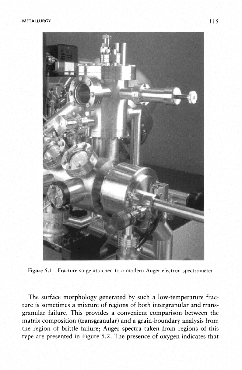
METALLURGY 115
Figure
5.1
Fracture stage attached
to a
modern Auger electron spectrometer
The
surface
morphology generated
by
such
a
low-temperature
frac-
ture
is
sometimes
a
mixture
of
regions
of
both intergranular
and
trans-
granular
failure.
This provides
a
convenient comparison between
the
matrix
composition (transgranular)
and a
grain-boundary analysis
from
the
region
of
brittle failure; Auger spectra taken from regions
of
this
type
are
presented
in
Figure 5.2.
The
presence
of
oxygen indicates that
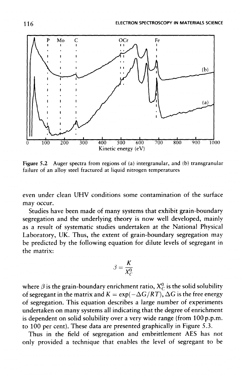
116
ELECTRON
SPECTROSCOPY
IN
MATERIALS
SCIENCE
100 200 300 400 500 600
Kinetic
energy (eV)
700 800 900
1000
Figure
5.2
Auger
spectra
from
regions
of (a)
intergranular,
and (b)
transgranular
failure
of an
alloy
steel
fractured
at
liquid
nitrogen
temperatures
even under clean
UHV
conditions some contamination
of the
surface
may
occur.
Studies have been made
of
many systems that exhibit grain-boundary
segregation
and the
underlying theory
is now
well developed,
mainly
as
a
result
of
systematic studies undertaken
at the
National Physical
Laboratory,
UK.
Thus,
the
extent
of
grain-boundary segregation
may
be
predicted
by the
following equation
for
dilute
levels
of
segregant
in
the
matrix:
i
— _r\
X*
where
(3
is the
grain-boundary enrichment ratio,
X
0
C
is the
solid solubility
of
segregant
in the
matrix
and K =
exp(- AG/RT),
AG is the
free
energy
of
segregation.
This
equation describes
a
large number
of
experiments
undertaken
on
many systems
all
indicating
that
the
degree
of
enrichment
is
dependent
on
solid solubility over
a
very wide range
(from
100 p.
p.m.
to 100 per
cent).
These
data
are
presented graphically
in
Figure 5.3.
Thus
in the field of
segregation
and
embrittlement
AES has not
only
provided
a
technique that enables
the
level
of
segregant
to be