Berg J.M., Tymoczko J.L., Stryer L. Biochemistry
Подождите немного. Документ загружается.

altered and elaborated in the Golgi complex. The O-linked sugar units are fashioned there, and the N-linked sugars,
arriving from the ER as a component of a glycoprotein, are modified in many different ways. Second, the Golgi complex
is the major sorting center of the cell. Proteins proceed from the Golgi complex to lysosomes, secretory granules (as is
the case for the elastase zymogen), or the plasma membrane, according to signals encoded within their amino acid
sequences and three-dimensional structures (Figure 11.24).
The Golgi complex of a typical mammalian cell has 3 or 4 membranous sacs (cisternae), and those of many plant cells
have about 20. The Golgi complex is differentiated into (1) a cis compartment, the receiving end, which is closest to the
ER; (2) medial compartments; and (3) a trans compartment, which exports proteins to a variety of destinations. These
compartments contain different enzymes and mediate distinctive functions.
The N-linked carbohydrate units of glycoproteins are further modified in each of the compartments of the Golgi
complex. In the cis Golgi compartment, three mannose residues are removed from the oligosaccharide chains of proteins
destined for secretion or for insertion in the plasma membrane. The carbohydrate units of glycoproteins targeted to the
lumen of lysosomes are further modified. In the medial Golgi compartments of some cells, two more mannose residues
are removed, and two N- acetylglucosamine residues and a fucose residue are added. Finally, in the trans Golgi, another
N-acetylglucosamine residue can be added, followed by galactose and sialic acid, to form a complex oligosaccharide
unit. The sequence of N-linked oligosaccharide units of a glycoprotein is determined both by (1) the sequence and
conformation of the protein undergoing glycosylation and by (2) the glycosyltransferases present in the Golgi
compartment in which they are processed. Note that, despite all of this processing, N-glycosylated proteins have in
common a pentasaccharide core (see Figure 11.19). Carbohydrate processing in the Golgi complex is called terminal
glycosylation to distinguish it from core glycosylation, which takes place in the ER. Tremendous structural
diversification can occur as a result of the terminal glycosylation process.
11.3.5. Mannose 6-phosphate Targets Lysosomal Enzymes to Their Destinations
A carbohydrate marker directs certain proteins from the Golgi complex to lysosomes. A clue to the identity of this
marker came from analyses of I-cell disease (also called mucolipidosis II), a lysosomal storage disease. Lysosomes
are organelles that degrade and recycle damaged cellular components or material brought into the cell by endocytosis.
Patients with I-cell disease suffer severe psychomotor retardation and skeletal deformities. Their lysosomes contain large
inclusions of undigested glycosaminoglycans (Section 11.2.4) and glycolipids (Section 12.2.3) hence the "I" in the
name of the disease. These inclusions are present because at least eight acid hydrolases required for their degradation are
missing from affected lysosomes. In contrast, very high levels of the enzymes are present in the blood and urine. Thus,
active enzymes are synthesized, but they are exported instead of being sequestered in lysosomes. In other words, a whole
series of enzymes is mislocated in I-cell disease. Normally, these enzymes contain a mannose 6-phosphate residue, but,
in I-cell disease, the attached mannose is unmodified (Figure 11.25). Mannose 6-phosphate is in fact the marker that
normally directs many hydrolytic enzymes from the Golgi complex to lysosomes. I-cell patients are deficient in the
phosphotransferase catalyzing the first step in the addition of the phosphoryl group; the consequence is the mistargeting
of eight essential enzymes.
11.3.6. Glucose Residues Are Added and Trimmed to Aid in Protein Folding
The oligosaccharide precursors added to proteins may play a role in protein folding as well as in protein targeting. As we
have seen, before a glycoprotein leaves the ER, two glucosidases cleave the three glucose residues of the oligosaccharide
in a step-by-step fashion. If the protein is properly folded, it moves to the Golgi complex for further processing (Section
11.3.3). However, if the protein is sufficiently unfolded that the oligosaccharide can act as a substrate for
glucosyltransferase, another enzyme residing in the lumen of the ER, a glucose residue will be reattached (Figure 11.26).
This residue, in turn, is bound by one of two chaperone proteins called calnexin and calretic-ulin. Calnexin, the more
fully understood of the two proteins, is membrane bound, whereas calreticulin is a soluble component of the ER lumen.
Unfolded proteins held by these carbohydrate-binding proteins (lectins, Section 11.4) cannot leave the ER, giving the
unfolded proteins time to fold properly. When a chaperone releases the bound protein, the glucose residue will be
cleaved by a glucosidase. If the folding is correct, the protein moves to the Golgi complex. Otherwise, the protein will
repeat another cycle of glucose addition and binding until the glucose-free (and, hence, properly folded) protein can be
translocated to the Golgi complex. This qualitycontrol system reveals an important principle: carbohydrates carry
information. Here, the availability of carbohydrates to specific glycosyltransferases conveys information about the
folding state of the protein. Moreover, we see the reiteration of a theme in the control of protein folding: other chaperone
proteins rely on the same essential mechanism of allowing misfolded proteins multiple attempts to reach a folded state
(Section 3.6), even though carbohydrate modification is not a part of their reaction cycles.
11.3.7. Oligosaccharides Can Be "Sequenced"
Given the large diversity of oligosaccharide structures and the many possible points of attachment to most proteins, how
is it possible to determine the structure of a glycoprotein? Most approaches are based on the use of enzymes that cleave
oligosaccharides at specific types of linkages. For example, N-linked oligosaccharides can be released from proteins by
an enzyme such as Peptide N-glycosidase F, which cleaves the N-glycosidic bonds linking the oligosaccharide to the
protein. The oligosaccharides can then be isolated and analyzed. Through the use of MALDI-TOF or other mass
spectrometric techniques (Section 4.1.7), the mass of an oligosaccharide fragment can be determined. However, given
the large number of potential monosaccharide combinations, many possible oligosaccharide structures are consistent
with a given mass. More complete information can be obtained by cleaving the oligosaccharide with enzymes of varying
specificities. For example, β-1,4-galactosidase cleaves β-glycosidic bonds exclusively at galactose residues. The
products can again be analyzed by mass spectrometry (Figure 11.27). The repetition of this process with the use of an
array of enzymes of different specificity will eventually reveal the structure of the oligosaccharide.
The points of oligosaccharide attachment can be determined through the use of proteases. Cleavage of a protein by
applying specific proteases yields a characteristic pattern of peptide fragments that can be analyzed chromatographically
(Section 4.2.1). The chromatographic properties of peptides attached to oligosaccharides will change on glycosidase
treatment. Mass spectrometric analysis or direct peptide sequencing can reveal the identity of the peptide in question and,
with additional effort, the exact site of oligosaccharide attachment.
Posttranslational modifications such as glycosylation greatly increase the complexity of the proteome. A given protein
with several potential glycosidation sites can have many different glycosylated forms (sometimes called glycoforms),
each of which may be generated only in a specific cell type or developmental stage. Now that the sequencing of the
human genome is essentially complete, the characterization of the much more complex proteome, including the
biological roles of specifically modified proteins, can begin in earnest.
I. The Molecular Design of Life 11. Carbohydrates 11.3. Carbohydrates Can Be Attached to Proteins to Form Glycoproteins
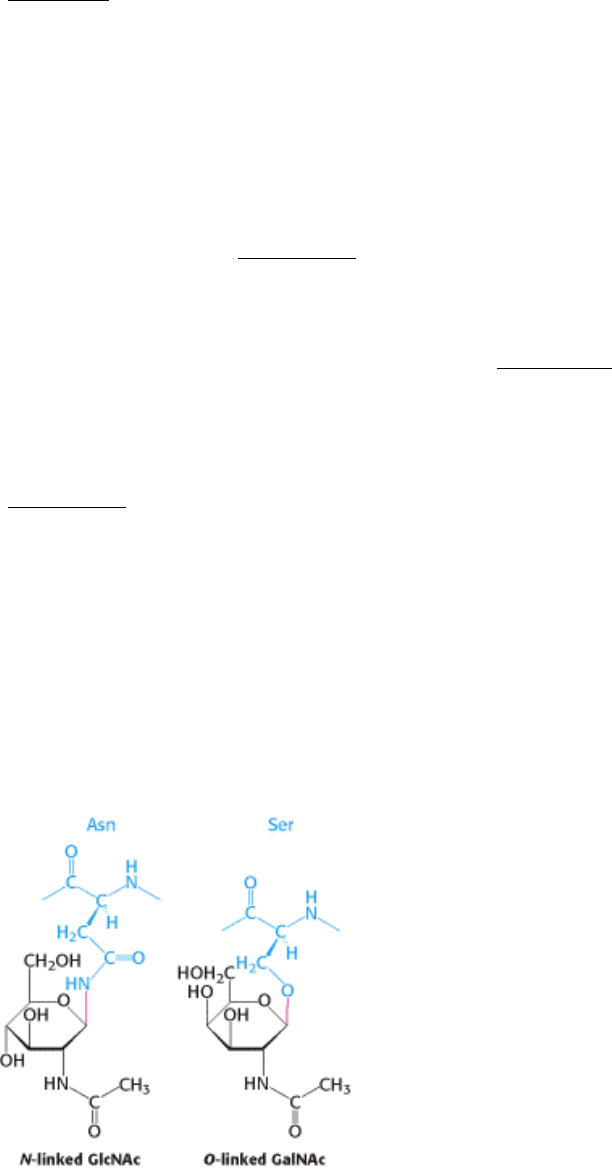
Figure 11.18. Glycosidic Bonds between Proteins and Carbohydrates. A glycosidic bond links a carbohydrate to the
side chain of asparagine (N-linked) or to the side chain of serine or threonine (O-linked). The glycosidic bonds are
shown in red.
I. The Molecular Design of Life 11. Carbohydrates 11.3. Carbohydrates Can Be Attached to Proteins to Form Glycoproteins
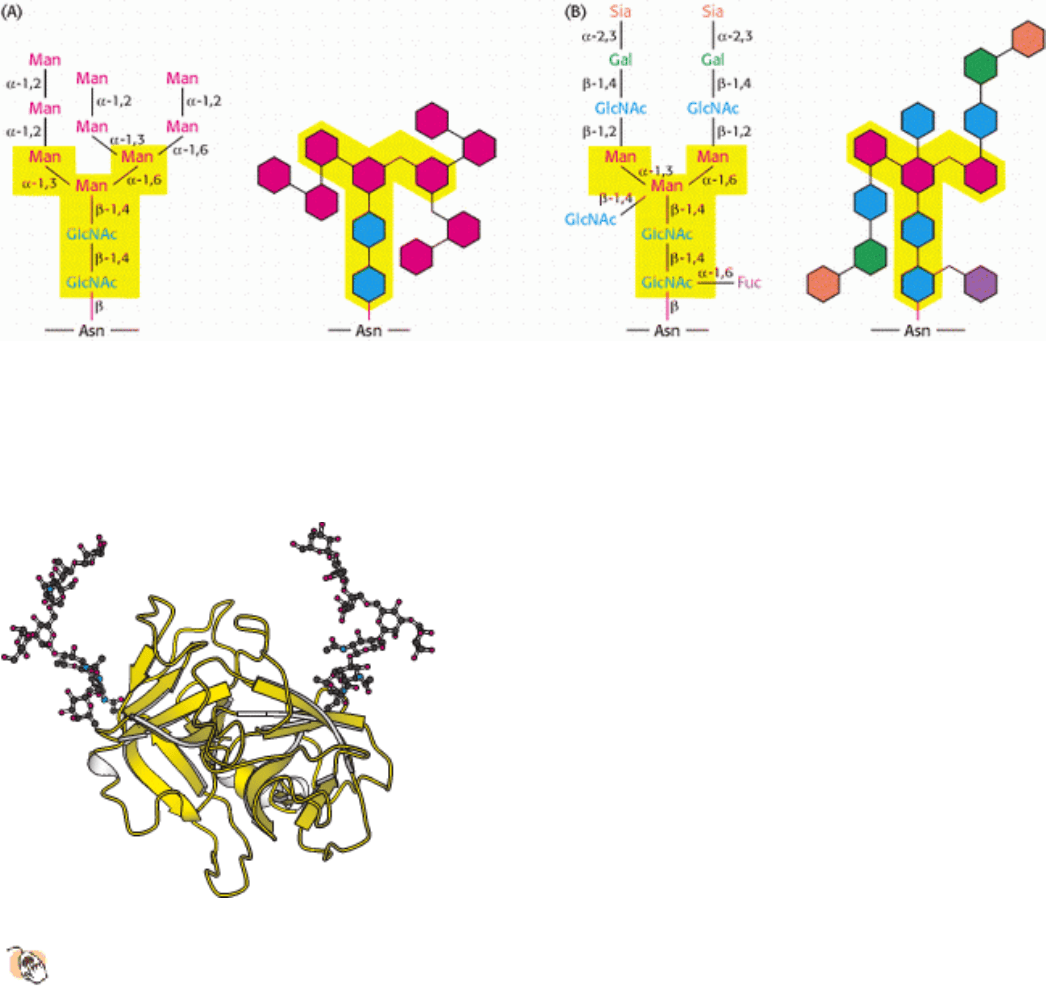
Figure 11.19. N -linked oligosaccharides. A pentasaccharide core (shaded yellow) is common to all N-linked
oligosaccharides and serves as the foundation for a wide variety of N-linked oligosaccharides, two of which are
illustrated: (A) high-mannose type; (B) complex type. Detailed chemical formulas and schematic structures are shown
for each type.
I. The Molecular Design of Life 11. Carbohydrates 11.3. Carbohydrates Can Be Attached to Proteins to Form Glycoproteins
Figure 11.20. Elastase, a Secreted Glycoprotein, Showing Linked Carbohydrates on Its Surface.
Elastase is a
protease found in serum. Note that the oligosaccharide chains have substantial size even for this protein, which has
a relatively low level of glycosylation.
I. The Molecular Design of Life 11. Carbohydrates 11.3. Carbohydrates Can Be Attached to Proteins to Form Glycoproteins
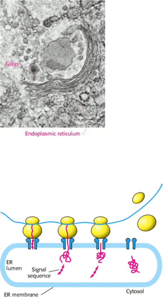
Figure 11.21. Golgi Complex and Endoplasmic Reticulum. The electron micrograph shows the Golgi complex and
adjacent endoplasmic reticulum. The black dots on the cytoplasmic surface of the ER membrane are ribosomes.
[Micrograph courtesy of Lynne Mercer.]
I. The Molecular Design of Life 11. Carbohydrates 11.3. Carbohydrates Can Be Attached to Proteins to Form Glycoproteins
Figure 11.22. Transport Into the Endoplasmic Reticulum. As translation takes place, a signal sequence on membrane
and secretory proteins directs the nascent protein through channels in the ER membrane and into the lumen. In most
cases, the signal sequence is subsequently cleaved and degraded.
I. The Molecular Design of Life 11. Carbohydrates 11.3. Carbohydrates Can Be Attached to Proteins to Form Glycoproteins
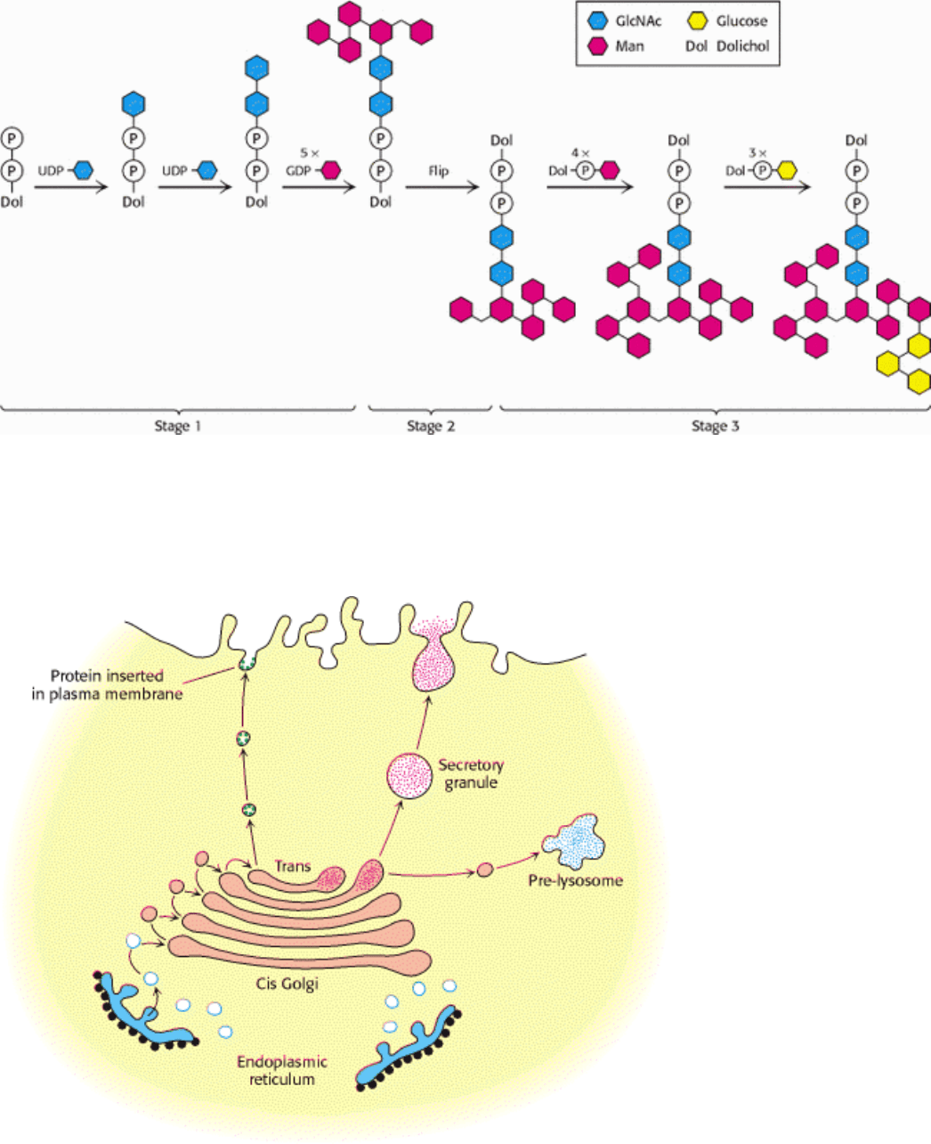
Figure 11.23. Assembly of an N -linked oligosaccharide precursor on dolichol phosphate. The first stage of
oligosaccharide synthesis takes place in the cytoplasm on the exposed phosphate of a membrane-embedded dolichol
molecule. Synthesis of the precursor is completed in the lumen of the ER after flipping of the dolichol phosphate and
attached oligosaccharide.
I. The Molecular Design of Life 11. Carbohydrates 11.3. Carbohydrates Can Be Attached to Proteins to Form Glycoproteins
Figure 11.24. Golgi Complex as Sorting Center. The Golgi complex is the sorting center in the targeting of proteins to
lysosomes, secretory vesicles, and the plasma membrane. The cis face of the Golgi complex receives vesicles from the
ER, and the trans face sends a different set of vesicles to target sites. Vesicles also transfer proteins from one
compartment of the Golgi complex to another. [Courtesy of Dr. Marilyn Farquhar.]
I. The Molecular Design of Life 11. Carbohydrates 11.3. Carbohydrates Can Be Attached to Proteins to Form Glycoproteins
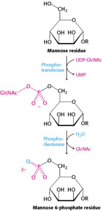
Figure 11.25. Formation of a Mannose 6-Phosphate Marker. A glycoprotein destined for delivery to lysosomes
acquires a phosphate marker in the cis Golgi compartment in a two-step process. First, a phosphotransferase adds a
phospho-N-acetylglucosamine unit to the 6-OH group of a mannose, and then a phosphodiesterase removes the added
sugar to generate a mannose 6-phosphate residue in the core oligosaccharide.
I. The Molecular Design of Life 11. Carbohydrates 11.3. Carbohydrates Can Be Attached to Proteins to Form Glycoproteins
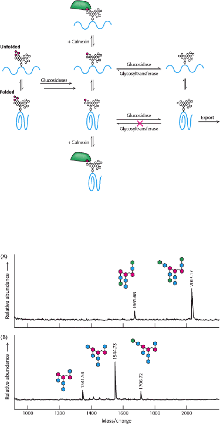
Figure 11.26. Quality-Control System for Protein Folding in the ER. A properly folded glycoprotein will move to the
Golgi complex after the removal of glucose moieties (shown in red). An unfolded or misfolded protein will receive a
glucose residue, through the action of a glucosyltransferase. Such glucosylated glycoproteins bind to calnexin (or the
related protein calreticulin), which serves as a chaperone to allow multiple attempts to attain correct folding. Properly
folded proteins are not reglucosylated.
I. The Molecular Design of Life 11. Carbohydrates 11.3. Carbohydrates Can Be Attached to Proteins to Form Glycoproteins

Figure 11.27. Mass Spectrometric "Sequencing" of Oligosaccharides. Carbohydrate-cleaving enzymes were used to
release and specifically cleave the oligosaccharide component of the glycoprotein fetuin from bovine serum. Parts A and
B show the masses obtained with MALDI-TOF spectrometry as well as the corresponding structures of the
oligosaccharide digestion products (using the same scheme as that in Figure 11.19): (A) digestion with Peptide N-
glycosidase F (to release the oligosaccharide from the protein) and neuraminidase; (B) digestion with Peptide N-
glycosidase F, neuraminidase, and β-1,4-galactosidase. Knowledge of the enzyme specificities and the masses of the
products permits the characterization of the oligosaccharide. [After A. Varki, R. Cummings, J. Esko, H. Freeze, G. Hart,
and J. Marth (Eds.), Essentials of Glycobiology (Cold Spring Harbor Laboratory Press, 1999), p. 596.]
I. The Molecular Design of Life 11. Carbohydrates
11.4. Lectins Are Specific Carbohydrate-Binding Proteins
The diverse carbohydrate structures displayed on cell surfaces are well suited to serve as interaction sites between cells
and their environments. Proteins termed lectins (from the Latin legere, "to select") are the partners that bind specific
carbohydrate structures. Lectins are ubiquitous, being found in animals, plants, and microorganisms. We have already
seen that some lectins, such as calnexin, function as chaperones in protein folding (Section 11.3.6).
11.4.1. Lectins Promote Interactions Between Cells
The chief function of lectins in animals is to facilitate cell-cell contact. A lectin usually contains two or more binding
sites for carbohydrate units; some lectins form oligomeric structures with multiple binding sites. The binding sites of
lectins on the surface of one cell interact with arrays of carbohydrates displayed on the surface of another cell. Lectins
and carbohydrates are linked by a number of relatively weak interactions that ensure specificity yet permit unlinking as
needed. The interactions between one cell surface with carbohydrates and another with lectins resemble the action of
Velcro; each interaction is relatively weak but the composite is strong.
The exact role of lectins in plants is unclear, although they can serve as potent insecticides. Castor beans contain so
much lectin that they are toxic to most organisms. The binding specificities of lectins from plants have been well
characterized (Figure 11.28). Bacteria, too, contain lectins. Escherichia coli bacteria are able to adhere to epithelial cells
of the gastrointestinal tract because lectins on the E. coli surface recognize oligosaccharide units on the surfaces of target
cells. These lectins are located on slender hairlike appendages called fimbriae (pili).
Lectins can be divided into classes on the basis of their amino acid sequences and biochemical properties. One large
class is the C type (for calcium-requiring) found in animals. These proteins have in common a domain of 120 amino
acids that is responsible for carbohydrate binding. The structure of one such domain bound to a carbohydrate target is
shown in Figure 11.29. A calcium ion acts as a bridge between the protein and the sugar through direct interactions with
sugar hydroxyl groups. In addition, two glutamate residues in the protein bind to both the calcium ion and the sugar,
while other protein side chains form hydrogen bonds with other hydroxyl groups on the carbohydrate. Changes in the
amino acid residues that interact with the carbohydrate alter the carbohydrate-binding specificity of the lectin.
Proteins termed selectins are members of the C-type family. Selectins bind immune-system cells to the sites of
injury in the inflammatory response (Figure 11.30). The L, E, and P forms of selectins bind specifically to
carbohydrates on lymph-node vessels, endothelium, or activated blood platelets, respectively. New therapeutic agents
that control inflammation may emerge from a deeper understanding of how selectins bind and distinguish different
carbohydrates.
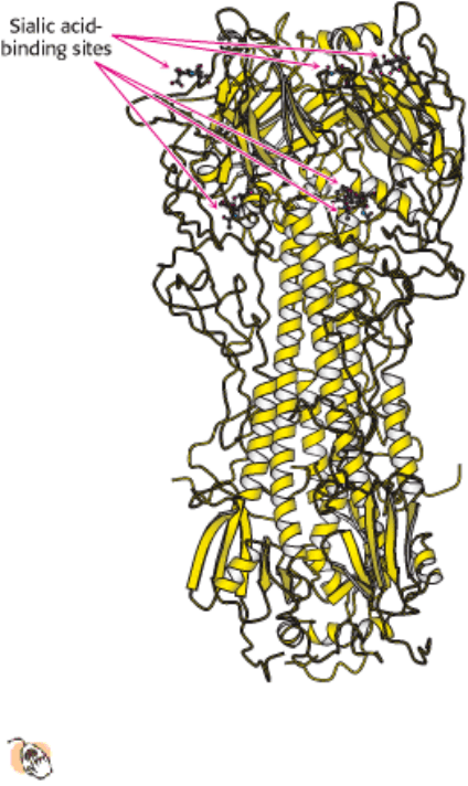
11.4.2. Influenza Virus Binds to Sialic Acid Residues
The ability of viruses to infect specific cell types is dictated in part by the ability of these viruses to bind to
particular structures or receptors on the surfaces of cells. In some cases, these receptors are carbohydrates. For
example, influenza virus recognizes sialic acid residues present on cell-surface glycoproteins. The viral protein that binds
to these sugars is called hemagglutinin(Figure 11.31).
After these surface interactions have taken place and the virus has been taken into the cell, another viral protein,
neuramidase, cleaves the glycosidic bonds to the sialic acid residues, freeing the virus to infect the cell. Inhibitors of this
enzyme are showing some promise as anti-influenza agents.
I. The Molecular Design of Life 11. Carbohydrates 11.4. Lectins Are Specific Carbohydrate-Binding Proteins
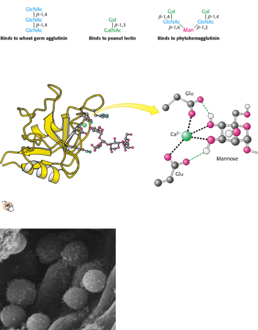
Figure 11.28. Binding Selectivities of Plant Lectins. The plant lectins wheat germ agglutinin, peanut lectin, and
phytohemagglutinin recognize different oligosaccharides.
I. The Molecular Design of Life 11. Carbohydrates 11.4. Lectins Are Specific Carbohydrate-Binding Proteins
Figure 11.29. Structure of a C-Type Carbohydrate-Binding Domain from an Animal Lectin.
A calcium ion links a
mannose residue to the lectin. Selected interactions are shown, with some hydrogen atoms omitted for clarity.
I. The Molecular Design of Life 11. Carbohydrates 11.4. Lectins Are Specific Carbohydrate-Binding Proteins
Figure 11.30. Selectins Mediate Cell-Cell Interactions. The scanning electron micrograph shows lymphocytes
adhering to the endothelial lining of a lymph node. The L selectins on the lymphocyte surface bind specifically to
carbohydrates on the lining of the lymph-node vessels. [Courtesy of Dr. Eugene Butcher.]
I. The Molecular Design of Life 11. Carbohydrates 11.4. Lectins Are Specific Carbohydrate-Binding Proteins
Figure 11.31. Structure of a Part of Influenza Hemagglutinin.
This viral protein has multiple binding sites for linking
to sialic acid residues on the target-cell surface.
I. The Molecular Design of Life 11. Carbohydrates
Summary
Monosaccharides Are Aldehydes or Ketones with Multiple Hydroxyl Groups
An aldose is a carbohydrate with an aldehyde group (as in glyceraldehyde and glucose), whereas a ketose contains a keto
group (as in dihydroxyacetone and fructose). A sugar belongs to the
d series if the absolute configuration of its
asymmetric carbon farthest from the aldehyde or keto group is the same as that of d-glyceraldehyde. Most naturally
occurring sugars belong to the d series. The C-1 aldehyde in the open-chain form of glucose reacts with the C-5 hydroxyl
group to form a six-membered pyranose ring. The C-2 keto group in the open-chain form of fructose reacts with the C-5
hydroxyl group to form a five-membered furanose ring. Pentoses such as ribose and deoxyribose also form furanose
rings. An additional asymmetric center is formed at the anomeric carbon atom (C-1 in aldoses and C-2 in ketoses) in
these cyclizations. The hydroxyl group attached to the anomeric carbon atom is below the plane of the ring (viewed in
the standard orientation) in the α anomer, whereas it is above the ring in the β anomer. Not all the atoms in the rings lie
in the same plane. Rather, pyranose rings usually adopt the chair conformation, and furanose rings usually adopt the
envelope conformation. Sugars are joined to alcohols and amines by glycosidic bonds from the anomeric carbon atom.
For example, N-glycosidic bonds link sugars to purines and pyrimidines in nucleotides, RNA, and DNA.
Complex Carbohydrates Are Formed by Linkage of Monosaccharides
Sugars are linked to one another in disaccharides and polysaccharides by O-glycosidic bonds. Sucrose, lactose, and
maltose are the common disaccharides. Sucrose (common table sugar), obtained from cane or beet, consists of α -
glucose and β -fructose joined by a glycosidic linkage between their anomeric carbon atoms. Lactose (in milk) consists