Givan A.L. Flow Cytometry. First Principles
Подождите немного. Документ загружается.

International Society for Analytical
Cytometry (ISAC), 232
Interrogation point, 247
see also Analysis point
Intracellular proteins
cytokeratin, 121, 121f
estrogen receptor, 121, 121f
interferon-g, 119, 120f
staining for, 115±122
di½culties in, 115±116, 117f
permeabilization of cells prior to,
116±118
Isotype control, 92±94, 118
Jet-in-air con®guration, 23
Jett, James, 218
Kamentsky, Louis, 3, 6, 175
Karyotyping, 147, 148f, 149f, 150, 150f
Keyhole limpet hemocyanin, 92, 94
Kinetics, 198±202, 227
lacZ gene, 207, 208f
Laminar ¯ow, 6, 21
Lasers
air-cooled vs water-cooled, 65
argon ion, 63±64, 64f, 64t, 67, 125
beam pro®le from, 16±17, 17f, 26
coherence of, 63f
electrical hazard potential, 18
general description of gas lasers, 61±
63
helium neon and red diode, 63, 64t,
72
multilaser systems, 18, 72
options for wavelengths and
appropriate ¯uorochromes, 64t
plasma tube, 62
restricted choice of ¯uorochromes, 67
Laser scanning cytometry (LSC), 189,
227
Leukemia/lymphoma, 101f, 111, 177,
179±180, 181f
Leukocytes
cytokine secretion by, 119
®xation of, 178
gating on, 100±110
light scatter signals from, 81, 84±86,
85f
in AIDS, 181
in HLA-B27 typing, 184
in malignancies, 179
size and concentration in blood, 82±
84, 83f, 84t
suited for ¯ow analysis, 7, 81
surface proteins with CD
classi®cation, 87
with activation markers, 196
see also Lymphocytes
Light
from ¯uorochromes as ¯uorescence,
65±72
from lasers, 61±65
illumination of stream, 16±18
partitioning of light signal from cells,
72±80
scattered by cells, 27±29, 81, 84±86
Light scatter
forward (FSC), 27±28
refractive index and, 27
side (SSC), 28±29
to distinguish leukocytes, 81, 84±86
see also Forward scatter and Side
scatter
Lineage in®delity/promiscuity, 179
Linear ampli®cation, 31±34, 37, 38f,
126, 248
List mode, 44±45, 248
Livermore Laboratory, 7, 165, 214±
215
Logarithmic ampli®cation, 31±34, 37,
38f, 248
Los Alamos Laboratory, 4, 6, 7, 44,
165, 214±215, 226
Lymphocytes, 249
back-gating on, 105±108
CD3 on, 91
CD4 on, 108
CD5 on B lymphocytes, 93±94, 93f,
170±171
CD8 on, 100, 119, 120f, 145f
CD45/SSC gate for, 109
change in light scatter after
activation, 102, 102f
e¨ect of gate size on enumeration and
phenotype, 103t
®rst sorting by ¯ow into puri®ed
state, 7
gating on scatter from, 100±102
hematopoiesis, 180f
Index 269
Lymphocytes (cont.)
microscopy vs ¯ow cytometry in
study of, 55, 86±88
scatter signals from, 85±87, 85f
scatter from dead vs live, 155f
size and concentration in blood, 82,
84t
staining for cytokines synthesized by,
119±121, 120f
T and B, distinguished by
monoclonal antibodies, 87
see also Leukocytes
Magnetic beads, 170
Major histocompatibility (MHC)
antigens, 183
Malignancy/cancer
DNA analysis for, 126±129, 127f,
130f, 187
S phase as prognostic indicator,
141±142, 188±189
breast tumors, 121, 121f, 127f, 130f,
145, 187
leukemia/lymphomas, 179
transplantation after irradiation for,
184
Marker/cursor, 249
de®ning percent positive, 46, 47f, 51,
94
Mathematical algorithms for cell cycle
analysis, 135±137, 136f, 251,
254, 255
Mean, 46±48, 47f, 94, 197, 249
Median, 46±48, 47f, 94, 197, 249
Meiosis, 126
MESF (mean equivalents of soluble
¯uorochrome), 96, 97f
Melamed, MR, 3
Microbiological applications, 211±213
gel microdroplets, 212, 213f, 214f
plankton and the aquatic
environment, 202±206
Microdroplets. See Gel microdroplets
Microscopes
comparison with ¯ow methodology,
27, 41, 55, 86±88, 90, 95, 99±
102, 129, 147±148, 150, 182,
189±190
¯ow cytometrists' need for, 5, 13, 227
for study of leukocytes, 82±84, 86
historical antecedents to ¯ow, 1±3
optical path in ¯uorescence
microscope, 2f
Microspheres. See Beads
Mirrors, 73±74, 74f, 76±77
Mitosis, 126
Mode, 46±48, 47f, 94, 250
Moldavan, Andrew, 2±3
Molecular biological applications, 213±
218
chromosome sorting, 213±215, 216f
DNA fragment analysis, 215±217
DNA sequencing, 218
Monensin, 119, 121
Monoclonal antibodies
controls for, 91±93
for leukocyte surface antigens, 81, 87
historical development of, 2
routine use in hematology, 179
staining cells with, 87±90
see also Antibodies
Monocytes, 82, 84t, 91, 103±105, 250
Multiplex cytometry, 218±220
Multiploidy, 126
Necrosis, 150, 154±157, 250
Neutrophils, 84, 84t
Nonradiative energy transfer, 71
Nozzle, 21±23, 160±165
see also Flow cell
NP40, 118
Nucleic acid stains, 123±125, 124t
see also DNA analysis and individual
stains
Obscuration bar, 27, 30, 250
Observation point. See Analysis point
Operator, 11±12, 163, 168
Optical bench, 25±26, 25f, 250
Optical zapping, 172, 215, 215t, 225,
251
Orbitals, 59±62, 60f
excited and ground state, 60±62,
60f
in ¯uorochromes, 65±67, 66f
in lasers, 61±62, 65
Ori®ce, 251
size, 21±24
in drop formation, 160±165, 161f
Orthogonal orientation, 6, 30
Orthogonal light scatter. See Side
scatter
Flow Cytometry270
Para½n blocks, 128
Parameter, 30, 251
di½culties with multiparameter, 55±
56, 56f
relationship to size of data ®le, 41±
42
single parameter data storage, 45
supermultiparameter ¯ow and vision
of future, 226
Particle, 251
acceptable concentration for ¯ow,
19±20
acceptable size range for ¯ow, 20±21
coincidence in laser beam, 20f
Pathology applications, 186±189
S phase fraction determinations, 188±
189
tumor ploidy analysis, 186±188
PerCP, 64t, 69t, 71
Peridinin, 71
Peridinium,71
Permeabilization
for DNA staining, 126, 144, 147
for intracellular protein staining,
115±118
in necrosis, 151, 154
Phosphatidyl serine, 151
Photochemical principles, 59±61
Photodetectors, 27±35, 252
ampli®cation of signals from, 31±35
conversion of light signal to electrical
signal by, 31
mirrors and ®lters to direct light to,
29±30, 73±80, 74f
photodiodes, 26±27, 252
photomultiplier tubes (PMTs), 27,
29±31, 252
relative color blindness of, 29
Photodiode. See Photodetectors
Photomultiplier tube. See Photo-
detectors
Photons, 60±62, 66
Phototoxic dyes, 172
Phycoerythrin (PE), 64t, 69t, 252
from algae for conjugation to
antibodies, 70±71
in tandem dyes, 71
on calibration beads, 96
providing auto¯uorescent signatures
for ¯ow analysis of algae, 203,
204f, 205f
requirement for compensation, 76±79,
93
use in dual-parameter analysis with
¯uorescein, 71±72, 75±76
Phytoplankton. See Plankton
PKH dyes, 198
Plankton, 20, 202±206, 252
Plasma tube, 62
Platelets, 84, 84t, 85f, 108
hyperactive platelets, 183
platelet-associated immunoglobulin,
183
Ploidy analysis, 125±131, 186±189
see also DNA analysis
Poisson distribution of cells in drops,
164
Precursor frequency, 198, 199f, 252
Prenatal diagnosis, 220±221
Probe, 252
Prochlorophytes, 206
Programmed cell death. See Apoptosis
Propidium iodide, 64t, 67, 69t, 70, 124t,
253
for analysis of
apoptosis, 151±154
cell cycle, 133±134, 147
chromosomes, 147, 148f
necrosis, 154±157
ploidy, 126, 127f
speci®city of, 125
to exclude dead cells, 156±157
with BrdU, 140±141, 140f
P-selectin (CD62), 183
Pulse processing, 137±138
Purity
of gate, 104±108
of sort, 166±168, 170
Quadrants, 51±52, 53f, 253
Quality control, 176±177, 193, 228
Quantitation, 95±98, 97f
Rare event analysis, 177, 185, 186f, 221,
253
Ratiometric analysis, 200, 201f
Red cells. See erythrocytes
Red diode laser, 63, 64t
Refractive index, 6, 27±28, 65, 155
Region, 253
combination into a gate, 53±54
for sorting, 163
Index 271
Region (cont.)
lymphocyte region by FSC and SSC,
86±87
see also Gate
Relapse/remission, 179±180, 181f
Reporter molecules, 207±211
Reproductive technology, 220±222
determining sex, 221±222
prenatal diagnosis, 220±221
Research applications, 195±222
aquatic studies, 202±206
functional assays, 196±202
microbiology, 211±213
molecular biology, 213±217
multiplex cytometry for soluble
analytes, 217±220
reporter molecules, 207±211
reproductive technology, 220±222
Resonance energy transfer, 71±72
Restriction enzyme digestion, 215±217,
217f
Reticulocyte counts, 183
Reverse sorting, 172
Rh-incompatibility, 185, 220
RNA
in cell cycle, 143, 196f
in reticulocytes, 183
staining of, by some DNA
¯uorochromes, 124t, 125
dual parameter analysis, with DNA,
143, 143f
RNase, 126, 144
Robotic systems, 228
Rube Goldberg, 11, 254
S phase, 131, 134±136, 139, 141±142,
146, 188, 254
correlated to clinical prognosis, 141±
142, 188±189
Safety
electrical, 18
infectious precautions, 177±178
Sample
cell concentration in, 19±20
coincidence of multiple cells in laser
beam, 17, 20
diameter of sample core, 22, 22f
joining with sheath stream, 18
size of particles in, 20±21
uniformity of alignment, 18
Saponin, 118
Scatter. See Forward scatter and Side
scatter
Seaweed. See Algae
Sensitivity
of chromosome analysis for clinical
diagnosis, 150
of detection of activation markers,
197
of light detection in general, 98,
99f
of transplant cross-match, 192
Sheath ¯uid, 18, 21, 23f, 254
in sorting, 171±173
Side scatter (SSC), 28±29, 254
for gating, 109, 109f, 110f
of blood cells, 81, 85±86, 85f
Shapiro, Howard, 248
Signatures, 254
of bacteria, 217
of plankton, 203
Single nucleotide polymorphisms
(SNPs), 218
Software, 43±46
for aggregate subtraction, 137
for cell cycle analysis, 135±137
for spectral compensation, 79
Soluble analytes, 218±220
Sorting, 159±173
accuracy of, 166, 168
alternate methods for, 170±172
batch, 170±171
break-o¨ point, 161±163
e½ciency of, 168±170, 169f
for prenatal diagnosis, 221
high speed, 165±166, 169±170, 172,
215, 225
of chromosomes, 213±215
of four or more populations at once,
226
of sperm, 221±222
purity of, 166±170
rate of, 168±169, 169f
sheath ¯uid for, 171±173
sterility of, 172
time delay computation for, 163
viability after, 172±173
whole animal (WACS), 207
Sort matrix, 163
Sperm sorting, 221±222
Flow Cytometry272
Steinkamp, John, 226
Sterility, 172
Stokes shift, 67, 71, 255
Stream velocity, 160, 165
SubG0/G1 peak, 154
Surface antigens
CD nomenclature for leukocytes, 87
direct staining of, 88
indirect staining of, 88±90
in hematopoiesis, 180f
monoclonal antibodies against, 87
Sweet, RG, 5
Talbot, David, 190
Tandem dyes, 71±72, 255
Tape back-up, 43
Temporal resolution, 17
Terminal deoxynucleotidyl transferase
(TdT), 153
Tetraploidy, 130±131, 255
Thiazole orange, 69t, 124t, 183
Threshold, 37, 85, 90, 131, 155, 183,
255
Time, 255
in kinetic analysis, 200±202
in sorting, 164±166, 164t, 168, 169f
Time delay calculation for sorting,
163
Tracking dyes, 198
Transplantation
solid organ, 189±192
stem cell, 184
Tritiated thymidine, 133
Trotter, Joseph, 44
Tsien, Roger, 200
Tumors, 110, 119, 121, 121f, 127f, 128,
130, 145, 186±189, 187f
see also Malignancy/cancer
TUNEL assay, 153±154, 154f, 256
Ultraviolet (UV)
¯uorochromes absorbing, 69t, 124,
124t
lasers emitting, 63, 64t
Van Dilla, Marvin, 6
Velocity of stream, 160, 165
Viability, 155, 171±173
see also Cell death
Volume, 256
not directly related to forward scatter,
27±28
relationship to Coulter signal, 28
WACS (whole animal cell sorting), 207,
208f, 209f
Wavelength (of light), 60, 61f, 256
¯uorochrome absorption/emission
spectra, 68f, 69t, 124t
from lasers, 64f, 64t
Wavelength (between drops), 159, 256
White blood cells. See Leukocytes and
Lymphocytes
X-chromosomes, 221±222
YFP, 64t, 70t, 210
Y-chromosomes, 221±222
Zip cartridges, 42±43, 42t, 227
Index 273

1
The Past as Prologue
Flow cytometry, like most scienti®c developments, has roots ®rmly
grounded in history. In particular, ¯ow technology ®nds intellectual
antecedents in microscopy, in blood cell counting instruments, and
in the ink jet technology that was, in the 1960s, being developed for
computer printers. It was the coming together of these three strands
of endeavor that provided the basis for the development of the ®rst
¯ow cytometers. Because thorough accounts of the history of ¯ow
cytometry have been written elsewhere (and make a fascinating story
for those interested in the history of science), I cover past history here
in just enough detail to give readers a perspective as to why current
instruments have developed as they have.
Microscopes have, since the seventeenth century, been used to
examine cells and tissue sections. Particularly since the end of the
nineteenth century, stains have been developed that make various
cellular constituents visible; in the 1940s and 1950s, ¯uorescence mi-
croscopy began to be used in conjunction with ¯uorescent stains for
nucleic acids in order to detect malignant cells. With the advent of
antibody technology and the work of Albert Coons in linking anti-
bodies with ¯uorescent tags, the use of ¯uorescent stains gained wider
and more speci®c applications. In particular, cell suspensions or tissue
sections are now routinely stained with antibodies speci®c for anti-
genic markers of cell type or function. The antibodies are either di-
rectly or indirectly conjugated to ¯uorescent molecules (most usually
¯uorescein or rhodamine). The cellular material can then be exam-
ined on a glass slide under a microscope ®tted with an appropriate
lamp and ®lters so that the ¯uorescence of the cells can be excited
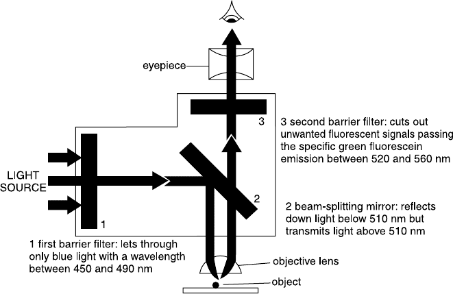
and observed (Fig. 1.1). The ¯uorescence microscope allows us to see
cells, to identify them in terms of both their physical structure and
1
Flow Cytometry: First Principles, Second Edition. Alice Longobardi Givan
Copyright
2001 by Wiley-Liss, Inc.
ISBNs 0-471-38224-8 (Paper); 0-471-22394-8 (Electronic)
their orientation within tissues, and then to determine whether and in
what pattern they ¯uoresce when stained with one or another of the
speci®c stains available. In addition, a microscope can also be ®tted
with a camera or photodetector, which will then record the intensity
of ¯uorescence arising from the ®eld in view. The logical extension of
this technique is image analysis cytometry, digitizing the output to
allow precise quantitation of ¯uorescence intensity patterns in detail
(pixel by pixel) within that ®eld of view. The development of mono-
clonal antibody technology (for which Ko
È
hler and Milstein were
awarded the Nobel Prize) led to a vast increase in the number of
cellular components that can be speci®cally stained and that can be
used to classify cells. Whereas monoclonal antibody techniques are
not directly related to the development of ¯ow technology, their inven-
tion was a serendipitous event that had great impact on the poten-
tial utility of ¯ow cytometric systems.
In 1934, Andrew Moldavan in Montreal took a ®rst step from static
microscopy toward a ¯owing system. He suggested the development
of an apparatus to count red blood cells and neutral-red±stained
Fig. 1.1. The optical path of a ¯uorescence microscope. In this example, the ®lters
and mirrors are set for detection of ¯uorescein ¯uorescence. From Alberts et al.
(1989).
Flow Cytometry2
yeast cells as they were forced through a capillary on a microscope
stage. A photodetector attached to the microscope eyepiece would
register each passing cell. Although it is unclear from Moldavan's
paper whether he actually ever built this cytometer, the development
of staining procedures over the next 30 years made it obvious that the
technique he suggested could be useful not simply for counting the
number of cells but also for quantitating their characteristics.
In the mid-1960s, Louis Kamentsky took his background in optical
character recognition and applied it to the problem of automated
cervical cytology screening. He developed a microscope-based spec-
trophotometer (on the pattern of the one suggested by Moldavan)
that measured and recorded ultraviolet absorption and the scatter of
blue light (``as an alternative to mimicking the complex scanning
methods of the human microscopist'') from cells ¯owing ``at rates
exceeding 500 cells per second'' past a microscope objective. Then, in
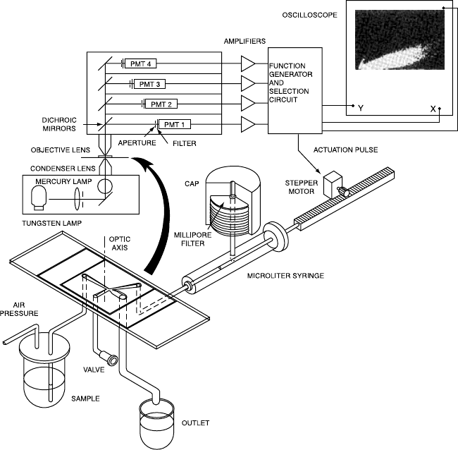
1967, Kamentsky and Melamed elaborated this design into a sorting
instrument (Fig. 1.2) that provided for the electronic actuation of a
syringe to pull cells with high absorption/scatter ratios out of the
¯ow stream. These ``suspicious'' cells could then be subjected to de-
tailed microscopic analysis. In 1969, Dittrich and Go
È
hde in Mu
È
nster,
Germany, described a ¯ow chamber for a microscope-based system
whereby ¯uorescence intensity histograms could be generated based on
the ethidium bromide ¯uorescence from the DNA of alcohol-®xed cells.
During this period of advances in ¯ow microscopy, so-called
Coulter technology had been developed by Wallace Coulter for anal-
ysis of blood cells. In the 1950s, instruments were produced that
counted cells as they ¯owed in a liquid stream; analysis was based on
the amount by which the cells increased electrical resistance as they
displaced isotonic saline solution while ¯owing through an ori®ce.
Cells were thereby classi®ed more or less on the basis of their volume
because larger cells have greater electrical resistance. These Coulter
counters soon became essential equipment in hospital hematology
laboratories, allowing the rapid and automated counting of white and
red blood cells. They actually incorporated many of the features of
analysis that we now think of as being typical of ¯ow cytometry: the
rapid ¯ow of single cells in ®le through an ori®ce, the electronic
detection of signals from those cells, and the automated analysis of
those signals.
At the same time as Kamentsky's work on cervical screening,
The Past as Prologue 3
Mack Fulwyler at the Los Alamos Laboratory in New Mexico had
decided to investigate a problem well known to everyone looking at
red blood cells in Coulter counters. Red cells were known to show a
bimodal distribution of their electrical resistance (``Coulter volume'').
Anyone looking at erythrocytes under the microscope cannot help
but be impressed by the remarkable structural uniformity of these
cells; Fulwyler wondered if the bimodal Coulter volume distribution
represented di¨erences between two classes of these apparently very
uniform cells or, alternatively, whether the bimodal pro®le was simply
an artifact based on some quirky aspect of the electronic resistance
measurements. The most direct way of testing these two alternatives
Fig. 1.2. A diagram of Kamentsky's original ¯ow sorter. From Kamentsky and
Melamed (1967). Science 156:1364±1365. Copyright AAAS.
Flow Cytometry4
was to separate erythrocytes according to their electronic resistance
signals and then to determine whether the separated classes remained
distinct when they were re-analyzed.
The technique that Fulwyler developed for sorting the erythrocytes
combined Coulter methodology with the ink jet technology being
developed at Stanford University by RG Sweet for running computer
printers. Ink jet technology involves the vibration of a nozzle so as
to generate a stream that breaks up into discrete drops and then the
charging and grounding of that stream at appropriate times so as to
leave indicated drops, as they break o¨, carrying an electrical charge.
For purposes of printing, those charged drops of ink can then be
de¯ected to positions on the paper as required by the computer print
messages. Fulwyler took the intellectual leap of combining this
methodology with Coulter ¯ow technology; he developed an instru-
ment that would charge drops containing suspended cells, thereby
allowing de¯ection of the cells (within the drops) as dictated by sig-
nals based on the cell's measured Coulter volume.
The data from this limited but pioneering experiment led to a con-
clusion that with hindsight seems obvious: Erythrocytes are indeed
uniform. When red cells are sorted according to their electrical resis-
tance, the resulting cells from one class or the other still show a
bimodal distribution when re-analyzed for their electrical resistance
pro®le. The bimodal ``volume'' signal from erythrocytes was there-
fore artifactualÐresulting in part from the discoid (nonspherical)
shape of the cells. The technology developed for this landmark experi-
ment is the essence of all the technology required for ¯ow sorting as
we now know it. That experiment also, unwittingly, emphasized an
aspect of ¯ow cytometry that has remained with us to this day: Flow
cytometrists still need to be continually vigilant (and know how to
use a microscope) because signals from cells (particularly signals that
are related to cell volume) are subject to artifactual in¯uences and may
not be what they seem. (Fulwyler's 1965 paper actually describes the
separation of mouse from human erythrocytes and the separation of
a large component from a population of mouse lymphoma cells; the
experiments on the bimodal signals from red cells have been relegated
to ¯ow folk history.)
In 1953, PJ Crosland-Taylor, working at the Middlesex Hospital
in London, noted that attempts to count small particles suspended in
¯uid ¯owing through a tube had not hitherto been very successful.
With particles such as red blood cells, the experimenter must choose
The Past as Prologue 5