Raven P.H., Johnson G.B., Mason K.A. Biology (Ninth Edition)
Подождите немного. Документ загружается.

Apago PDF Enhancer
Plasma
membrane
Protein
Cell interior
0.054 μm
TABLE 4.1
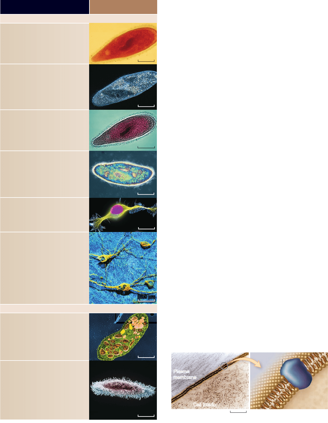
Microscopes
LIGHT MICROSCOPES
Bright-fi eld microscope:
Light is transmitted through a specimen, giving
little contrast. Staining specimens improves
contrast but requires that cells be xed (not
alive), which can distort or alter components.
Dark-fi eld microscope:
Light is directed at an angle toward the specimen.
A condenser lens transmits only light re ected o
the specimen. The eld is dark, and the specimen
is light against this dark background.
Phase-contrast microscope:
Components of the microscope bring light
waves out of phase, which produces di erences
in contrast and brightness when the light waves
recombine.
Diff erential-interference–contrast
microscope:
Polarized light is split into two beams that have
slightly di erent paths through the sample.
Combining these two beams produces greater
contrast, especially at the edges of structures.
Fluorescence microscope:
Fluorescent stains absorb light at one
wavelength, then emit it at another. Filters
transmit only the emitted light.
Confocal microscope:
Light from a laser is focused to a point and
scanned across the uorescently stained
specimen in two directions. This produces clear
images of one plane of the specimen. Other
planes of the specimen are excluded to prevent
the blurring of the image. Multiple planes can
be used to reconstruct a 3-D image.
ELECTRON MICROSCOPES
Transmission electron microscope:
A beam of electrons is passed through the
specimen. Electrons that pass through are
used to expose lm. Areas of the specimen that
scatter electrons appear dark. False coloring
enhances the image.
Scanning electron microscope:
An electron beam is scanned across the surface
of the specimen, and electrons are knocked
o the surface. Thus, the topography of the
specimen determines the contrast and the
content of the image. False coloring enhances
the image.
28.4 μm
67.7 μm
32.8 μm
26.6 μm
10.2 μm
25.0 μm
bind to cellular structures that contain the target molecule and
can be seen with light microscopy. This approach has been used
extensively in the analysis of cell structure and function .
All cells exhibit basic structural similarities
The general plan of cellular organization varies between differ-
ent organisms, but despite these modifications, all cells resem-
ble one another in certain fundamental ways. Before we begin a
detailed examination of cell structure, let’s first summarize four
major features all cells have in common: (1) a nucleoid or nu-
cleus where genetic material is located, (2) cytoplasm, (3) ribo-
somes to synthesize proteins, and (4) a plasma membrane .
Centrally located genetic material
Every cell contains DNA, the hereditary molecule. In pro karyotes,
the simplest organisms, most of the genetic material lies in a single
circular molecule of DNA. It typically resides near the center of
the cell in an area called the nucleoid. This area is not segregated,
however, from the rest of the cell’s interior by membranes.
By contrast, the DNA of eukaryotes, which are more
complex organisms, is contained in the nucleus, which is sur-
rounded by a double-membrane structure called the nuclear
envelope. In both types of organisms, the DNA contains the
genes that code for the proteins synthesized by the cell. (Details
of nucleus structure are described later in the chapter.)
The cytoplasm
A semifluid matrix called the cytoplasm fills the interior of the
cell. The cytoplasm contains all of the sugars, amino acids, and
proteins the cell uses to carry out its everyday activities. Al-
though it is an aqueous medium, cytoplasm is more like jello
than water due to the high concentration of proteins and other
macromolecules. We call any discrete macromolecular struc-
ture in the cytoplasm specialized for a particular function an
organelle. The part of the cytoplasm that contains organic
molecules and ions in solution is called the cytosol to distin-
guish it from the larger organelles suspended in this fluid .
The plasma membrane
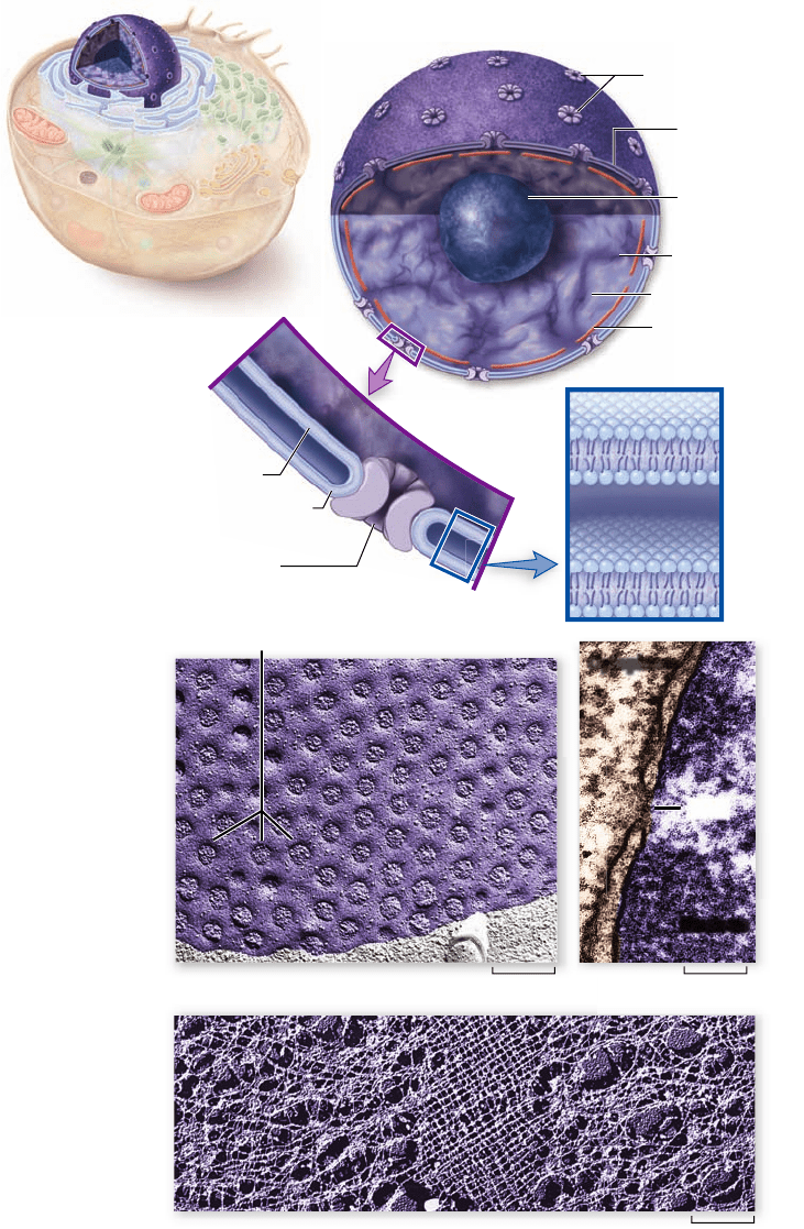
The plasma membrane encloses a cell and separates its con-
tents from its surroundings. The plasma membrane is a phos-
pholipid bilayer about 5 to 10 nm (5 to 10 billionths of a meter)
thick, with proteins embedded in it. Viewed in cross section
with the electron microscope, such membranes appear as two
dark lines separated by a lighter area. This distinctive appear-
ance arises from the tail-to-tail packing of the phospholipid
molecules that make up the membrane (see chapter 5 ) .
2.56 μm
6.76 μm
62
part
II
Biology of the Cell
rav32223_ch04_059-087.indd 62rav32223_ch04_059-087.indd 62 11/6/09 12:23:19 PM11/6/09 12:23:19 PM
Apago PDF Enhancer
Flagellum
Cell wall
Plasma membrane
Capsule
Ribosomes
Nucleoid (DNA)
Pilus
Cytoplasm
Pili
0.3 μm
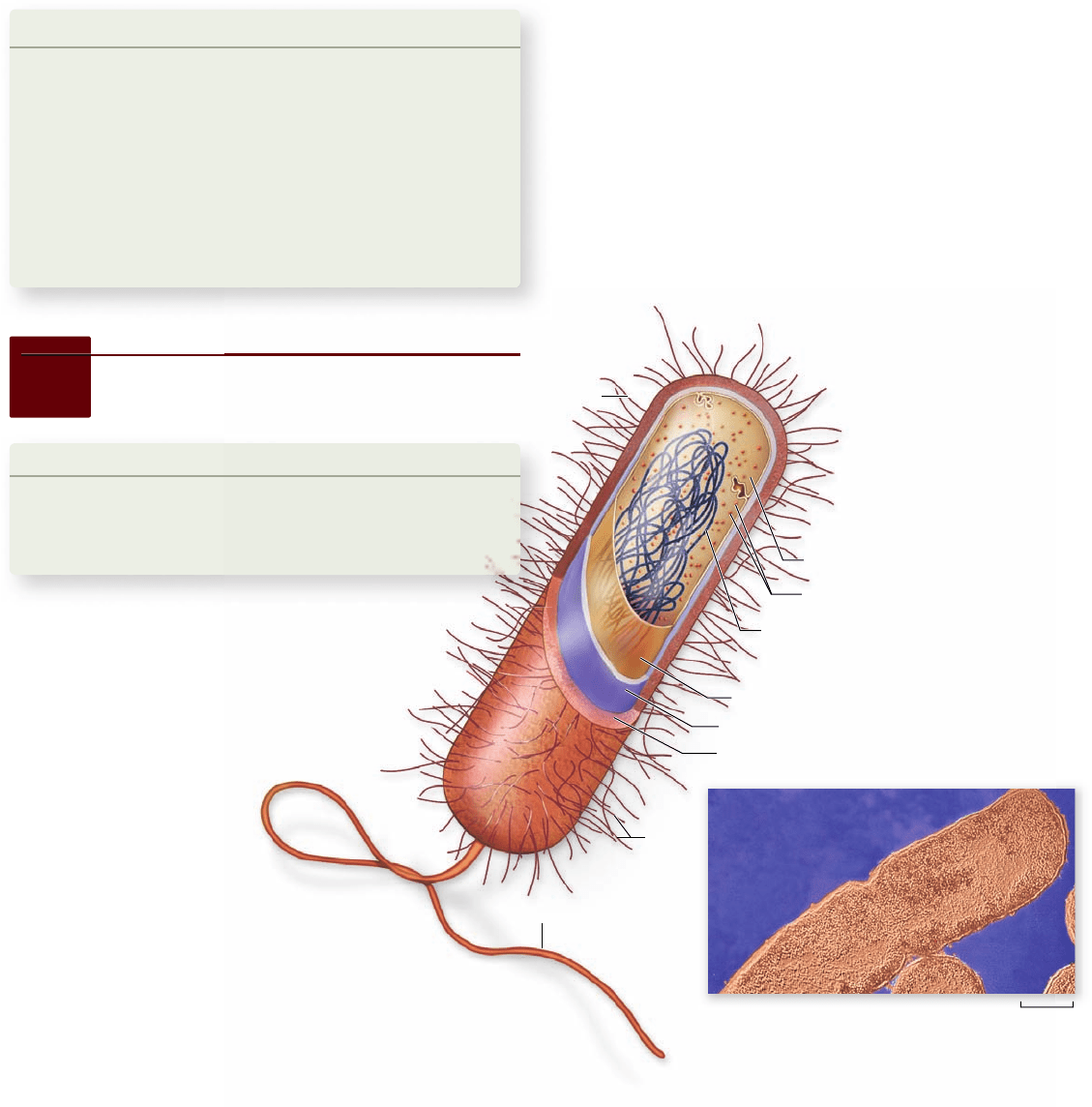
Figure 4.3
Structure of a
prokaryotic cell.
Generalized cell
organization of a
prokaryote. The
nucleoid is visible as a
dense central region
segregated from the
cytoplasm. Some
prokaryotes have
hairlike growths
(called pili [singular,
pilus]) on the outside
of the cell.
The proteins of the plasma membrane are generally re-
sponsible for a cell’s ability to interact with the environment.
Transport proteins help molecules and ions move across the plas-
ma membrane, either from the environment to the interior of
the cell or vice versa. Receptor proteins induce changes within the
cell when they come in contact with specific molecules in the
environment, such as hormones, or with molecules on the sur-
face of neighboring cells. These molecules can function as
markers that identify the cell as a particular type. This interac-
tion between cell surface molecules is especially important in
multicellular organisms, whose cells must be able to recognize
one another as they form tissues.
We’ll examine the structure and function of cell mem-
branes more thoroughly in chapter 5 .
Learning Outcomes Review 4.1
All organisms are single cells or aggregates of cells, and all cells arise from
preexisting cells. Cell size is limited primarily by the effi ciency of diff usion
across the plasma membrane. As a cell becomes larger, its volume increases
more quickly than its surface area. Past a certain point, diff usion cannot
support the cell’s needs . All cells are bounded by a plasma membrane and fi lled
with cytoplasm. The genetic material is found in the central portion of the cell;
and in eukaryotic cells, it is contained in a membrane-bounded nucleus.
■ Would finding life on Mars change our view of
cell theory?
4.2
Prokaryotic Cells
Learning Outcomes
1. Describe the organization of prokaryotic cells.
2. Distinguish between bacterial and archaeal
cell types.
When cells were visualized with microscopes, two
basic cellular architectures were recognized:
eukaryotic and prokaryotic. These terms refer
to the presence or absence, respectively, of
a membrane-bounded nucleus that
contains genetic material. We have
already mentioned that in
addition to lacking a nucleus,
prokaryotic cells do not have
an internal membrane system
or numerous membrane-
bounded organelles.
Prokaryote cells have relatively
simple organization
Prokaryotes are the simplest organisms. Prokaryotic cells are
small. They consist of cytoplasm surrounded by a plasma mem-
brane and are encased within a rigid cell wall. They have no
distinct interior compartments (figure 4.3). A prokaryotic cell is
like a one-room cabin in which eating, sleeping, and watching
TV all occur.
Prokaryotes are very important in the ecology of living
organisms. Some harvest light by photosynthesis, others break
down dead organisms and recycle their components. Still oth-
ers cause disease or have uses in many important industrial
processes . There are two main domains of prokaryotes: ar-
chaea and bacteria. Chapter 28 covers prokaryotic diversity in
more detail.
Although prokaryotic cells do contain organelles like
ribosomes, which carry out protein synthesis, most lack the
membrane-bounded organelles characteristic of eukaryotic
cells. It was long thought that prokaryotes also lack the
elaborate cytoskeleton found in eukaryotes, but we have
now found they have molecules related to both actin and
tubulin, which form two of the cytoskeletal elements de-
scribed later in the chapter. The actin-like proteins form
supporting fibrils near the surface of the cell, but the cyto-
plasm of a prokaryotic cell does not appear to have an ex-
tensive internal support structure. Consequently, the
strength of the cell comes primarily from its rigid cell wall
(see figure 4.3).
The plasma membrane of a prokaryotic cell carries out
some of the functions organelles perform in eukaryotic cells.
For example, some photosynthetic bacteria, such as the
chapter
4
Cell Structure
63www.ravenbiology.com
rav32223_ch04_059-087.indd 63rav32223_ch04_059-087.indd 63 11/6/09 12:23:26 PM11/6/09 12:23:26 PM

Apago PDF Enhancer
a. b. c.
0.5 μm
Plasma
membrane
Outer
membrane
Outer protein ring
Hook
Filament
Peptidoglycan
portion of
cell wall
Inner protein ring
H
+
H
+
Photosynthetic membranes
Nucleoid
Cytoplasm
Plasma
membrane
Cell wall
0.6 μm
Figure 4.5
Some prokaryotes
move by rotating their agella.
a. The photograph shows Vibrio
cholerae, the microbe that causes the
serious disease cholera. b. The
bacterial agellum is a complex
structure. The motor proteins,
powered by a proton gradient, are
anchored in the plasma membrane.
Two rings are found in the cell wall.
The motor proteins cause the entire
structure to rotate. c. As the agellum
rotates it creates a spiral wave down
the structure. This powers the
cell forward.
Figure 4.4
Electron micrograph of a photosynthetic
bacterial cell. Extensive folded photosynthetic membranes are
shown in green in this false color electron micrograph of a
Prochloron cell.
Inquiry question
?
What modifications would you include if you wanted to make
a cell as large as possible?
cyanobacterium Prochloron (figure 4.4), have an extensively
folded plasma membrane, with the folds extending into the
cell’s interior. These membrane folds contain the bacterial pig-
ments connected with photosynthesis. In eukaryotic plant cells,
photosynthetic pigments are found in the inner membrane of
the chloroplast.
Because a prokaryotic cell contains no membrane-bounded
organelles, the DNA, enzymes, and other cytoplasmic constituents
have access to all parts of the cell. Reactions are not compartmen-
talized as they are in eukaryotic cells, and the whole prokaryote
operates as a single unit.
Bacterial cell walls consist of peptidoglycan
Most bacterial cells are encased by a strong cell wall . This cell
wall is composed of peptidoglycan, which consists of a carbohy-
drate matrix (polymers of sugars) that is cross-linked by short
polypeptide units. Details about the structure of this cell wall
are discussed in chapter 28 . Cell walls protect the cell, maintain
its shape, and prevent excessive uptake or loss of water. The
exception is the class Mollicutes, which includes the common
genus Mycoplasma, which lack a cell wall. Plants, fungi, and most
protists also have cell walls but with a chemical structure differ-
ent from peptidoglycan.
The susceptibility of bacteria to antibiotics often depends
on the structure of their cell walls. The drugs penicillin and
vancomycin, for example, interfere with the ability of bacteria
to cross-link the peptides in their peptidoglycan cell wall. Like
removing all the nails from a wooden house, this destroys the
integrity of the structural matrix, which can no longer prevent
water from rushing in and swelling the cell to bursting.
Some bacteria also secrete a jelly-like protective capsule
of polysaccharide around the cell. Many disease-causing bac-
teria have such a capsule, which enables them to adhere to
teeth, skin, food—or to practically any surface that will sup-
port their growth.
Archaea lack peptidoglycan
We are still learning about the physiology and structure of ar-
chaea. Many of these organisms are difficult to culture in the
laboratory, and so this group has not yet been studied in detail.
More is known about their genetic makeup than about any
other feature.
The cell walls of archaea are composed of various chem-
ical compounds , including polysaccharides and proteins, and
possibly even inorganic components. A common feature
64
part
II
Biology of the Cell
rav32223_ch04_059-087.indd 64rav32223_ch04_059-087.indd 64 11/6/09 12:23:29 PM11/6/09 12:23:29 PM

Apago PDF Enhancer
distinguishing archaea from bacteria is the nature of their
membrane lipids. The chemical structure of archaeal lipids
is distinctly different from that of lipids in bacteria and can
include saturated hydrocarbons that are covalently attached
to glycerol at both ends, such that their membrane is a mono-
layer. These features seem to confer greater thermal stability
to archaeal membranes, although the tradeoff seems to be
an inability to alter the degree of saturation of the
hydrocarbons—meaning that archaea with this characteristic
cannot adapt to changing environmental temperatures.
The cellular machinery that replicates DNA and syn-
thesized proteins in archaea is more closely related to eukary-
otic systems than to bacterial systems. Even though they share
a similar overall cellular architecture with prokaryotes, ar-
chaea appear to be more closely related on a molecular basis
to eukaryotes.
Some prokaryotes move by means
of rotating agella
Flagella (singular, flagellum) are long, threadlike structures
protruding from the surface of a cell that are used in locomo-
tion. Prokaryotic flagella are protein fibers that extend out
from the cell. There may be one or more per cell, or none,
depending on the species. Bacteria can swim at speeds of up
to 70 cell lengths per second by rotating their flagella like
screws (figure 4.5). The rotary motor uses the energy stored
in a gradient that transfers protons across the plasma mem-
brane to power the movement of the flagellum. Interestingly,
the same principle, in which a proton gradient powers the
rotation of a molecule, is used in eukaryotic mitochondria
and chloroplasts by an enzyme that synthesizes ATP (see
chapters 7 and 8 ) .
Learning Outcomes Review 4.2
Prokaryotes are small cells that lack complex interior organization. The
two domains of prokaryotes are archaea and bacteria. The cell wall of
bacteria is composed of peptidoglycan, which is not found in archaea.
Archaea have cell walls made from a variety of polysaccharides and
peptides, as well as membranes containing unusual lipids. Some bacteria
move using a rotating fl agellum.
■ What features do bacteria and archaea share?
4.3
Eukaryotic Cells
Learning Outcomes
1. Compare the organization of eukaryotic and
prokaryotic cells.
2. Discuss the role of the nucleus in eukaryotic cells.
3. Describe the role of ribosomes in protein synthesis.
Eukaryotic cells (figures 4.6 and 4.7) are far more complex than
prokaryotic cells. The hallmark of the eukaryotic cell is com-
partmentalization. This is achieved through a combination of
an extensive endomembrane system that weaves through the
cell interior and by numerous organelles. These organelles in-
clude membrane-bounded structures that form compartments
within which multiple biochemical processes can proceed si-
multaneously and independently.
Plant cells often have a large, membrane-bounded sac
called a central vacuole, which stores proteins, pigments, and
waste materials. Both plant and animal cells contain vesicles—
smaller sacs that store and transport a variety of materials. In-
side the nucleus, the DNA is wound tightly around proteins
and packaged into compact units called chromosomes.
All eukaryotic cells are supported by an internal protein
scaffold, the cytoskeleton. Although the cells of animals and
some protists lack cell walls, the cells of fungi, plants, and many
protists have strong cell walls composed of cellulose or chitin
fibers embedded in a matrix of other polysaccharides and pro-
teins. Through the rest of this chapter, we will examine the in-
ternal components of eukaryotic cells in more detail.
The nucleus acts as the information center
The largest and most easily seen organelle within a eukaryotic
cell is the nucleus (Latin, “kernel” or “nut”), first described by
the botanist Robert Brown in 1831. Nuclei are roughly spheri-
cal in shape, and in animal cells, they are typically located in
the central region of the cell (figure 4.8a). In some cells, a net-
work of fine cytoplasmic filaments seems to cradle the nucleus
in this position.
The nucleus is the repository of the genetic information
that enables the synthesis of nearly all proteins of a living eu-
karyotic cell. Most eukaryotic cells possess a single nucleus, al-
though the cells of fungi and some other groups may have
several to many nuclei. Mammalian erythrocytes (red blood
cells) lose their nuclei when they mature. Many nuclei exhibit a
dark-staining zone called the nucleolus, which is a region
where intensive synthesis of ribosomal RNA is taking place.
The nuclear envelope
The surface of the nucleus is bounded by two phospholipid bi-
layer membranes, which together make up the nuclear enve-
lope (see figure 4.8). The outer membrane of the nuclear
envelope is continuous with the cytoplasm’s interior membrane
system, called the endoplasmic reticulum (described later).
Scattered over the surface of the nuclear envelope are
what appear as shallow depressions in the electron micro-
graph but are in fact structures called nuclear pores (see
figure 4.8b, c). These pores form 50 to 80 nm apart at locations
where the two membrane layers of the nuclear envelope pinch
together. They have a complex structure with a cytoplasmic
face, a nuclear face, and a central ring embedded in the mem-
brane. The proteins that make up this nuclear pore complex are
arranged in a circle with a large central hole. The complex al-
lows small molecules to diffuse freely between nucleoplasm and
cytoplasm while controlling the passage of proteins and RNA–
protein complexes. Passage is restricted primarily to two kinds
chapter
4
Cell Structure
65www.ravenbiology.com
rav32223_ch04_059-087.indd 65rav32223_ch04_059-087.indd 65 11/6/09 12:23:30 PM11/6/09 12:23:30 PM
Apago PDF Enhancer
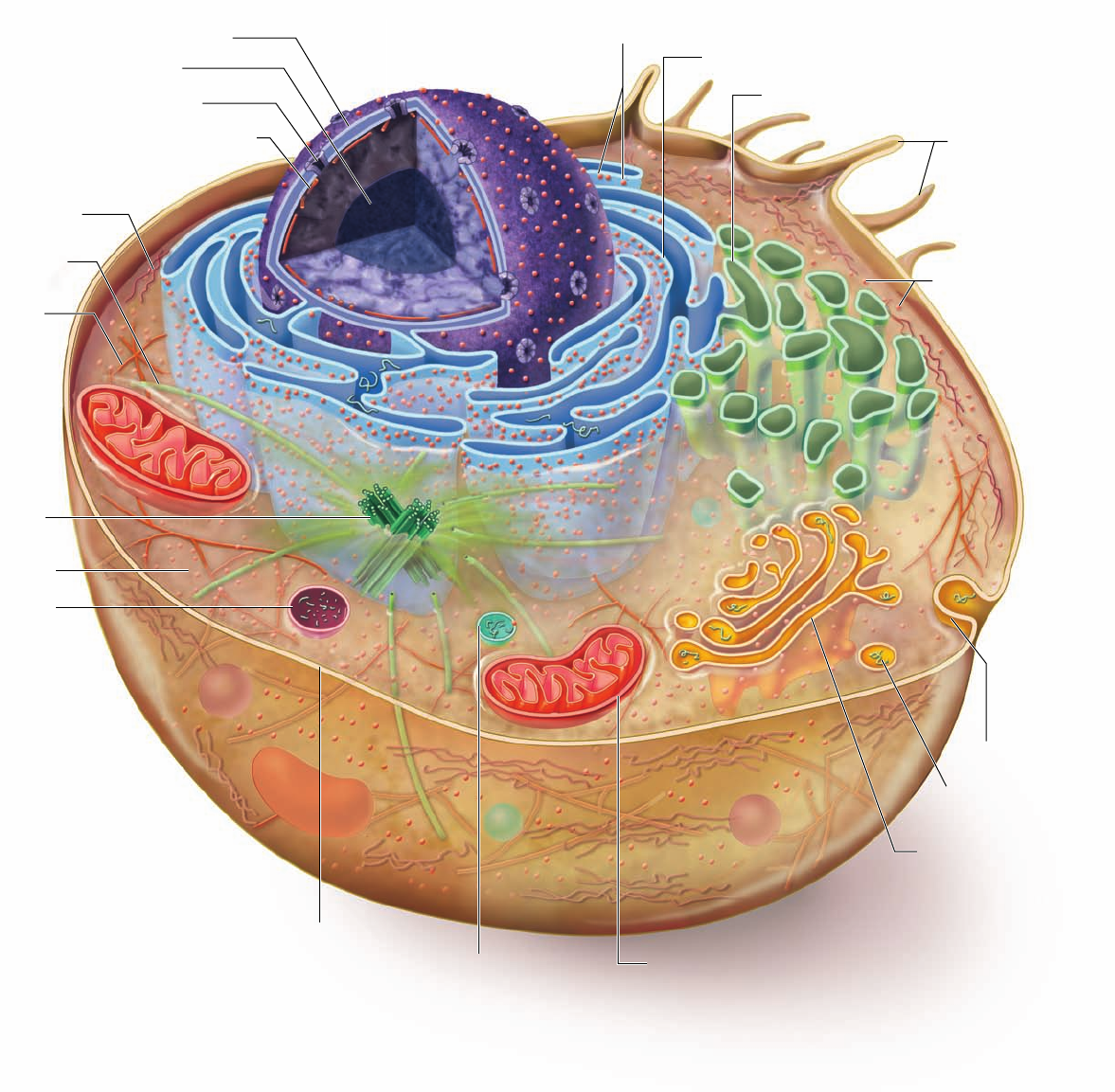
Nucleus
Nucleolus
Nuclear pore
Intermediate filament
Ribosomes
Ribosomes
Cytoplasm
Cytoskeleton
Intermediate
filament
Microtubule
Actin filament
(microfilament)
Lysosome
Centriole
Plasma membrane
Mitochondrion
Golgi apparatus
Exocytosis
Vesicle
Peroxisome
Smooth endoplasmic reticulum
Rough endoplasmic reticulum
Microvilli
Nuclear envelope
Figure 4.6
Structure of an animal cell. In this generalized diagram of an animal cell, the plasma
membrane encases the cell, which contains the cytoskeleton and various cell organelles and interior structures
suspended in a semi uid matrix called the cytoplasm. Some kinds of animal cells possess nger-like projections
called microvilli. Other types of eukaryotic cells—for example, many protist cells—may possess agella, which
aid in movement, or cilia, which can have many different functions.
66
part
II
Biology of the Cell
rav32223_ch04_059-087.indd 66rav32223_ch04_059-087.indd 66 11/6/09 12:23:31 PM11/6/09 12:23:31 PM
Apago PDF Enhancer
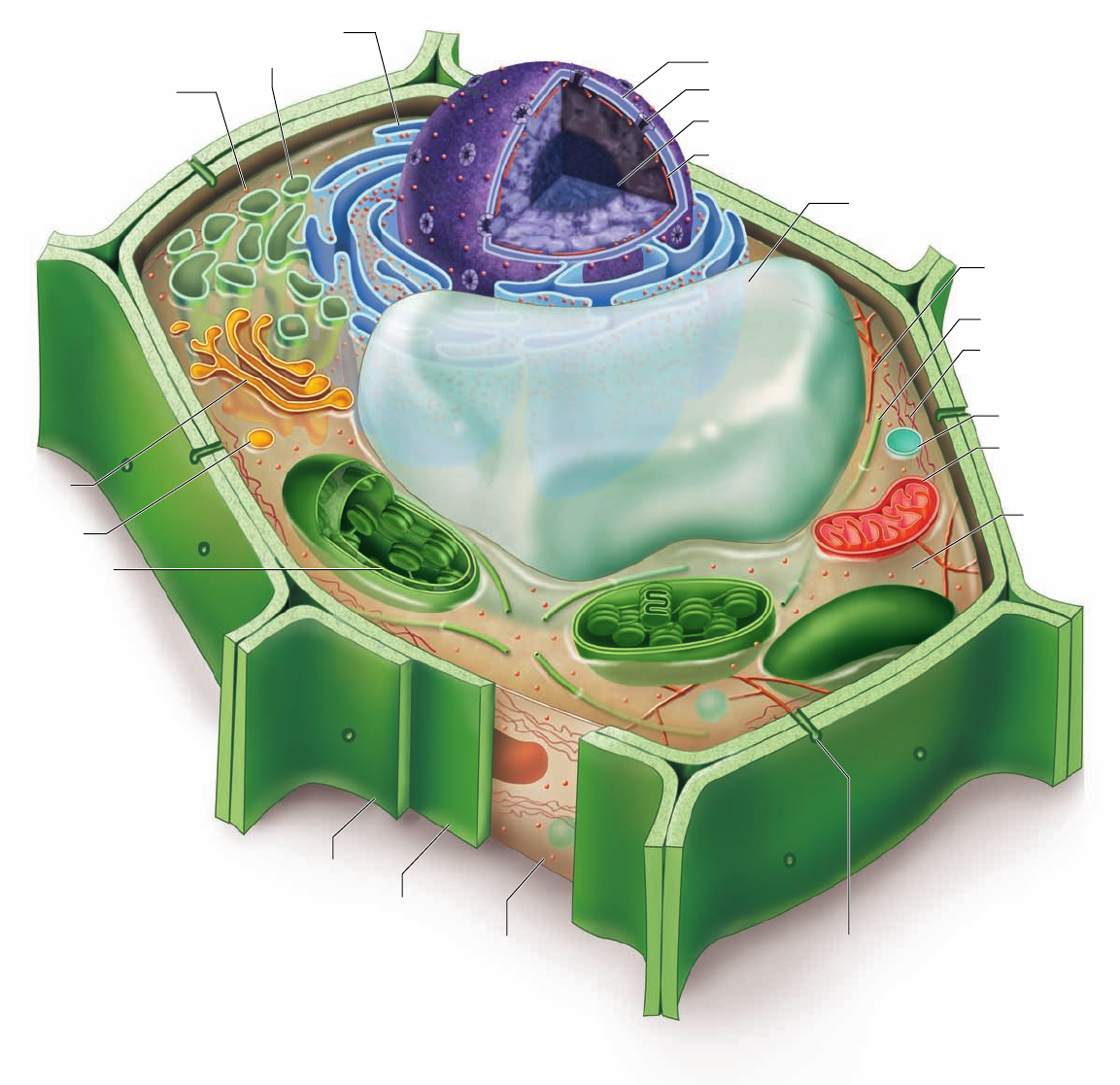
Nucleus
Nucleolus
Nuclear pore
Intermediate filament
Ribosome
Cytoplasm
Cytoskeleton
Intermediate
filament
Microtubule
Actin filament
(microfilament)
Plasma membrane
Mitochondrion
Golgi
apparatus
Vesicle
Peroxisome
Smooth endoplasmic reticulum
Rough endoplasmic reticulum
Nuclear envelope
Central vacuole
Plasmodesmata
Adjacent cell wall
Cell wall
Chloroplast
Figure 4.7
Structure of a plant cell. Most mature plant cells contain a large central vacuole, which
occupies a major portion of the internal volume of the cell, and organelles called chloroplasts, within which
photosynthesis takes place. The cells of plants, fungi, and some protists have cell walls, although the
composition of the walls varies among the groups. Plant cells have cytoplasmic connections to one another
through openings in the cell wall called plasmodesmata. Flagella occur in sperm of a few plant species, but are
otherwise absent from plant and fungal cells. Centrioles are also usually absent.
chapter
4
Cell Structure
67www.ravenbiology.com
rav32223_ch04_059-087.indd 67rav32223_ch04_059-087.indd 67 11/6/09 12:23:35 PM11/6/09 12:23:35 PM
Apago PDF Enhancer
b.
c.
d.
a.
Nuclear pores
Nuclear pore
Nuclear
envelope
Nucleoplasm
Inner
membrane
Nucleolus
Outer membrane
Chromatin
Nuclear
lamina
Cytoplasm
Nuc
leus
Nuclear pores
1
μ
m
0.069 μm
0.1 μm
Pore
Figure 4.8
The nucleus. a. The nucleus is composed of a double
membrane called the nuclear envelope, enclosing a uid- lled interior
containing chromatin. The individual nuclear pores extend through the
two membrane layers of the envelope. b. A freeze-fracture electron
micrograph (see gure 5.3 ) of a cell nucleus, showing many nuclear
pores. c. A transmission electron micrograph of the nuclear membrane
showing a single nuclear pore. The dark material within the pore is
protein, which acts to control access through the pore. d. The nuclear
lamina is visible as a dense network of bers made of intermediate
laments. The nucleus has been colored purple in the micrographs.
(b): © Dr. Richard Kessel & Dr. Gene Shih/Visuals Unlimited
of molecules: (1) proteins moving into the
nucleus to be incorporated into nuclear struc-
tures or to catalyze nuclear activities and
(2) RNA and RNA–protein complexes formed
in the nucleus and exported to the cytoplasm.
The inner surface of the nuclear envelope
is covered with a network of fibers that make up
the nuclear lamina (see figure 4.8d). This is com-
posed of intermediate filament fibers called nuclear
lamins. This structure gives the nucleus its shape and is
also involved in the deconstruction and reconstruction of
the nuclear envelope that accompanies cell division.
Chromatin: DNA packaging
In both prokaryotes and eukaryotes, DNA contains the heredi-
tary information specifying cell structure and function. In most
prokaryotes, the DNA is organized into a single circular chro-
mosome. In eukaryotes, the DNA is divided into multiple lin-
ear chromosomes. The DNA in these chromosomes is organized
with proteins into a complex structure called chromatin.
Chromatin is usually in a more extended form that allows
regulatory proteins to attach to specific nucleotide sequences
along the DNA and regulate gene expression. Without this
access, DNA could not direct the day-to-day activities of the
cell. When cells divide, the chromatin must be further com-
pacted into a more highly condensed form.
The nucleolus: Ribosomal subunit manufacturing
Before cells can synthesize proteins in large quantity, they must
first construct a large number of ribosomes to carry out this
synthesis. Hundreds of copies of the genes encoding the ribo-
somal RNAs are clustered together on the chromosome, facili-
tating ribsosome construction. By transcribing RNA molecules
from this cluster, the cell rapidly generates large numbers of the
molecules needed to produce ribosomes.
The clusters of ribosomal RNA genes, the RNAs they
produce, and the ribosomal proteins all come together within
the nucleus during ribosome production. These ribosomal as-
sembly areas are easily visible within the nucleus as one or more
dark-staining regions called nucleoli (singular, nucleolus). Nu-
cleoli can be seen under the light microscope even when the
chromosomes are uncoiled.
Ribosomes are the cell’s protein
synthesis machinery
Although the DNA in a cell’s nucleus encodes the amino acid
sequence of each protein in the cell, the proteins are not as-
sembled there. A simple experiment demonstrates this: If a brief
pulse of radioactive amino acid is administered to a cell, the
radioactivity shows up associated with newly made protein in
the cytoplasm, not in the nucleus. When investigators first car-
ried out these experiments, they found that protein synthesis is
associated with large RNA–protein complexes (called ribo-
somes) outside the nucleus.
Ribosomes are among the most complex molecular as-
semblies found in cells. Each ribosome is composed of two
68
part
II
Biology of the Cell
rav32223_ch04_059-087.indd 68rav32223_ch04_059-087.indd 68 11/6/09 12:23:38 PM11/6/09 12:23:38 PM

Apago PDF Enhancer
Small subunit
Large subunit
Ribosome
Figure 4.9
A ribosome. Ribosomes consist of a large and a
small subunit composed of rRNA and protein. The individual
subunits are synthesized in the nucleolus and then move
through the nuclear pores to the cytoplasm, where they
assemble to translate mRNA. Ribosomes serve as sites of
protein synthesis.
subunits (figure 4.9), and each subunit is composed of a com-
bination of RNA, called ribosomal RNA (rRNA), and pro-
teins. The subunits join to form a functional ribosome only
when they are actively synthesizing proteins. This complicated
process requires the two other main forms of RNA:
messenger RNA (mRNA), which carries coding information
from DNA, and transfer RNA (tRNA), which carries amino
acids. Ribosomes use the information in mRNA to direct the
synthesis of a protein. This process will be described in more
detail in chapter 15 .
Ribosomes are found either free in the cytoplasm or
associated with internal membranes, as described in the fol-
lowing section. Free ribosomes synthesize proteins that are
found in the cytoplasm, nuclear proteins, mitochondrial
proteins, and proteins found in other organelles not derived
from the endomembrane system. Membrane-associated ri-
bosomes synthesize membrane proteins, proteins found in
the endomembrane system, and proteins destined for export
from the cell.
Ribosomes can be thought of as “universal organelles”
because they are found in all cell types from all three do-
mains of life. As we build a picture of the minimal essential
functions for cellular life, ribosomes will be on the short list.
Life is protein-based, and ribosomes are the factories that
make proteins.
Learning Outcomes Review 4.3
In contrast to prokaryotic cells, eukaryotic cells exhibit
compartmentalization. Eukaryotic cells contain an endomembrane
system and organelles that carry out specialized functions. The nucleus,
composed of a double membrane connected to the endomembrane
system, contains the cell’s genetic information. Material moves between
the nucleus and cytoplasm through nuclear pores. Ribosomes translate
mRNA, which is transcribed from DNA in the nucleus, into polypeptides
that make up proteins. Ribosomes are a universal organelle found in all
known cells.
■ Would you expect cells in different organs in complex
animals to have the same structure?
4.4
The Endomembrane System
Learning Outcomes
Identify the different parts of the endomembrane system. 1.
Contrast the different functions of internal membranes 2.
and compartments.
Evaluate the importance of each step in the protein 3.
processing pathway.
The interior of a eukaryotic cell is packed with membranes so
thin that they are invisible under the low resolving power of
light microscopes. This endomembrane system fills the cell, di-
viding it into compartments, channeling the passage of mole-
cules through the interior of the cell, and providing surfaces for
the synthesis of lipids and some proteins. The presence of these
membranes in eukaryotic cells marks one of the fundamental
distinctions between eukaryotes and prokaryotes.
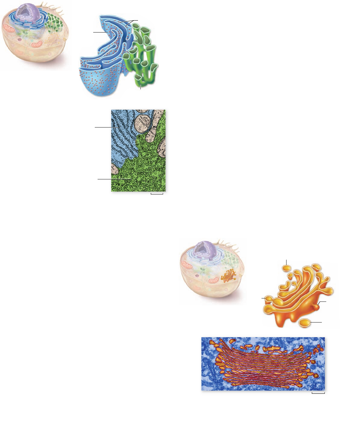
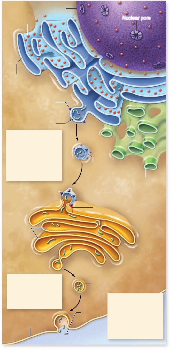
The largest of the internal membranes is called the
endoplasmic reticulum (ER). Endoplasmic means “within
the cytoplasm,” and reticulum is Latin for “a little net.” Like the
plasma membrane, the ER is composed of a phospholipid bi-
layer embedded with proteins. It weaves in sheets through the
interior of the cell, creating a series of channels between its
folds (figure 4.10). Of the many compartments in eukaryotic
cells, the two largest are the inner region of the ER, called the
cisternal space or lumen, and the region exterior to it, the
cytosol, which is the fluid component of the cytoplasm contain-
ing dissolved organic molecules such as proteins and ions.
The rough ER is a site of protein synthesis
The rough ER (RER) gets its name from its surface appear-
ance, which is pebbly due to the presence of ribosomes. The
RER is not easily visible with a light microscope, but it can be
seen using the electron microscope. It appears to be composed
of flattened sacs, the surfaces of which are bumpy with ribo-
somes (see figure 4.10).
The proteins synthesized on the surface of the RER are
destined to be exported from the cell, sent to lysosomes or vac-
uoles (described in a later section), or embedded in the plasma
membrane. These proteins enter the cisternal space as a first
step in the pathway that will sort proteins to their eventual des-
tinations. This pathway also involves vesicles and the Golgi ap-
paratus, described later. The sequence of the protein being
synthesized determines whether the ribosome will become as-
sociated with the ER or remain a cytoplasmic ribosome.
In the ER, newly synthesized proteins can be modified
by the addition of short-chain carbohydrates to form
glycoproteins. Those proteins destined for secretion are sep-
arated from other products and later packaged into vesicles.
The ER also manufactures membranes by producing mem-
brane proteins and phospholipid molecules. The membrane
chapter
4
Cell Structure
69www.ravenbiology.com
rav32223_ch04_059-087.indd 69rav32223_ch04_059-087.indd 69 11/6/09 12:23:43 PM11/6/09 12:23:43 PM
Apago PDF Enhancer
Transport vesicle
Secretory
vesicle
trans face
cis face
Forming
vesicle
Fusing
vesicle
0.9 μm
Ribosomes
0.08 μm
Rough
endoplasmic
reticulum
Rough
endoplasmic
reticulum
Smooth
endoplasmic
reticulum
Smooth
endoplasmic
reticulum
Figure 4.10
The
endoplasmic reticulum.
Rough ER (RER), blue in
the drawing, is composed
more of attened sacs and
forms a compartment
throughout the cytoplasm.
Ribosomes associated with
the cytoplasmic face of the
RER extrude newly made
proteins into the interior, or
lumen. The smooth ER
(SER), green in the drawing,
is a more tubelike structure
connected to the RER. The
micrograph has been colored
to match the drawing .
Figure 4.11
The Golgi apparatus. The Golgi apparatus is a
smooth, concave, membranous structure. It receives material for
processing in transport vesicles on the cis face and sends the
material packaged in transport or secretory vesicles off the trans
face. The substance in a vesicle could be for export out of the cell or
for distribution to another region within the same cell.
proteins are inserted into the ER’s own membrane, which can
then expand and pinch off in the form of vesicles to be trans-
ferred to other locations.
The smooth ER has multiple roles
Regions of the ER with relatively few bound ribosomes are re-
ferred to as smooth ER (SER). The SER appears more like a
network of tubules than the flattened sacs of the RER. The
membranes of the SER contain many embedded enzymes. En-
zymes anchored within the ER, for example, catalyze the syn-
thesis of a variety of carbohydrates and lipids. Steroid hormones
are synthesized in the SER as well. The majority of membrane
lipids are assembled in the SER and then sent to whatever parts
of the cell need membrane components.
The SER is used to store Ca
2+
in cells. This keeps the
cytoplasmic level low, allowing Ca
2+
to be used as a signaling
molecule. In muscle cells, for example, Ca
2+
is used to trigger
muscle contraction. In other cells, Ca
2+
release from SER stores
is involved in diverse signaling pathways.
The ratio of SER to RER depends on a cell’s function. In
multicellular animals such as ourselves, great variation exists in
this ratio. Cells that carry out extensive lipid synthesis, such as
those in the testes, intestine, and brain, have abundant SER.
Cells that synthesize proteins that are secreted, such as anti-
bodies, have much more extensive RER.
Another role of the SER is the modification of foreign
substances to make them less toxic. In the liver, the enzymes
of the SER carry out this detoxification. This action can in-
clude neutralizing substances that we have taken for a thera-
peutic reason, such as penicillin. Thus, relatively high doses
are prescribed for some drugs to offset our body’s efforts to
remove them. Liver cells have extensive SER as well as en-
zymes that can process a variety of substances by chemically
modifying them.
The Golgi apparatus sorts
and packages proteins
Flattened stacks of membranes, often interconnected with
one another, form a complex called the Golgi body . These
structures are named for Camillo Golgi, the 19th-century
physician who first identified them. The number of stacked
membranes within the Golgi body ranges from 1 or a few in
protists, to 20 or more in animal cells and to several hundred
in plant cells. They are especially abundant in glandular cells,
which manufacture and secrete substances. The Golgi body is
often referred to as the Golgi apparatus (figure 4.11).
The Golgi apparatus functions in the collection, packag-
ing, and distribution of molecules synthesized at one location
and used at another within the cell or even outside of it. A Golgi
body has a front and a back, with distinctly different membrane
compositions at these opposite ends. The front, or receiving
end, is called the cis face and is usually located near ER. Materi-
als move to the cis face in transport vesicles that bud off the ER.
These vesicles fuse with the cis face, emptying their contents
into the interior, or lumen, of the Golgi apparatus. The ER-
synthesized molecules then pass through the channels of the
Golgi apparatus until they reach the back, or discharging end,
70
part
II
Biology of the Cell
rav32223_ch04_059-087.indd 70rav32223_ch04_059-087.indd 70 11/6/09 12:23:44 PM11/6/09 12:23:44 PM
Apago PDF Enhancer
Nucleus
Nuclear pore
Rough
endoplasmic
reticulum
Smooth
endoplasmic
reticulum
Ribosome
Membrane
protein
Golgi membrane protein
Newly
synthesized
protein
Transport
vesicle
cis face
trans face
Cisternae
Secretory vesicle
Secreted
protein
Cell membrane
Extracellular fluid
1. Vesicle containing
proteins buds from
the rough endo-
plasmic reticulum,
diffuses through the
cell, and fuses to
the cis face of the
Golgi apparatus.
2. The proteins are
modified and
packaged into
vesicles for
transport.
3. The vesicle may
travel to the plasma
membrane,
releasing its
contents to the
extracellular
environment.
Golgi
Apparatus
Figure 4.12
Protein transport through the
endomembrane system. Proteins synthesized by ribosomes on
the RER are translocated into the internal compartment of the ER.
These proteins may be used at a distant location within the cell or
secreted from the cell. They are transported within vesicles that
bud off the rough ER. These transport vesicles travel to the cis face
of the Golgi apparatus. There they can be modi ed and packaged
into vesicles that bud off the trans face of the Golgi apparatus.
Vesicles leaving the trans face transport proteins to other locations
in the cell, or fuse with the plasma membrane, releasing their
contents to the extracellular environment.
called the trans face, where they are discharged in secretory
vesicles (figure 4.12).
Proteins and lipids manufactured on the rough and
smooth ER membranes are transported into the Golgi appara-
tus and modified as they pass through it. The most common
alteration is the addition or modification of short sugar chains,
forming glycoproteins and glycolipids. In many instances, en-
zymes in the Golgi apparatus modify existing glycoproteins and
glycolipids made in the ER by cleaving a sugar from a chain or
by modifying one or more of the sugars.
The newly formed or altered glycoproteins and glyco-
lipids collect at the ends of the Golgi bodies in flattened,
stacked membrane folds called cisternae (Latin, “collecting
vessels”). Periodically, the membranes of the cisternae push
together, pinching off small, membrane-bounded secretory
vesicles containing the glycoprotein and glycolipid molecules.
These vesicles then diffuse to other locations in the cell, dis-
tributing the newly synthesized molecules to their appropri-
ate destinations.
Another function of the Golgi apparatus is the synthesis
of cell wall components. Noncellulose polysaccharides that
form part of the cell wall of plants are synthesized in the Golgi
apparatus and sent to the plasma membrane where they can be
added to the cellulose that is assembled on the exterior of the
cell. Other polysaccharides secreted by plants are also synthe-
sized in the Golgi apparatus.
Lysosomes contain digestive enzymes
Membrane-bounded digestive vesicles, called lysosomes, are
also components of the endomembrane system. They arise
from the Golgi apparatus. They contain high levels of degrad-
ing enzymes, which catalyze the rapid breakdown of proteins,
nucleic acids, lipids, and carbohydrates. Throughout the lives
of eukaryotic cells, lysosomal enzymes break down old organ-
elles and recycle their component molecules. This makes room
for newly formed organelles. For example, mitochondria are
replaced in some tissues every 10 days.
The digestive enzymes in the lysosome are optimally
active at acid pH. Lysosomes are activated by fusing with a
food vesicle produced by phagocytosis (a specific type of endo-
cytosis; see chapter 5 ) or by fusing with an old or worn-out
organelle. The fusion event activates proton pumps in the
lysosomal membrane, resulting in a lower internal pH. As the
interior pH falls, the arsenal of digestive enzymes contained
in the lysosome is activated. This leads to the degradation of
macromolecules in the food vesicle or the destruction of the
old organelle.
A number of human genetic disorders, collectively called
lysosomal storage disorders, affect lysosomes. For example, the
genetic abnormality called Tay–Sachs disease is caused by the
loss of function of a single lysosomal enzyme. This enzyme is
necessary to break down a membrane glycolipid found in nerve
cells. Accumulation of glycolipid in lysosomes affects nerve cell
function, leading to a variety of clinical symptoms such as sei-
zures and muscle rigidity.
In addition to breaking down organelles and other
structures within cells, lysosomes eliminate other cells that
chapter
4
Cell Structure
71www.ravenbiology.com
rav32223_ch04_059-087.indd 71rav32223_ch04_059-087.indd 71 11/6/09 12:23:47 PM11/6/09 12:23:47 PM