Raven P.H., Johnson G.B., Mason K.A. Biology (Ninth Edition)
Подождите немного. Документ загружается.

Apago PDF Enhancer
Lysosome aiding in the
breakdown of an old organelle
Lysosome aiding in the
digestion of phagocytized particles
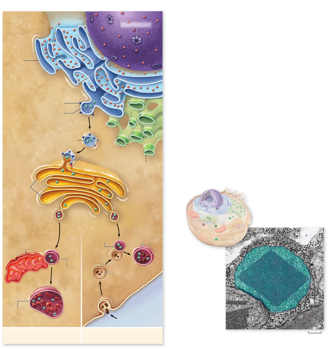
Nucleus
Nuclear pore
Rough
endoplasmic
reticulum
Smooth
endoplasmic
reticulum
Ribosome
Membrane protein
Golgi membrane protein
Hydrolytic enzyme
Transport vesicle
cis face
trans face
Cisternae
Golgi
Apparatus
Old or damaged
organelle
Lysosome
Breakdown
of organelle
Phagocytosis
Food vesicle
Lysosome
Digestion
0.21 μm
Figure 4.14
A peroxisome. Peroxisomes are spherical
organelles that may contain a large crystal structure composed of
protein. Peroxisomes contain digestive and detoxifying enzymes
that produce hydrogen peroxide as a by-product. A peroxisome has
been colored green in the electron micrograph.
Figure 4.13
Lysosomes. Lysosomes are formed from vesicles
budding off the Golgi. They contain hydrolytic enzymes that digest
particles or cells taken into the cell by phagocytosis, and break
down old organelles.
the cell has engulfed by phagocytosis. When a white blood
cell, for example, phagocytizes a passing pathogen, lyso-
somes fuse with the resulting “food vesicle,” releasing their
enzymes into the vesicle and degrading the material within
(figure 4.13).
Microbodies are a diverse
category of organelles
Eukaryotic cells contain a variety of enzyme-bearing, membrane-
enclosed vesicles called microbodies. These are found in the
cells of plants, animals, fungi, and protists. The distribution of
enzymes into microbodies is one of the principal ways eukaryotic
cells organize their metabolism.
Peroxisomes: Peroxide utilization
An important type of microbody is the peroxisome (figure
4.14), which contains enzymes involved in the oxidation of
fatty acids. If these oxidative enzymes were not isolated within
microbodies, they would tend to short-circuit the metabolism
of the cytoplasm, which often involves adding hydrogen at-
oms to oxygen. Because many peroxisomal proteins are syn-
thesized by cytoplasmic ribosomes, the organelles themselves
were long thought to form by the addition of lipids and pro-
teins, leading to growth. As they grow larger, they divide to
produce new peroxisomes. Although division of peroxisomes
still appears to occur, it is now clear that peroxisomes can
form from the fusion of ER-derived vesicles. These vesicles
then import peroxisomal proteins to form a mature peroxi-
some. Genetic screens have isolated some 32 genes that en-
code proteins involved in biogenesis and maintenance of
peroxisomes. The human genetic diseases called peroxisome
biogenesis disorders (PBDs) appear to be caused by mutations
in some of these genes.
Peroxisomes get their name from the hydrogen peroxide
produced as a by-product of the activities of oxidative enzymes.
Hydrogen peroxide is dangerous to cells because of its violent
72
part
II
Biology of the Cell
rav32223_ch04_059-087.indd 72rav32223_ch04_059-087.indd 72 11/6/09 12:23:51 PM11/6/09 12:23:51 PM
Apago PDF Enhancer
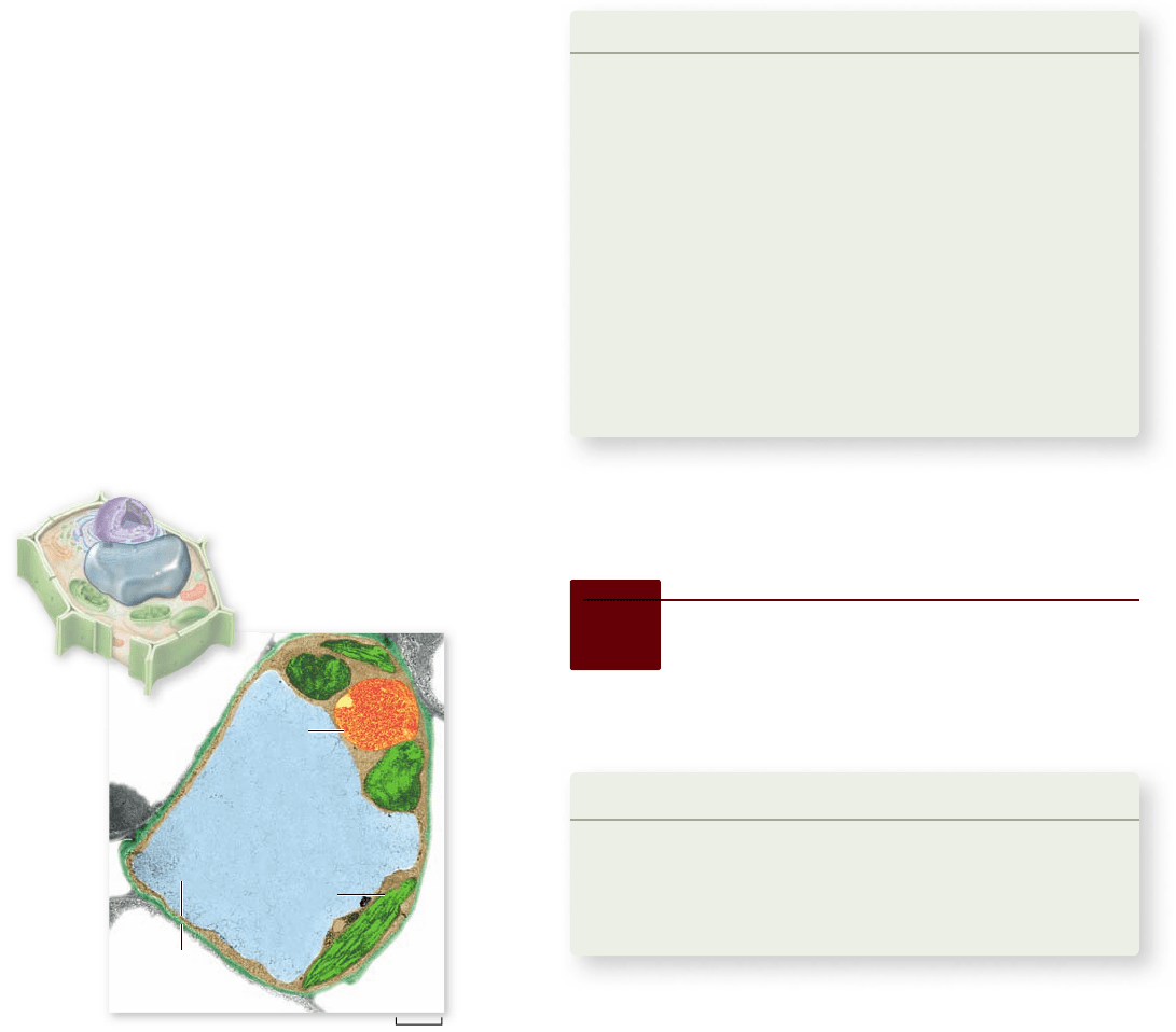
0.9 μm
Cell
wall
Central
vacuole
Nucleus
Chloroplast
Tonoplast
Figure 4.15
The central vacuole. A plant’s central vacuole
stores dissolved substances and can expand in size to increase the
tonicity of a plant cell. Micrograph shown with false color.
chemical reactivity. However, peroxisomes also contain the en-
zyme catalase, which breaks down hydrogen peroxide into its
harmless constituents—water and oxygen.
Plants use vacuoles for storage
and water balance
Plant cells have specialized membrane-bounded structures
called vacuoles. The most conspicuous example is the large
central vacuole seen in most plant cells (figure 4.15). In fact,
vacuole actually means blank space, referring to its appearance
in the light microscope. The membrane surrounding this vacu-
ole is called the tonoplast because it contains channels for wa-
ter that are used to help the cell maintain its tonicity, or osmotic
balance (see osmosis in chapter 5 ).
For many years biologists assumed that only one type of
vacuole existed and that it served multiple functions. The func-
tions assigned to this vacuole included water balance and stor-
age of both useful molecules (such as sugars, ions and pigments)
and waste products. The vacuole was also thought to store en-
zymes involved in the breakdown of macromolecules and those
used in detoxifying foreign substances. Old textbooks of plant
physiology referred to vacuoles as the attic of the cell for the
variety of substances thought to be stored there.
Studies of tonoplast transporters and the isolation of vac-
uoles from a variety of cell types have led to a more complex
view of vacuoles. These studies have made it clear that different
vacuolar types can be found in different cells. These vacuoles
are specialized, depending on the function of the cell.
The central vacuole is clearly important for a number
of roles in all plant cells. The central vacuole and the water
channels of the tonoplast maintain the tonicity of the cell,
allowing the cell to expand and contract depending on con-
ditions. The central vacuole is also involved in cell growth
by occupying most of the volume of the cell. Plant cells grow
by expanding the vacuole, rather than by increasing cyto-
plasmic volume.
Vacuoles with a variety of functions are also found in
some types of fungi and protists. One form is the contrac-
tile vacuole, found in some protists, which can pump water
and is used to maintain water balance in the cell. Other vac-
uoles are used for storage or to segregate toxic materials
from the rest of the cytoplasm. The number and kind of
vacuoles found in a cell depends on the needs of the par-
ticular cell type.
Learning Outcomes Review 4.4
The endoplasmic reticulum (ER) is an extensive system of folded
membranes that spatially organize the cell’s biosynthetic activities.
Smooth ER (SER) is the site of lipid and membrane synthesis and is used
to store Ca
2+
. Rough ER (RER) is covered with ribosomes and is a site of
protein synthesis. Proteins from the RER are transported by vesicles to
the Golgi apparatus where they are modified, packaged, and distributed
to their final location. Lysosomes are vesicles that contain digestive
enzymes used to degrade materials such as invaders or worn-out
components. Peroxisomes carry out oxidative metabolism that generates
peroxides. Vacuoles are membrane-bounded structures that have roles
ranging from storage to cell growth in plants. They are also found in
some fungi and protists.
■ How do ribosomes on the RER differ from cytoplasmic
ribosomes ?
4.5
Mitochondria
and Chloroplasts:
Cellular Generators
Learning Outcomes
Describe the structure of mitochondria and chloroplasts.1.
Compare the function of mitochondria and chloroplasts.2.
Explain the probable origin of mitochondria 3.
and chloroplasts.
Mitochondria and chloroplasts share structural and functional
similarities. Structurally, they are both surrounded by a double
membrane, and both contain their own DNA and protein
synthesis machinery. Functionally, they are both involved in
energy metabolism, as we will explore in detail in later chapters
on energy metabolism and photosynthesis.
chapter
4
Cell Structure
73www.ravenbiology.com
rav32223_ch04_059-087.indd 73rav32223_ch04_059-087.indd 73 11/6/09 12:23:56 PM11/6/09 12:23:56 PM
Apago PDF Enhancer
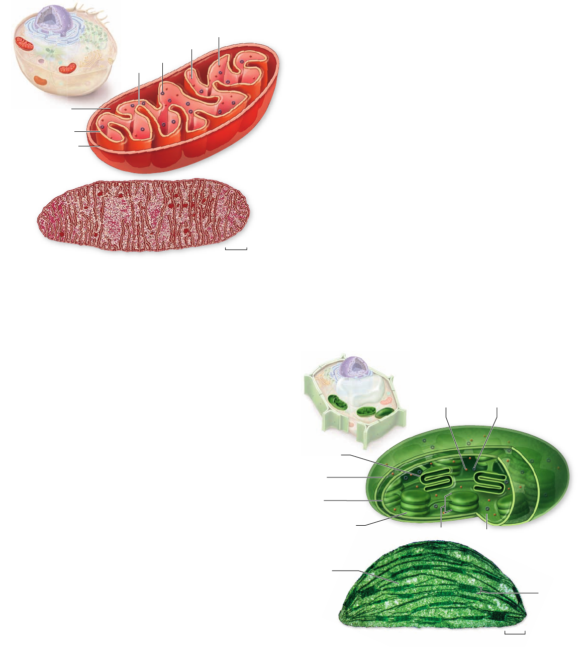
Intermembrane
space
Inner membrane
Outer membrane
Matrix
Crista
0.2 μm
Ribosome
DNA
Outer
membrane
Ribosome DNA
Inner
membrane
Granum
Thylakoid disk
Stroma
Stroma
Granum
1.5 μm
Thylakoid
membrane
Figure 4.16
Mitochondria. The inner membrane of a
mitochondrion is shaped into folds called cristae that greatly increase
the surface area for oxidative metabolism. A mitochondrion in cross
section and cut lengthwise is shown colored red in the micrograph.
Figure 4.17
Chloroplast structure. The inner membrane of
a chloroplast surrounds a membrane system of stacks of closed
chlorophyll-containing vesicles called thylakoids, within which
photosynthesis occurs. Thylakoids are typically stacked one on top
of the other in columns called grana. The chloroplast has been
colored green in the micrograph.
Mitochondria metabolize sugar
to generate ATP
Mitochondria (singular, mitochondrion) are typically tubular or
sausage-shaped organelles about the size of bacteria that are
found in all types of eukaryotic cells (figure 4.16). Mitochon-
dria are bounded by two membranes: a smooth outer mem-
brane, and an inner folded membrane with numerous contiguous
layers called cristae (singular, crista).
The cristae partition the mitochondrion into two compart-
ments: a matrix, lying inside the inner membrane; and an outer
compartment, or intermembrane space, lying between the two
mitochondrial membranes. On the surface of the inner mem-
brane, and also embedded within it, are proteins that carry out
oxidative metabolism, the oxygen-requiring process by which en-
ergy in macromolecules is used to produce ATP (chapter 7 ) .
Mitochondria have their own DNA; this DNA contains
several genes that produce proteins essential to the mitochon-
drion’s role in oxidative metabolism. Thus, the mitochondrion,
in many respects, acts as a cell within a cell, containing its own
genetic information specifying proteins for its unique functions.
The mitochondria are not fully autonomous, however, because
most of the genes that encode the enzymes used in oxidative me-
tabolism are located in the cell nucleus.
A eukaryotic cell does not produce brand-new mitochon-
dria each time the cell divides. Instead, the mitochondria them-
selves divide in two, doubling in number, and these are partitioned
between the new cells. Most of the components required for mi-
tochondrial division are encoded by genes in the nucleus and are
translated into proteins by cytoplasmic ribosomes. Mitochondrial
replication is, therefore, impossible without nuclear participation,
and mitochondria thus cannot be grown in a cell-free culture.
Chloroplasts use light to generate
ATP and sugars
Plant cells and cells of other eukaryotic organisms that carry
out photosynthesis typically contain from one to several hun-
dred chloroplasts. Chloroplasts bestow an obvious advantage
on the organisms that possess them: They can manufacture
their own food. Chloroplasts contain the photosynthetic pig-
ment chlorophyll that gives most plants their green color.
The chloroplast, like the mitochondrion, is surrounded by
two membranes (figure 4.17). However, chloroplasts are larger and
more complex than mitochondria. In addition to the outer and in-
ner membranes, which lie in close association with each other, chlo-
roplasts have closed compartments of stacked membranes called
grana (singular, granum), which lie inside the inner membrane.
A chloroplast may contain a hundred or more grana, and
each granum may contain from a few to several dozen disk-
shaped structures called thylakoids. On the surface of the thy-
lakoids are the light-capturing photosynthetic pigments, to be
discussed in depth in chapter 8 . Surrounding the thylakoid is a
fluid matrix called the stroma. The enzymes used to synthesize
glucose during photosynthesis are found in the stroma.
Like mitochondria, chloroplasts contain DNA, but many
of the genes that specify chloroplast components are also located
in the nucleus. Some of the elements used in photosynthesis, in-
cluding the specific protein components necessary to accomplish
the reaction, are synthesized entirely within the chloroplast.
74
part
II
Biology of the Cell
rav32223_ch04_059-087.indd 74rav32223_ch04_059-087.indd 74 11/6/09 12:23:57 PM11/6/09 12:23:57 PM
Apago PDF Enhancer
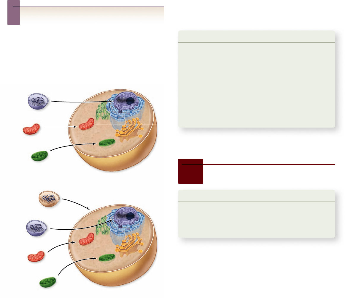
Nucleus
Nucleus
Mitochondrion
Chloroplast
Mitochondr
ion
Chloroplast
Modern Eukaryote
Modern Eukaryote
Unknown Bacterium
Unknown Archaeon
Protobacterium
Cyanobacterium
Unknown Archaeon
Protobacterium
Cyanobacterium
Figure 4.18
Possible origins of eukaryotic cells. Both
mitochondria and chloroplasts are thought to have arisen by
endosymbiosis where a free-living cell is taken up but not digested.
The nature of the engul ng cell is unknown. Two possibilities are
The engul ng cell (top) is an archaeon that gave rise to the nuclear
genome and cytoplasmic contents. The engul ng cell (bottom)
consists of a nucleus derived from an archaeon in a bacterial cell.
This could arise by a fusion event or by engulfment of the archaeon
by the bacterium.
Other DNA-containing organelles in plants, called leuco-
plasts, lack pigment and a complex internal structure. In root
cells and some other plant cells, leucoplasts may serve as starch
storage sites. A leucoplast that stores starch (amylose) is some-
times termed an amyloplast. These organelles—chloroplasts,
leucoplasts, and amyloplasts—are collectively called plastids.
All plastids are produced by the division of existing plastids.
Inquiry question
?
Mitochondria and chloroplasts both generate ATP. What
structural features do they share?
Mitochondria and chloroplasts
arose by endosymbiosis
Symbiosis is a close relationship between organisms of differ-
ent species that live together. As noted in chapter 29 , the theory
of endosymbiosis proposes that some of today’s eukaryotic or-
ganelles evolved by a symbiosis arising between two cells that
were each free-living. One cell, a prokaryote, was engulfed by
and became part of another cell, which was the precursor of
modern eukaryotes (figure 4.18).
According to the endosymbiont theory, the engulfed
prokaryotes provided their hosts with certain advantages associ-
ated with their special metabolic abilities. Two key eukaryotic
organelles are believed to be the descendants of these endosym-
biotic prokaryotes: mitochondria, which are thought to have
originated as bacteria capable of carrying out oxidative metabo-
lism, and chloroplasts, which apparently arose from photosyn-
thetic bacteria. This is discussed in detail in chapter 29 .
Learning Outcomes Review 4.5
Mitochondria and chloroplasts have similar structures, with an outer
membrane and an extensive inner membrane compartment. Both
mitochondria and chloroplasts have their own DNA, but both also depend on
nuclear genes for some functions. Mitochondria and chloroplasts are both
involved in energy conversion: Mitochondria metabolize sugar to produce
ATP, whereas chloroplasts harness light energy to produce ATP and synthesize
sugars. Endosymbiosis theory proposes that both mitochondria and
chloroplasts arose as prokaryotic cells engulfed by a eukaryotic precursor.
■ Many proteins in mitochondria and chloroplasts
are encoded by nuclear genes. In light of the
endosymbiont hypothesis, how might this come about?
4.6
The Cytoskeleton
Learning Outcomes
Contrast the structure and function of different fibers in 1.
the cytoskeleton.
Illustrate the role of microtubules in intracellular 2.
transport.
The cytoplasm of all eukaryotic cells is crisscrossed by a network
of protein fibers that supports the shape of the cell and anchors
organelles to fixed locations. This network, called the cytoskele-
ton, is a dynamic system, constantly assembling and disassem-
bling. Individual fibers consist of polymers of identical protein
subunits that attract one another and spontaneously assemble
into long chains. Fibers disassemble in the same way, as one sub-
unit after another breaks away from one end of the chain.
Three types of bers compose
the cytoskeleton
Eukaryotic cells may contain the following three types of
cytoskeletal fibers, each formed from a different kind of sub-
unit: (1) actin filaments, sometimes called microfilaments,
(2) microtubules, and (3) intermediate filaments.
chapter
4
Cell Structure
75www.ravenbiology.com
rav32223_ch04_059-087.indd 75rav32223_ch04_059-087.indd 75 11/6/09 12:24:01 PM11/6/09 12:24:01 PM
Apago PDF Enhancer
Microtubule
Intermediate filament
Actin filament
Cell membrane
a. Actin filaments
b. Microtubules
c. Intermediate filament
Figure 4.19
Molecules that make up the cytoskeleton.
a. Actin laments: Actin laments, also called micro laments, are
made of two strands of the globular protein actin twisted together.
They are often found in bundles or in a branching network. Actin
laments in many cells are concentrated below the plasma
membrane in bundles known as stress bers, which may have a
contractile function. b. Microtubules: Microtubules are composed of
α- and β-tubulin protein subunits arranged side by side to form a
tube. Microtubules are comparatively stiff cytoskeletal elements and
have many functions in the cell including intracellular transport
and the separation of chromosomes during mitosis. c. Intermediate
laments: Intermediate laments are composed of overlapping
staggered tetramers of protein. These tetramers are then bundled
into cables. This molecular arrangement allows for a ropelike
structure that imparts tremendous mechanical strength to the cell.
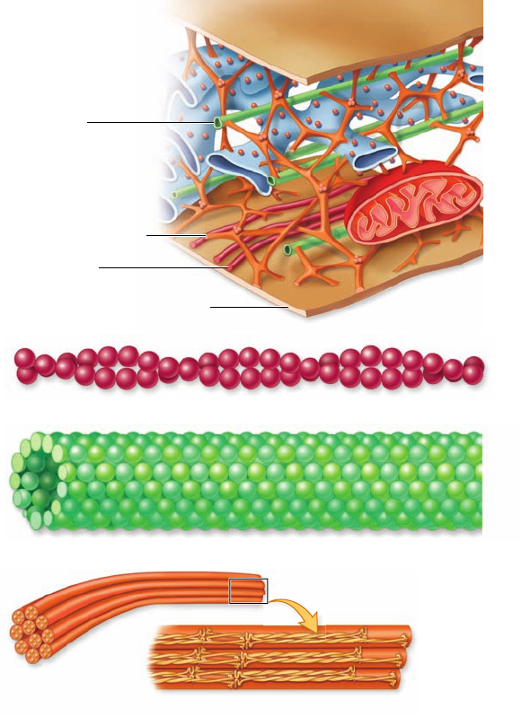
Actin filaments (microfilaments)
Actin filaments are long fibers about 7 nm in diameter. Each
filament is composed of two protein chains loosely twined to-
gether like two strands of pearls (figure 4.19). Each “pearl,” or
subunit, on the chain is the globular protein actin. Actin fila-
ments exhibit polarity, that is, they have plus (+) and minus (–)
ends. These designate the direction of growth of the filaments.
Actin molecules spontaneously form these filaments, even in a
test tube.
Cells regulate the rate of actin polymerization through
other proteins that act as switches, turning on polymerization
when appropriate. Actin filaments are responsible for cellular
movements such as contraction, crawling, “pinching” during
division, and formation of cellular extensions.
Microtubule
Microtubules, the largest of the cytoskeletal elements, are hol-
low tubes about 25 nm in diameter, each composed of a ring of
13 protein protofilaments (see figure 4.19). Globular proteins
consisting of dimers of α- and β-tubulin subunits polymerize to
form the 13 protofilaments. The protofilaments are arrayed
side by side around a central core, giving the microtubule its
characteristic tube shape.
In many cells, microtubules form from nucleation centers
near the center of the cell and radiate toward the periphery.
They are in a constant state of flux, continually polymerizing
and depolymerizing. The average half-life of a microtubule
ranges from as long as 10 minutes in a nondividing animal cell
to as short as 20 seconds in a dividing animal cell. The ends of
the microtubule are designated as plus (+) (away from the nu-
cleation center) or minus (–) (toward the nucleation center).
Along with facilitating cellular movement, microtubules
organize the cytoplasm and are responsible for moving materi-
als within the cell itself, as described shortly.
Intermediate filaments
The most durable element of the cytoskeleton in animal cells
is a system of tough, fibrous protein molecules twined together
in an overlapping arrangement (see figure 4.19). These
intermediate filaments are characteristically 8 to 10 nm in
diameter—between the size of actin filaments and microtu-
bules. Once formed, intermediate filaments are stable and usu-
ally do not break down.
Intermediate filaments constitute a mixed group of cy-
toskeletal fibers. The most common type, composed of protein
subunits called vimentin, provides structural stability for many
kinds of cells. Keratin, another class of intermediate filament, is
found in epithelial cells (cells that line organs and body cavities)
and associated structures such as hair and fingernails. The inter-
mediate filaments of nerve cells are called neurofilaments.
Centrosomes are microtubule-
organizing centers
Centrioles are barrel-shaped organelles found in the cells of
animals and most protists. They occur in pairs, usually located
at right angles to each other near the nuclear membranes
(figure 4.20). The region surrounding the pair in almost all ani-
mal cells is referred to as a centrosome. Surrounding the centri-
oles in the centrosome is the pericentriolar material, which
contains ring-shaped structures composed of tubulin. The peri-
centriolar material can nucleate the assembly of microtubules
in animal cells. Structures with this function are called
microtubule-organizing centers. The centrosome is also respon-
sible for the reorganization of microtubules that occurs during
76
part
II
Biology of the Cell
rav32223_ch04_059-087.indd 76rav32223_ch04_059-087.indd 76 11/6/09 12:24:02 PM11/6/09 12:24:02 PM
Apago PDF Enhancer
Microtubule triplet
Vesicle
Dynactin
complex
Microtubule
Dynein
Hypothesis: Kinesin molecules can act as molecular motors and move
along microtubules using energy from ATP.
Test: A microscope slide is covered with puried kinesin. Puried microtubules
are added in a buer containing ATP. The microtubules are monitored under
a microscope using a video recorder to capture any movement.
Result: Over time, the movement of individual microtubules can be
observed in the microscope. This is shown schematically in the gure by
the movement of specic microtubules shown in color.
Conclusion: Kinesin acts as a molecular motor moving along (in this
case actually moving) microtubules.
Further Experiments: Are there any further controls that are not shown
in this experiment? What additional conclusions could be drawn by
varying the amount of kinesin sticking to the slide?
SCI ENT I FI C THINKING
Frame 1 Frame 2 Frame 3
Figure 4.20
Centrioles. Each centriole is composed of nine
triplets of microtubules. Centrioles are usually not found in plant
cells. In animal cells they help to organize microtubules.
Figure 4.21
Molecular motors.
Vesicles can be
transported along
microtubules using motor
proteins that use ATP to
generate force. The
vesicles are attached to
motor proteins by
connector molecules,
such as the dynactin
complex shown here.
The motor protein
dynein moves the
connected vesicle
along microtubules.
Figure 4.22
Demonstration of kinesin as molecular
motor. Microtubules can be observed moving over a slide coated
with kinesin.
cell division. The centrosomes of plants and fungi lack centri-
oles, but still contain microtubule-organizing centers. You will
learn more about the actions of the centrosomes when we de-
scribe the process of cell division in chapter 10 .
The cytoskeleton helps move
materials within cells
Actin filaments and microtubules often orchestrate their activi-
ties to affect cellular processes. For example, during cell repro-
duction (see chapter 10 ) , newly replicated chromosomes move
to opposite sides of a dividing cell because they are attached to
shortening microtubules. Then, in animal cells, a belt of actin
pinches the cell in two by contracting like a purse string.
Muscle cells also use actin filaments, which slide along
filaments of the motor protein myosin when a muscle contracts .
The fluttering of an eyelash, the flight of an eagle, and the awk-
ward crawling of a baby all depend on these cytoskeletal move-
ments within muscle cells.
Not only is the cytoskeleton responsible for the cell’s shape
and movement, but it also provides a scaffold that holds certain
enzymes and other macromolecules in defined areas of the cyto-
plasm. For example, many of the enzymes involved in cell metabo-
lism bind to actin filaments, as do ribosomes. By moving and
anchoring particular enzymes near one another, the cytoskeleton,
like the endoplasmic reticulum, helps organize the cell’s activities.
Molecular motors
All eukaryotic cells must move materials from one place to an-
other in the cytoplasm. One way cells do this is by using the
channels of the endoplasmic reticulum as an intracellular high-
way. Material can also be moved using vesicles loaded with
cargo that can move along the cytoskeleton like a railroad track.
For example, in a nerve cell with an axon that may extend far
from the cell body, vesicles can be moved along tracks of micro-
tubules from the cell body to the end of the axon.
Four components are required to move material along
microtubules: (1) a vesicle or organelle that is to be transported,
(2) a motor protein that provides the energy-driven motion,
(3) a connector molecule that connects the vesicle to the motor
molecule, and (4) microtubules on which the vesicle will ride
like a train on a rail (figure 4.21).
The direction a vesicle is moved depends on the type of
motor protein involved and the fact that microtubules are orga-
nized with their plus ends toward the periphery of the cell. In
one case, a protein called kinectin binds vesicles to the motor
protein kinesin. Kinesin uses ATP to power its movement to-
ward the cell periphery, dragging the vesicle with it as it travels
along the microtubule toward the plus end (figure 4.22). As na-
ture’s tiniest motors, these proteins pull the transport vesicles
along the microtubular tracks. Another set of vesicle proteins,
called the dynactin complex, binds vesicles to the motor protein
dynein (see figure 4.22), which directs movement in the opposite
chapter
4
Cell Structure
77www.ravenbiology.com
rav32223_ch04_059-087.indd 77rav32223_ch04_059-087.indd 77 11/6/09 12:24:04 PM11/6/09 12:24:04 PM
Apago PDF Enhancer
TABLE 4.2
Eukaryotic Cell Structures and their Functions
Structure Description Function
Plasma membrane Phospholipid bilayer with embedded proteins Regulates what passes into and out of cell; cell-to-cell
recognition; connection and adhesion; cell communication
Nucleus Structure (usually spherical) that contains chromosomes and is
surrounded by double membrane
Instructions for protein synthesis and cell reproduction;
contains genetic information
Chromosomes Long threads of DNA that form a complex with protein Contain hereditary information used to direct synthesis of
proteins
Nucleolus Site of genes for rRNA synthesis Synthesis of rRNA and ribosome assembly
Ribosomes Small, complex assemblies of protein and RNA, often bound
to ER
Sites of protein synthesis
Endoplasmic
reticulum (ER)
Network of internal membranes Intracellular compartment forms transport vesicles;
participates in lipid synthesis and synthesis of membrane or
secreted proteins
Golgi apparatus Stacks of attened vesicles Packages proteins for export from cell; forms secretory
vesicles
Lysosomes Vesicles derived from Golgi apparatus that contain hydrolytic
digestive enzymes
Digest worn-out organelles and cell debris; digest material
taken up by endocytosis
Microbodies Vesicles that are formed from incorporation of lipids and
proteins and that contain oxidative and other enzymes
Isolate particular chemical activities from rest of cell
Mitochondria Bacteria-like elements with double membrane “Power plants” of the cell; sites of oxidative metabolism
Chloroplasts Bacteria-like elements with double membrane surrounding
a third, thylakoid membrane containing chlorophyll, a
photosynthetic pigment
Sites of photosynthesis
Cytoskeleton Network of protein laments Structural support; cell movement; movement of vesicles
within cells
Flagella (cilia) Cellular extensions with 9 + 2 arrangement of pairs of
microtubles
Motility or moving uids over surfaces
Cell wall Outer layer of cellulose or chitin; or absent Protection; support
78
part
II
Biology of the Cell
rav32223_ch04_059-087.indd 78rav32223_ch04_059-087.indd 78 11/6/09 12:24:05 PM11/6/09 12:24:05 PM
Apago PDF Enhancer
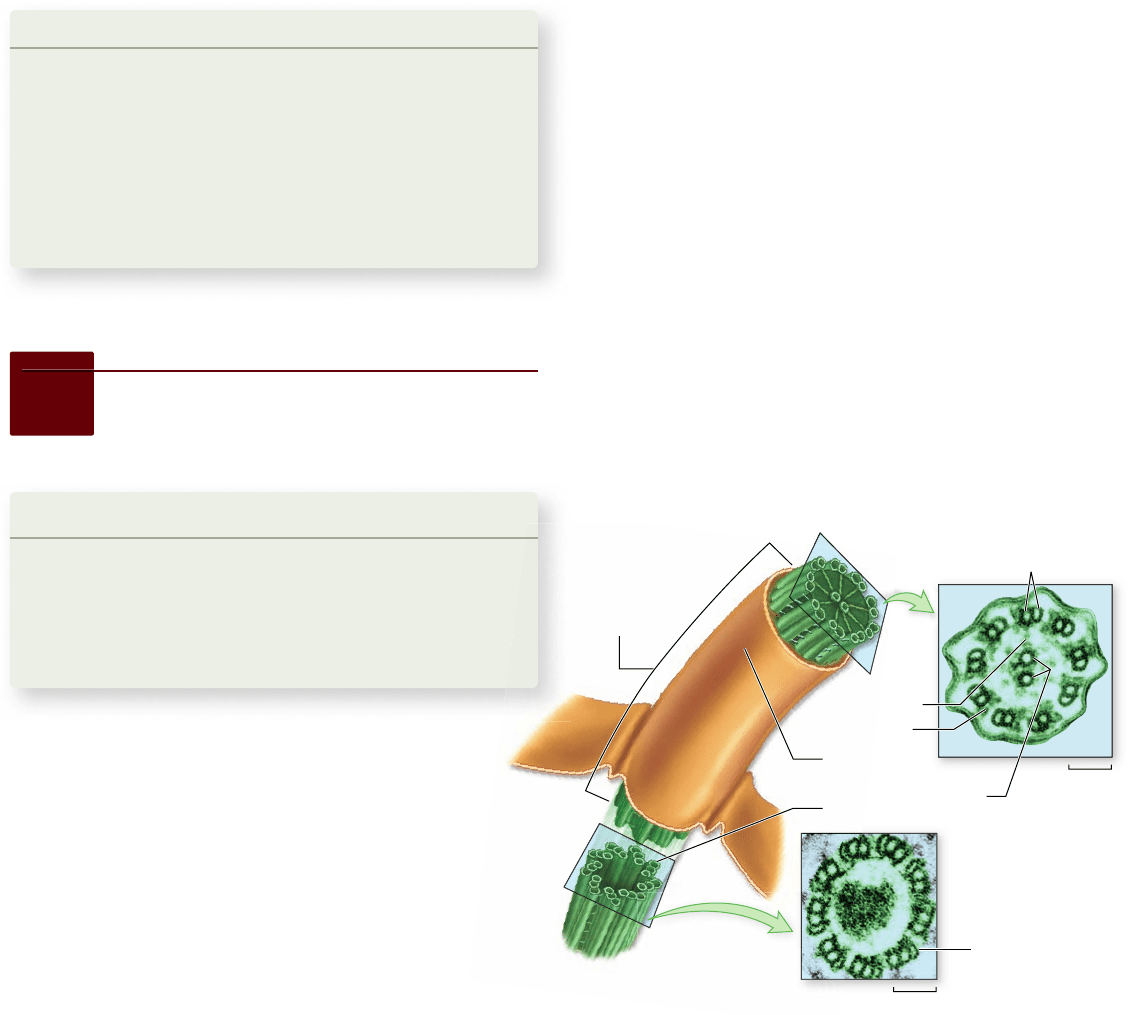
Outer microtubule pair
Microtubule
triplet
Flagellum
Plasma
membrane
Dynein arm
Radial spoke
Central
microtubule pair
Basal body
0.1 μm
0.1 μm
Figure 4.23
Flagella and cilia. A eukaryotic agellum
originates directly from a basal body. The agellum has two
microtubules in its core connected by radial spokes to an outer ring
of nine paired microtubules with dynein arms (9 + 2 structure). The
basal body consists of nine microtubule triplets connected by short
protein segments. The structure of cilia is similar to that of agella,
but cilia are usually shorter.
direction along microtubules toward the minus end, inward to-
ward the cell’s center. (Dynein is also involved in the movement
of eukaryotic flagella, as discussed later.) The destination of a
particular transport vesicle and its content is thus determined
by the nature of the linking protein embedded within the vesi-
cle’s membrane.
The major eukaryotic cell structures and their respective
functions are summarized in table 4.2.
Learning Outcomes Review 4.6
The three principal fi bers of the cytoskeleton are actin fi laments
(microfi laments), microtubules, and intermediate fi laments. These fi bers
interact to modulate cell shape and permit cell movement. They also act to
move materials within the cytoplasm. Material is also moved in large cells
using vesicles and molecular motors. The motor proteins move vesicles along
tracks of microtubules.
■ What advantage does the cytoskeleton give to large
eukaryotic cells?
4.7
Extracellular Structures
and Cell Movement
Learning Outcomes
Describe how cells move.1.
Identify the different cytoskeletal elements involved in 2.
cell movement.
Classify the elements of extracellular matrix in 3.
animal cells.
Essentially all cell motion is tied to the movement of actin
filaments, microtubules, or both. Intermediate filaments
act as intracellular tendons, preventing excessive
stretching of cells. Actin filaments play a major role in
determining the shape of cells. Because actin filaments can
form and dissolve so readily, they enable some cells to change
shape quickly.
Some cells crawl
The arrangement of actin filaments within the cell cytoplasm
allows cells to crawl, literally! Crawling is a significant cellular
phenomenon, essential to such diverse processes as inflamma-
tion, clotting, wound healing, and the spread of cancer. White
blood cells in particular exhibit this ability. Produced in the
bone marrow, these cells are released into the circulatory sys-
tem and then eventually crawl out of venules and into the tis-
sues to destroy potential pathogens.
At the leading edge of a crawling cell, actin filaments rap-
idly polymerize, and their extension forces the edge of the cell
forward. This extended region is stabilized when microtubules
polymerize into the newly formed region. Overall forward move-
ment of the cell is then achieved through the action of the pro-
tein myosin, which is best known for its role in muscle contraction.
Myosin motors along the actin filaments contract, pulling the
contents of the cell toward the newly extended front edge.
Cells crawl when these steps occur continuously, with a
leading edge extending and stabilizing, and then motors con-
tracting to pull the remaining cell contents along. Receptors on
the cell surface can detect molecules outside the cell and stimu-
late extension in specific directions, allowing cells to move to-
ward particular targets.
Flagella and cilia aid movement
Earlier in this chapter, we described the structure of prokary-
otic flagella. Eukaryotic cells have a completely different kind
of flagellum, consisting of a circle of nine microtubule pairs
surrounding two central microtubules. This arrangement is re-
ferred to as the 9 + 2 structure (figure 4.23).
As pairs of microtubules move past each other using arms
composed of the motor protein dynein, the eukaryotic flagel-
lum undulates, rather than rotates. When examined carefully,
each flagellum proves to be an outward projection of the cell’s
interior, containing cytoplasm and enclosed by the plasma
membrane. The microtubules of the flagellum are derived from
a basal body, situated just below the point where the flagellum
protrudes from the surface of the cell.
The flagellum’s complex microtubular apparatus evolved
early in the history of eukaryotes. Today the cells of many
chapter
4
Cell Structure
79www.ravenbiology.com
rav32223_ch04_059-087.indd 79rav32223_ch04_059-087.indd 79 11/6/09 12:24:06 PM11/6/09 12:24:06 PM
Apago PDF Enhancer
a.
b.
40 μm
66.6 μm
Plant cell
Plasmodesmata
Middle
lamella
Primary wall
Plasma
membrane
Secondary wall
Cell 1
Cell 2
Primary wall
Secondary wall
Plasma membrane
Middle lamella
0.4 μm
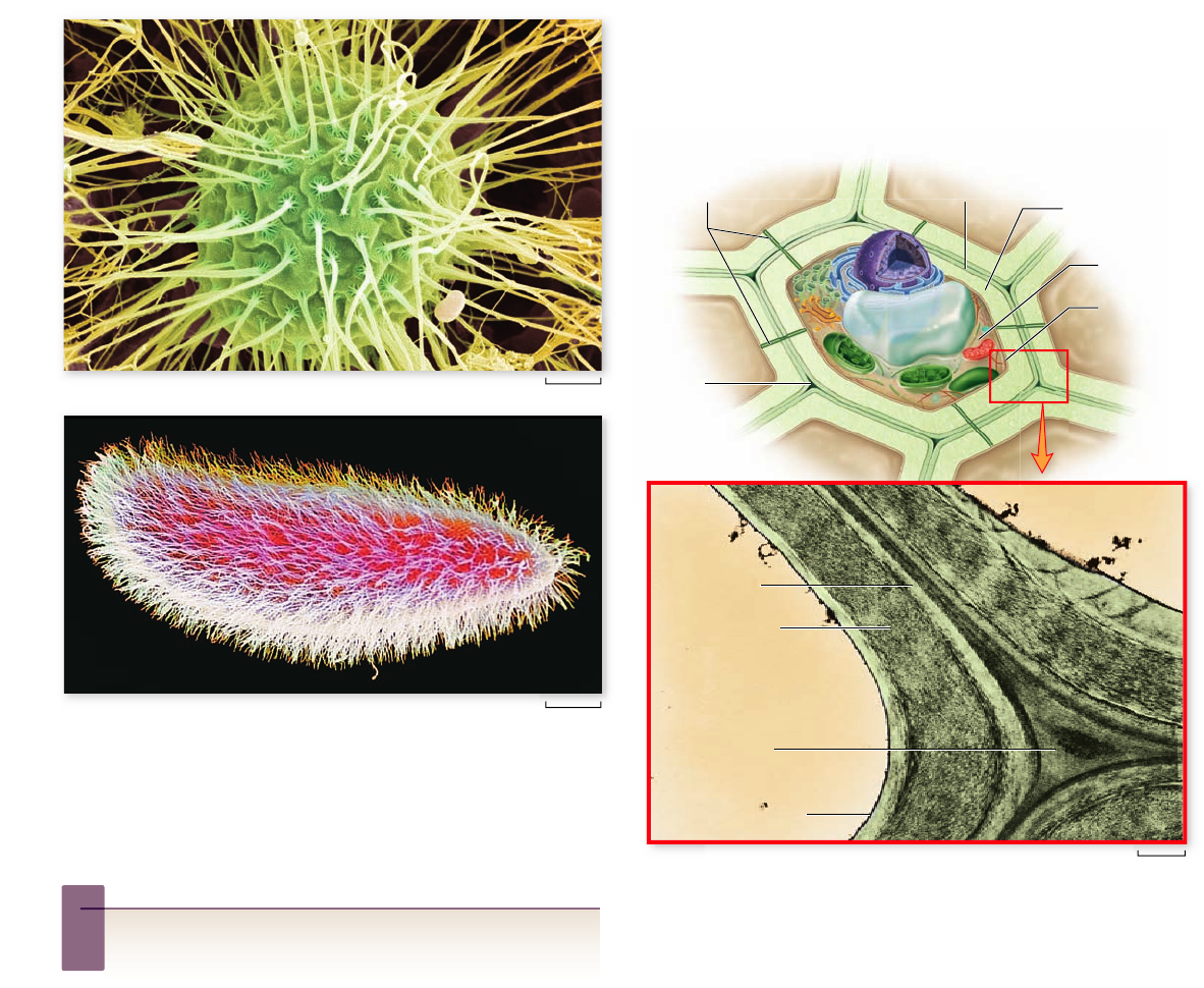
Figure 4.25
Cell walls in plants. Plant cell walls are thick,
strong, and rigid. Primary cell walls are laid down when the cell is
young. Thicker secondary cell walls may be added later when the
cell is fully grown.
Figure 4.24
Flagella and cilia. a. A green alga with
numerous agella that allow it to move through the water.
b. Paramecia are covered with many cilia, which beat in unison to
move the cell. The cilia can also be used to move uid into the
paramecium’s mouth to ingest material.
Inquiry question
?
The passageways of the human trachea (the path of airflow
into and out of the lungs) are known to be lined with ciliated
cells. What function could these cilia perform?
multicellular and some unicellular eukaryotes no long er pos-
sess flagella and are nonmotile. Other structures, called cilia
(singular, cilium), with an organization similar to the 9 + 2 ar-
rangement of microtubules can still be found within them. Cilia
are short cellular projections that are often organized in rows.
They are more numerous than flagella on the cell surface, but
have the same internal structure.
In many multicellular organisms, cilia carry out tasks far
removed from their original function of propelling cells
through water. In several kinds of vertebrate tissues, for ex-
ample, the beating of rows of cilia move water over the tissue
surface. The sensory cells of the vertebrate ear also contain
conventional cilia surrounded by actin-based stereocilia;
sound waves bend these structures and provide the initial sen-
sory input for hearing. Thus, the 9 + 2 structure of flagella and
cilia appears to be a fundamental component of eukaryotic
cells (figure 4.24).
Plant cell walls provide protection
and support
The cells of plants, fungi, and many types of protists have cell
walls, which protect and support the cells. The cell walls of
these eukaryotes are chemically and structurally different from
prokaryotic cell walls. In plants and protists, the cell walls are
composed of fibers of the polysaccharide cellulose, whereas in
fungi, the cell walls are composed of chitin.
In plants, primary walls are laid down when the cell is
still growing. Between the walls of adjacent cells a sticky sub-
stance, called the middle lamella, glues the cells together
(figure 4.25). Some plant cells produce strong secondary
walls, which are deposited inside the primary walls of fully
expanded cells.
Animal cells secrete an
extracellular matrix
Animal cells lack the cell walls that encase plants, fungi, and
most protists. Instead, animal cells secrete an elaborate mix-
ture of glycoproteins into the space around them, forming
80
part
II
Biology of the Cell
rav32223_ch04_059-087.indd 80rav32223_ch04_059-087.indd 80 11/6/09 12:24:10 PM11/6/09 12:24:10 PM
Apago PDF Enhancer
Cytoplasm
Actin filament
Integrin
Fibronectin
Collagen Elastin
Proteoglycan
Figure 4.26
The extracellular matrix. Animal cells are
surrounded by an extracellular matrix composed of various
glycoproteins that give the cells support, strength, and resilience.
TABLE 4.3
A Comparison of Prokaryotic, Animal, and Plant Cells
Prokaryote Animal Plant
EXTERIOR STRUCTURES
Cell wall Present (protein-polysaccharide) Absent Present (cellulose)
Cell membrane Present Present Present
Flagella/cilia Flagella may be present May be present (9 + 2 structure) Absent except in sperm of a few species
(9 + 2 structure)
INTERIOR STRUCTURES
Endoplasmic reticulum Absent Usually present Usually present
Ribosomes Present Present Present
Microtubules Absent Present Present
Centrioles Absent Present Absent
Golgi apparatus Absent Present Present
Nucleus Absent Present Present
Mitochondria Absent Present Present
Chloroplasts Absent Absent Present
Chromosomes Single; circle of DNA Multiple; DNA–protein complex Multiple; DNA–protein complex
Lysosomes Absent Usually present Present
Vacuoles Absent Absent or small Usually a large single vacuole
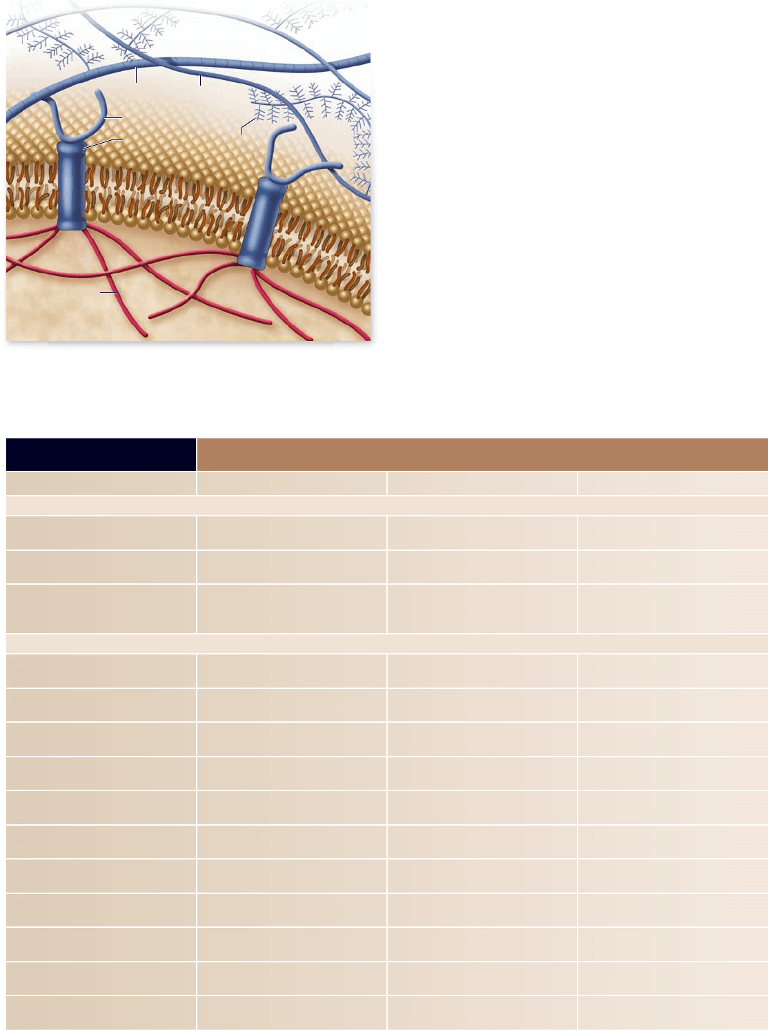
the extracellular matrix (ECM) (figure 4.26). The fibrous
protein collagen, the same protein found in cartilage, ten-
dons, and ligaments may be abundant in the ECM. Strong
fibers of collagen and another fibrous protein, elastin, are
embedded within a complex web of other glycoproteins,
called proteoglycans, that form a protective layer over the
cell surface.
The ECM of some cells is attached to the plasma mem-
brane by a third kind of glycoprotein, fibronectin. Fibronectin
molecules bind not only to ECM glycoproteins but also to pro-
teins called integrins. Integrins are an integral part of the plas-
ma membrane, extending into the cytoplasm, where they are
attached to the microfilaments and intermediate filaments of
the cytoskeleton. Linking ECM and cytoskeleton, integrins al-
low the ECM to influence cell behavior in important ways.
They can alter gene expression and cell migration patterns by a
combination of mechanical and chemical signaling pathways.
In this way, the ECM can help coordinate the behavior of all
the cells in a particular tissue.
Table 4.3 compares and reviews the features of three
types of cells.
chapter
4
Cell Structure
81www.ravenbiology.com
rav32223_ch04_059-087.indd 81rav32223_ch04_059-087.indd 81 11/6/09 12:24:15 PM11/6/09 12:24:15 PM